49(4) : 305∼ 311 (2018)
305
인간 유래 연골세포에서 꽃송이버섯 추출물의 염증성 매개인자 억제 효과
김은남·정길생*
계명대학교 약학대학
Sparassis crispa (Wulf.) Extract Inhibits IL-1β Stimulated Inflammatory Mediators Production on SW1353 Human Chondrocytes
Eun-Nam Kim and Gil-Saeng Jeong*
College of Pharmacy, Keimyung University, Daegu, Dalgubeol-daero 1095, (42601), Korea
Abstract − Osteoarthritis (OA) is the most common form of joint disease, characterized by articular cartilage, osteonecrosis, and osteochondral bone erosion. It is an early, progressive disease that combines joint stiffness and joint pain and reduces car- tilage function and condition. Interleukin-1 beta (IL-1β) is thought to be important to the pathogenesis of OA and significantly increases the expression of matrix metalloproteinases (MMPs), which play an important role in cartilage degradation in OA.
Sparassis crispa (Wulf.) is an edible / medicinal mushroom that has been reported to variety of biological activities. In this study, investigated the Anti-inflammatory effect of Sparassis crispa (Wulf.) ethanol extract (SCE) on IL-1β stimulated SW1353 chondrocytes. SCE decreased the expression and activity of MMPs by IL-1β and decreased the levels of cyclooxygenase-2 (COX-2) and inducible nitric oxide synthase (iNOS) associated with the inhibition of prostaglandin E2(PGE2) in IL-1β stim- ulated SW-1353 chondrocytes. In addition, SCE inhibits the expression of MAPK (mitogen-activated protein kinase) and NF- κB (nuclear factor-kappa B) signaling in IL-1β stimulated SW-1353 cells, and SCE inhibits the production of reactive oxygen species (ROS) through heme oxygenase-1 (HO-1) expression. Thus, it is suggested that SCE has a potential as an anti-inflam- matory agent in osteoarthritis treatments.
Keywords − Sparassis crispa (Wulf.), Interleukin-1 beta (IL-1β), Mitogen-activated protein kinase(MAPK), NF-κB (nuclear factor-kappa B), Heme oxygenase-1 (HO-1), Reactive oxygen species (ROS)
골 관절염은 관절 외상과 노화에 관련된 가장 흔한 관절 질환으로 연골 및 골 형성의 침식을 특징으로 하는 질병이 며, 이러한 골 관절염의 다양한 원인 중 염증은 골의 항상 성 감소와 세포 외 기질의 분해를 촉진하는 연골 대사 경로 의 불균형에 의해 진행되며,1)활성 산소종(reactive oxygen species, ROS)과 같은 산화 스트레스의 과잉 생산으로 신호 전달 경로의 변화 등을 원인으로 보고있다.2,3) IL-1β (Interleukin-1 beta)는 골 관절염에서 연골 분해에 중요한 역 할을 하는 MMPs(matrix metalloproteinases)의 발현을 유 의하게 증가시키며,4) 이러한 IL-1β는 연골세포에서 활성 산소종의 생성과 산화 질소(nitric oxide, NO) 및 PGE2 (prostaglandin E2), COX-2(cyclooxygenase-2)와 같은 염증 매개체를 방출하여 골 관절염의 발병에 중추적인 역할을 한
다고 보고되었다.5,6) 이러한 염증반응에서 HO-1(Heme oxygenase-1)은 일산화탄소, 철 및 biliverdin과 bilirubin의 형성을 유도하며, Nrf2(Nuclear factor erythroid-derived 2- related factor 2)에 의해 조절되는 heme catabolism의 속도 제한 효소로 산화 스트레스에 의한 염증성 질환에서 매우 중요한 역할을 하고 활성 산소종과 친염증성 사이토카인의 생성과 발현을 억제하는 것으로 보고되었다.7-9) 또한 염증반 응에서 ERK(extracellular signal regulated kinase), p38 MAP kinases, JNK(c-Jun N-terminal kinase)을 포함한 MAPK(mitogenactivated protein kinase)와 NF-κB(nuclear factor-kappa B)의 활성화는 MMPs 발현을 증가시켜 연골파 괴와 사이토카인 조절에 중요한 역할을 하며, IL-1β에 의해 유도 된 MMPs, iNOS 및 COX-2 생성은 MAPK와 NF-κB 의 활성화에 기인한다.10,11) 따라서 MAPK와 NF-κB의 활성 을 억제하고 활성 산소종과 염증매개체의 생성과 발현의 억 제가 염증성 질환의 치료에서 매우 중요한 역할을 한다.12)
*교신저자(E-mail):[email protected] (Tel): +82-53-580-6649
꽃송이버섯(Sparassis crispa (Wulf.))은 민주름버섯목 꽃송 이버섯과(Sparassidaceae)에 속하는 버섯으로,13,14) 이전의 연 구에서 꽃송이버섯의 에탄올 추출물이 MIA(monosodium iodoacetate)로 골관절염이 유발된 랫드에 미치는 영향을 평 가하였다. 따라서 본 연구에서는 IL-1β로 자극된 SW1353 연골세포에서 꽃송이버섯의 에탄올 추출물(SCE)이 골 관절 염의 병인에 중요한 역할을 하는 활성 산소종의 생성에 대 한 영향과 연골 분해효소 MMPs와 염증 매개 인자 iNOS 및 COX-2의 생성과 발현 그리고 MAPK와 NF-κB의 활성 화에 미치는 영향을 평가하였다.
재료 및 방법
실험 재료 − 본 실험에 사용된 꽃송이버섯은 (주)경신바 이오에서 제공 받았다. 꽃송이버섯의 동정은 덕성여자대학 교 심상희 교수가 하였으며, 표본은 계명대학교 약학대학 생약학연구실에 보관하였다. 꽃송이버섯의 에탄올 추출물을 얻기 위해 에탄올 4 l와 꽃송이버섯 1 kg을 혼합하여 70~80oC 에서 3회 환류 냉각 추출 후 감압농축하여 185 g의 에탄올 추출물을 얻었다.
시약 및 기기 − 본 실험에 사용된 recombinant Human IL-1β는 PeproTech EC Ltd.(London, UK)사의 제품을 사용 하였으며, ECL western blotting detection reagents와 PVDF membranes, BCA protein assay kit는 Thermo Scientific (Rockford, IL, USA)에서 구입하였다. PGE2 parameter assay kit와 MMP-13 ELISA kit는 R&D system사의 제품을 사용하였다. Dulbecco’s modified Eagle’s medium(DMEM) 과 fetal bovine serum(FBS)은 Welgene Inc. (Gyeongsan, Korea)사의 제품을 사용하였다. 2′, 7′-dichlorofluorescin diacetate와 NE-PER kit는 Thermo Scientific(Rockford, IL USA)사의 제품을 사용하였으며, 3-(4,5-Dimethylthiazol-2- yl)-2,5 diphenyltetrazoliumbromide(MTT)시약은 Amresco Inc.(OH, USA)사의 제품을 사용하였다. 또한 phospho-JNK, JNK, phospho-ERK, ERK, phospho-p38, p-38, phospho- AKT, AKT, HO-1, Nrf2, β-actin의 1차 항체와 각각의 2차 항체는 Cell Signaling Technology Inc.(Beverly, MA, USA) 사의 제품을 사용하였으며, MMP-9, NF-κB p65, COX-2, p-IkBα, IkBα, Lamin B의 1차 항체와 각각의 2차 항체는 Santa Cruz Biotechnology(Santa Cruz, CA, USA)사의 제품 을 사용하였다. Western blot 단백질 분석을 위해 LAS 4000 (GE Healthcare Life Science, Tokyo, Japan) 감광 검출기를 사용하였으며, Infinite m200 pro 흡광도 측정기(Tecan, Männedorf, Switzerland), 냉장 초고속 원심분리기(Labogene, Seoul, Korea), BX-51 편광현미경(Olympus, Tokyo, Japan) 을 사용하였다.
세포 배양 및 MTT 분석 − SW1353 연골 세포는 American
Type Culture Collection(ATCC, Rockville, MD, USA)에서 구입하였으며, 10% FBS, 100 units/ml 페니실린 및 100 mg/ml streptomycin이 첨가된 DMEM 배지로 5% CO2 환경 에서 37oC로 배양 하였다. 그 후 SW1353 세포를 96 well plate에 5×10³ cells/ml의 농도로 24 hr 배양 후 SCE 10, 20, 50, 100 μg/ml로 1 hr 동안 처리하고 IL-1β(40 ng/ml)를 24 hr 처리하였다. 세포 생존율은 540 nm의 파장에서 MTT 분석을 하였다.
Nitric Oxide (NO) 측정 − NO의 생성은 Griess reaction assay를 통해 측정하였다. SW1353 세포를 24 well plate에 3×10⁵cells/ml로 분주한 후, 24시간 뒤 SCE 10, 20, 50, 100 μg/ml로 1 hr 동안 처리하고 IL-1β(40 ng/mL)를 24 hr 동안 처리 후 배양 상층액을 수거하였다. 상층액과 standard (NaNO2)를 100 μl씩 96 well에 분주하고, Griess reagent를 100 μl씩 첨가한 후, 540 nm의 파장에서 흡광도를 측정하였 다.
PGE2, MM-9 및 MMP-13 분석 − SW1353 세포를 24 well plate에 3×10⁵cells/ml로 분주한 후, 24 hr 뒤 SCE 10, 20, 50, 100 μg/ml로 1 hr 동안 처리하고 IL-1β(40 ng/ml)를 24 hr 처리 후, 배양 상층액을 수거하여 상층액 중의 PGE2 및 MMP-13 수준을 PGE2 parameter assay kit(R&D Systems, Inc. USA)와 MMP-13 ELISA(R&D Systems, Inc. USA) 키트를 사용하여 제조사의 지시에 따라 정량화 하였으며, Western blot analysis를 통해 MMP-9의 단백질 발현을 측정하였다.
Western Blot Analysis − SW1353 세포를 RIPA 완충액 에 용해시키고 14,000 rpm에서 30 min 원심 분리하였다. 단 백질 농도는 제조자의 키트(Bio-Rad, USA)를 사용하여 Brad-ford 방법으로 정량화하였다. 그 후 sodium dodecyl sulfate polyacrylamide gel electrophoresis(SDS-PAGE)로 분 리 하였다. 전기 영동 후 단백질을 PVDF 막을 사용하여 옮 겼다. Skim milk 5%를 함유 한 TBS-T 완충액으로 blocking 한 후, Membrane에 각각의 1차 항체를 12 hr 처리한 다음 PBS-T로 5분마다 5회 세척하고, 2차 항체를 사용하여 상온 에서 2 hr 반응시킨 후, PBS-T로 5분마다 5회 세척하였다.
그 후 ECL 용액을 처리하고 LAS 4000(GE Healthcare Life Science, Tokyo, Japan)에서 감광시켜 단백질 발현을 확인하 였다.
세포질 및 핵 단백질 추출 − SW1353세포를 1×10⁶ cells/
ml의 밀도로 60 mm 세포 배양 접시에 24hr 배양 후 SCE 10, 20, 50, 100 μg/ml의 농도로 1 hr 처리 후 Nrf2 검출을 위한 세포질 및 핵의 단백질 추출을 위해 Thermo Scientific.
(Rockford, IL USA)사의 NE-PER kit 제품을 제조사의 지 시 따라 사용하여 분리하였다.
활성 산소종 형성 검출 − SW1353 세포를 24 well plate 에 3×10⁵cells/ml로 분주한 후 24 hr 동안 배양시켰다. SCE
10, 20, 50, 100 μg/ml의 농도로 6 hr 처리한 후 SnPP(20 μM) 를 2 hr 처리 후 IL-1β(40 ng/ml)를 24 hr 하였다. 그 후 세 포를 4% 파라 포름알데히드(pH 7.4)로 20 분간 고정시키고 PBS로 세척 후 차광하여 2', 7'-dichlorofluorescein diacetate (DCF-DA) 시약과 함께 30분간 염색하여 micro plate reader (485 nm)의 파장에서 검출 하였다.
통계분석 − 모든 실험은 평균과 표준편차로 표시하였으 며, SPSS(Version 22.0, IBM, Armonk, NY, USA)를 사용 하여 one-way analysis of variance(ANOVA) test를 실시한 후 least-significant differences(LSD) test로 측정하였으며, 유 의수준 p-value<0.05에서 검증하였다.
결과 및 고찰
염증반응은 외부로부터의 무생물 또는 생물의 침입이나 조직손상에 대한 기능을 회복시키는 체내의 방어기전으로 류마티스 관절염 등을 포함한 여러 질환에서 염증반응에 관 여하는 물질을 찾아 제거하려는 많은 연구가 진행되고 있 다.15) 따라서 본 연구에서는 IL-1β에 의해 자극된 SW1353 연골세포에서 꽃송이버섯 에탄올 추출물이 골 관절염에 원
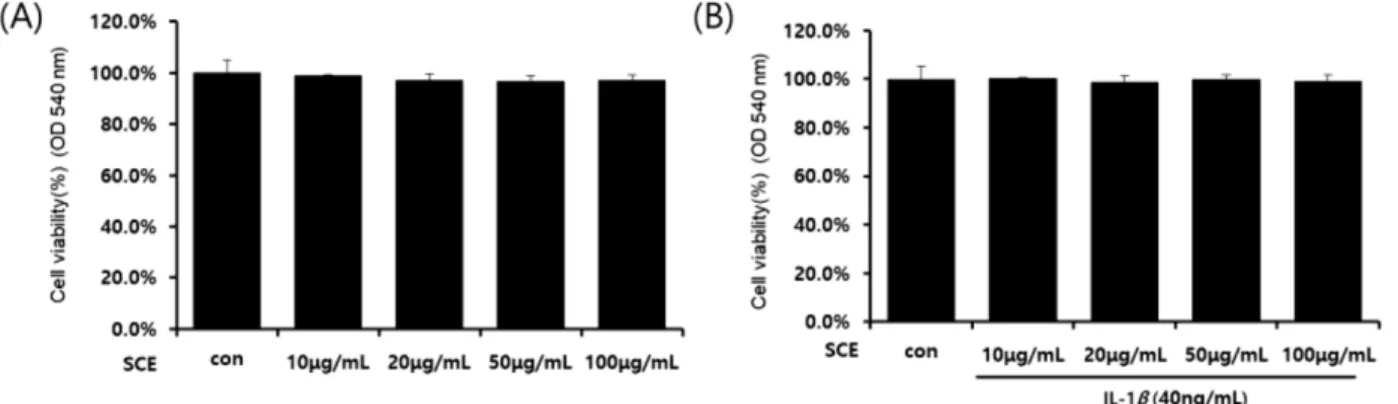
인이 되는 여러 염증성 매개인자의 생성과 발현에 대하여 연구하였다. 먼저 SW1353 연골세포에서 SCE 단독처리(Fig.
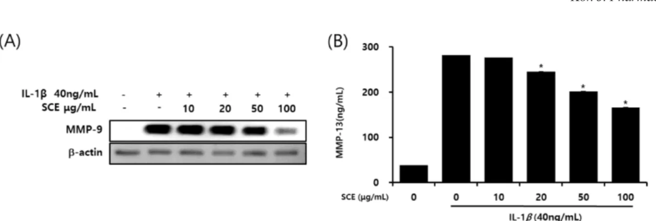
1A), IL-1β(40 ng/ml)와 SCE(10, 20, 50, 100 μg/ml)를 병용 처리 시(Fig. 1B) 세포독성에는 영향이 없는 것으로 확인되 었다. 따라서 이후의 실험은 IL-1β(40 ng/ml)와 SCE(10, 20, 50, 100 μg/ml)의 농도로 진행하였다. 골 관절염에서 IL-1β 가 매개하는 NO의 과잉 생산은 연골세포와 활막세포의 사 멸을 유도하여 골 관절염의 병인에 중요한 역할을 하는 염 증 매개체로 보고되었다.16) 또한 IL-1β는 COX-2의 발현을 자극하여 PGE2의 생성과 합성을 증가시키고 골 관절염에서 골 흡수와 관절 통증에 관여한다.17) IL-1β로 자극된 SW1353 연골세포에 SCE를 처리한 결과 COX-2, iNOS(Fig. 2A)와 PGE2, NO(Fig. 2B)의 생성과 발현을 농도 의존적으로 억제 였다. NO 및 PGE2의 생성은 연골 분해 효소 MMPs와 여 러 염증성 사이토카인의 생성을 유발할 수 있다.18)따라서 IL-1β로 염증반응이 유도된 SW1353 연골세포에서SCE의 영향을 평가하였으며, 그 결과 SCE는 연골 분해 효소 MMP- 9과 MMP-13의 생성을 농도 의존적으로 억제하였다. 또한 NF-kB는 SW1353 연골세포에서 IL-1β 신호 전달의 중요한 전사인자이며 염증매개체와 MMPs의 전사에 영향이 있다
Fig. 1. Effects of SCE and IL-1β on SW1353 cell viability. Cells were incubated with the indicated concentrations of SCE for 24hr (A), the indicated concentrations of SCE before 1h incubated, and IL-1β for 24hr (B).
Fig. 2. Effects of SCE on COX-2 and iNOS (A) expression and COX-2-derived PGE2 (B) and iNOS-derived NO (C) production in SW-1353 cells. Cells were pretreated with different concentrations of SCE (10, 20, 50 and 100 µg/mL) for 1hr before incubation with IL-1β (40 ng/mL) for 24hr. Western blot analyses for COX-2 and iNOS expression were performed. The nitrite content was measured using the griess reaction. The PGE2 concentration was measured in culture media using a commercial ELISA kit. *p- value<0.05 compared to the group treated with IL-1β.
고 보고되었다.19) 따라서 SCE의 MMPs, iNOS 및 COX-2 억제 효과가 NF-κB 신호의 억제에 의해 매개되는지 여부 를 확인하기 위해 NF-κB의 활성화를 평가하였으며, SCE는 NF-κB p65(Fig. 4A)와 인산화된 IkB-α(Fig. 4B)의 발현을 농도 의존적으로 억제하였다. PI3K/Akt와 MAPKs는 전 염 증성 사이토카인의 생성과 하류 신호 전달을 조절하여 이 화 작용에 의한 관절 파괴를 일으키며, MAPKs 신호 전달 경로에서 ERK와 JNK의 활성화는 MMPs, iNOS 및 COX- 2의 생성과 발현이 유도된다고 보고되었다.20,21) 따라서 본 연구에서이러한 신호전달 경로에 대한 SCE의 영향을 연구 하였다. SCE는 IL-1β로 자극된 SW1353 연골세포에서 ERK 및 PI3K/Akt 경로에 영향을 주지 않았으며, JNK 및 p38의 인산화를 억제하였다(Fig. 5). 따라서, SCE에 의한 JNK 및 p38 인산화의 억제는 IL-1β 자극된 세포에서의 연골 분해 효소 MMPs의 이화 반응 억제에 기여하는 것으로 추측된 다. 다양한 산화 자극에 의해 연골 세포가 손상되면 염증 인 자의 생산과 단백 분해 효소의 활성이 증가하고 활성 산소 종의 증가에 따른 세포 사멸의 개시가 촉진된다. 따라서 산 화 스트레스로부터 연골 세포를 보호하기 위한 항산화 물 질에 대한 많은 연구가 진행되고 있다.22,23) 연골 세포를 포 함한 많은 세포에서 과도하게 생성된 활성 산소종으로부터 손상을 제거하기 위한 내인성 방어 전략으로 핵 전사 인자
인 Nrf2 신호 전달 경로가 연구되고 있으며, 핵 전사 인자 인 Nrf2 신호 전달은 세포 내 산화 스트레스를 방어하기 위 Fig. 3. Effect of SCE on the expression of MMP-9 and MMP-13 in IL-1β stimulated SW1353 cells. Cells were pretreated with SCE (10, 20, 50 and 100 µg/mL) 1hr prior to incubation with IL-1β (40 ng/mL) for 24 hr. Western blots were performed using an antibody specific for MMP-9 (A). The culture supernatant was obtained and the amount of MMP-13 (B) produced was determined using a commercial ELISA kit. *p-value<0.05 compared to the group treated with IL-1β.
Fig. 4. Effect of SCE on the NF-κB pathway in IL-1β stimulated SW1353 cells. (A) Cells were treated with SCE (10, 20, 50 and 100 µg/mL) for 1hr before IL-1β treatment (40 ng/mL) for 1 hr. Total protein was subjected to 10% SDS-polyacrylamide gels fol- lowed by western blotting using anti-NF-κB p65 and IkBα, p-IkBα antibodies.
Fig. 5. Effect of SCE on IL-1β induced phosphorylation of Akt and MAPKs in SW1353 cells. Cells were treated with the indicated concentrations of SCE for 1hr before being incu- bated with IL-1β (40ng/mL) for 1 hr. Total protein was sub- jected to 10% SDS polyacrylamide gels followed by western blotting with antibodies specific for the total and phosphory- lated forms of Akt and MAPKs.
해 산화 환원 상태를 유지하는 중요한 항산화제 중 하나이
다.24,25) 이러한 Nrf2에 의해 조절되는 속도 제한 효소로서
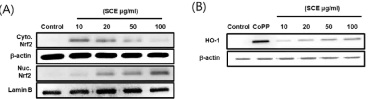
HO-1은 산화 스트레스로 인한 질병 예방에 매우 중요한 것 으로 보고되었으며, HO-1의 발현은 NF-κB의 활성화를 억 제하여 iNOS 발현 및 NO 생성을 억제한다.26,27) 따라서 본 연구에서는 SW1353 연골세포에서 SCE의 HO-1 발현과 Nrf2 활성을 평가 하였고, HO-1 억제제인 SnPP를 처리하 여 IL-1β에 의해 자극되어 생성된 활성 산소종의 생성의 변 화를 확인하였다. 그 결과 SCE는 Nrf2의 핵으로 전이를 농 도 의존적으로 증가시켰으며(Fig. 6A), HO-1의 발현 또한 증가시켰다(Fig. 6B). 또한 IL-1β에 의해 자극되어 생성된 활성 산소종에 대한 SCE의 영향을 평가하기 위해 SW1353 연골세포에 dichlorofluorescin 염색을 실시하여 활성 산소종 을 정량화 하였다(Fig. 7A). 그 결과 IL-1β에 의해 자극되어
생성된 활성 산소종은 SCE에 의해 농도 의존적으로 생성 이 감소하였으며, HO-1의 억제제 SnPP(Tin protoporphyrin IX)의 처리에 의해 활성 산소종의 생성이 증가됨을 확인 할 수 있었다(Fig. 7B). 이러한 결과는 IL-1β에 의해 자극된 SW1353 연골세포에서 SCE에 의한 HO-1 발현이 활성 산 소종의 생성을 억제한다고 판단된다. 염증반응에 관여하는 염증 매개 인자를 조절하기 위해 여러 약용버섯들이 연구 되었으며, 그중 상황버섯, 영지버섯과 노루궁뎅이버섯 추출 물은 RAW264.7 대식세포에서 iNOS, COX-2 및 TNF-α와 같은 염증 매개 인자를 조절함으로 항산화, 항염증 및 항알 레르기 효과를 나타내는 것으로 보고되었다.28-31) 본 연구 결 과는 꽃송이버섯 에탄올 추출물이 IL-1β에 의해 자극된 SW1353 인간유래 연골세포에서 염증 매개 인자와 활성 산 소종의 생성을 억제함으로 골 관절염 억제에 영향을 미치 Fig. 6. Effects of SCE on nuclear translocation of Nrf2, Nrf2 mediated HO1 expression in SW-1353 cells. SW-1353 cells were treated with SCE (10, 20, 50, 100 µg/mL) for 1 hr (A) or 24 hr (B). Nuclei were separated from the cytosol using NE-PER mam- malian protein extraction buffer. Protein was subjected to 10% SDS polyacrylamide gels followed by western blotting with anti- bodies specific for the total and phosphorylated forms of Nrf2 and HO-1.
Fig. 7. Effect of SCE on IL-1β induced ROS production in SW1353 cells. SW1353 cells were treated with SCE (10, 20, 50 and 100 µg/ml) for 6 hr after treated with SnPP 50 µM for 2 hr. And then treated with IL-1β (40ng/mL) for 24 hr. ROS production by incubation with 10 µM of the ROS-sensitive fluorophore DCF. *p-value<0.05 compared to the group treated with IL-1β.
는 것으로 판단되며, 이러한 결과는 꽃송이버섯 또한 염증 또는 골 관절염 억제에 효과가 있는 약용버섯으로서의 가 능성을 제시한다.
결 론
본 연구는 IL-1β에 의해 자극된 SW1353 연골세포에서 꽃송이버섯 에탄올 추출물이 미치는 영향을 평가하였으며, 꽃송이버섯 에탄올 추출물은 IL-1β에 의해 자극된 SW1353 연골세포에서 염증 매개 인자의 생성을 억제 하였으며, 연 골 분해 효소 MMPs의 발현을 유의하게 억제하였다. 또한 꽃송이버섯 에탄올 추출물은 NF-κB p65, IkB-α와 JNK, p38의 신호전달 경로의 활성을 억제하였으며, HO-1의 발현 을 통해 활성 산소종의 생성을 농도 의존적으로 억제 시켰 다. 따라서 꽃송이버섯 에탄올 추출물은 염증반응 억제에 영향을 미치는 것으로 판단된다.
사 사
본 논문은 농림축산식품부의 재원으로 농림수산식품기술 기획평가원의 고부가가치식품기술개발사업의 지원을 받아 연구되었음(116001-03-2-HD030).
인용문헌
1. Son, Y. O. and Chun, J. S. (2018) Estrogen-related receptor is a novel catabolic regulator of osteoarthritis pathogenesis.
BMB Rep. 51: 165-166.
2. Hochberg, M., Chevalier, X., Henrotin, Y., Hunter, D. J. and Uebelhart, D. (2013) Symptom and structure modification in osteoarthritis with pharmaceutical-grade chondroitin sulfate:
What’s the evidence. Curr Med Res Opin. 29: 259-267.
3. Martel, P. J., Boileau, C., Pelletier, J. P. and Roughley, P. J.
(2008) Cartilage in normal and osteoarthritis conditions. Best Pract. Res. Clin. Rheumatol. 22: 351-384.
4. Fernandes, J. C., Martel, P, J. and Pelletier, J. P. (2002) The role of cytokines in osteoarthritis pathophysiology. Biorhe- ology. 39: 237-246.
5. Abramson, S. B., Attur, M., Amin, A. R. and Clancy, R.
(2001) Nitric oxide and inflammatory mediators in the per- petuation of osteoarthritis. Curr Rheumatol. Rep. 3: 535-541.
6. Ding, Q. H., Cheng, Y., Chen, W. P., Zhong, H. M. and Wang, X. H. (2013) Celastrol, an inhibitor of heat shock protein 90_
potently suppresses the expression of matrix metalloprotein- ases, inducible nitric oxide synthase and cyclooxygenase-2 in primary human osteoarthritic chondrocytes. Eur. J. Phar- macol. 708: 1-7.
7. Ndisang, J. F. (2017) Synergistic interaction between heme
oxygenase (HO) and nuclear-factor E2-related factor-2 (Nrf2) against oxidative stress in cardiovascular related diseases.
Curr. Pharm. Des. 23: 1465-1470.
8. Loboda, A., Damulewicz, M., Pyza, E., Jozkowicz, A. and Dulak, J. (2016) Role of Nrf2/HO-1 system in development, oxidative stress response and diseases: An evolutionarily con- served mechanism. Cell. Mol. Life Sci. 73: 3221-3247.
9. Abraham, N. G. and Kappas, A. (2008) Pharmacological and clinical aspects heme oxygenase. Pharmcol. Rev. 60: 79-127.
10. Kumar, A., Takada, Y., Boriek, A. M. and Aggarwal, B. B.
(2004) Nuclear factorkappaB: its role in health and disease. J.
Mol. Med (Berl). 82: 434-448.
11. Thalhamer, T., McGrath, M. A. and Harnett, M. M. (2008) MAPKs and their relevance to arthritis and inflammation.
Rheumatology 47: 409-414.
12. Hemavathy, H., Ibrahim, J., Areeful, H. and Endang, K.
(2018) Phyllanthin from Phyllanthus amarus inhibits LPS- induced proinflammatory responses in U937 macrophages via downregulation of NF?κB/MAPK/PI3K?Akt signaling pathways. Phytother. Res. 32: 1-10.
13. Kirk, P. M., Cannon, P. F., David, J. C. and Stalpers, J. A.
(2003) Ainsworth & Bisby’s Dictionary of the Fungi.
Lichenologist. 35: 365-366.
14. Oh, D. S., Park, J. M., Park, H., Ka, K. H. and Chun, W.J.
(2009) Site characteristics and vegetation structure of the hab- itat of cauliflower mushroom (Sparassis crispa). Korean. J.
Mycol. 37: 33-40.
15. Ji, J. D., Lee, Y. Ho. and Song, G. G. (2004) Prostaglandin E2 (PGE2): Roles in Immune Responses and Inflammation. J.
Rheum. Dis. 11: 4.
16. Graham, S., Gamie, Z., Polyzois, I., Narvani, A. A., Tzafetta, K., Tsiridis, E., Helioti, M., Mantalaris, A. and Tsiridis, E.
(2009) Prostaglandin EP2 and EP4 receptor agonists in bone formation and bone healing: in vivo and in vitro evidence.
Expert. Opin. Investig. Drugs. 18: 746-766.
17. Sasaki, K., Hattori, T., Fujisawa, T., Takahashi, K., Inoue, H.
and Takigawa, M. (1998) Nitric oxide mediates interleukin-1- induced gene expression of matrix metalloproteinases and basic fibroblast growth factor in cultured rabbit articular chondrocytes. J. Biochem. 123: 431-439.
18. Tung, J. T., Arnold, C. E., Alexander, L. H., Yuzbasiyan, G.
V., Venta, P. J., Richardson, D. W. and Caron, J. P. (2002) Evaluation of the influence of prostaglandin E2 on recom- binant equine interleukin-1beta-stimulated matrix metallo- proteinases 1, 3, and 13 and tissue inhibitor of matrix metalloproteinase 1 expression in equine chondrocyte cul- tures. Am. J. Vet. Res. 63: 987-993.
19. Lu, Y. C., Jayakumar, T., Duann, Y. F., Chou, Y. C., Hsieh, C.
Y., Yu, S. Y., Sheu, J. R. and Hsiao, G. (2011) Chondro- protective role of sesamol by inhibiting MMPs expression via retaining NF-κB signaling in activated SW1353 cells. J.
Agric. Food Chem. 59: 4969-4978.
20. Sondergaard, B. C., Schultz, N., Madsen, S. H., Bay-Jensen, A. C., Kassem, M. and Karsdal, M. A. (2010) MAPKs are essential upstream signaling pathways in proteolytic cartilage degradation - divergence in pathways leading to aggrecanase and MMP-mediated articular cartilage degradation. Osteo- arthritis Cartilage 18: 279-288.
21. Liacini, A., Sylvester, J., Li, W. Q., Huang, W., Dehnade, F., Ahmad, M. and Zafarullah, M. (2003) Induction of matrix metalloproteinase-13 gene expression by TNF-alpha is medi- ated by MAP kinases, AP-1, and NF-kappaB transcription factors in articular chondrocytes. Exp. Cell Res. 288: 208- 217. 22. Lepetsos, P. and Papavassiliou, A. G. (2016) ROS/
oxidative stress signaling in osteoarthritis. Biochim. Biophys.
Acta. 1862: 576-591.
23. Li, D., Xie, G. and Wang, W. (2012) Reactive oxygen species:
The 2-edged sword of osteoarthritis. Am. J. Med. Sci. 344:
486-490.
24. Collins, J. A., Diekman, B. O. and Loeser, R. F. (2018) Tar- geting aging for disease modification in osteoarthritis. Curr.
Opin. Rheumatol. 30: 101-107.
25. Marchev, A. S., Dimitrova, P. A., Burns, A. J., Kostov, R. V., Dinkova-Kostova, A. T. and Georgiev, M. I. (2017) Oxi- dative stress and chronic inflammation in osteoarthritis: Can NRF2 counteract these partners in crime. Ann. N. Y. Acad.
Sci. 1401: 114-135.
26. Ndisang, J. F. (2017) Synergistic interaction between heme
oxygenase (HO) and nuclear-factor E2-related factor-2 (Nrf2) against oxidative stress in cardiovascular related diseases.
Curr. Pharm. Des. 23: 1465-1470.
27. Loboda, A., Damulewicz, M., Pyza, E., Jozkowicz, A. and Dulak, J. (2016) Role of Nrf2/HO-1 system in development, oxidative stress response and diseases: An evolutionarily con- served mechanism. Cell. Mol. Life Sci. 73: 3221-3247.
28. Baskaran, A., Chua, K. H., Sabaratnam, V., Ram, M. R. and Kuppusamy, U. R. (2017) Pleurotus giganteus (Berk. Karun
& Hyde), the giant oyster mushroom inhibits NO production in LPS/H2O2 stimulated RAW 264.7 cells via STAT 3 and COX-2 pathways. BMC Complement Altern Med. 17: 40.
29. Lee, C. B. (2012) Anti-inflammation Activity of Water Extracts from Hericium Erinacium among Medicinal Mush- rooms. Culi. Sci. & Hos. Res. 18: 233-242.
30. Marwa, M. A. S., Noha, H. H. and Wafa E. A. (2018) In vitro evaluation of the synergistic antioxidant and anti-inflamma- tory activities of the combined extracts from Malaysian Gan- oderma lucidum and Egyptian Chlorella vulgaris. BMC Complement Altern. Med. 18: 154.
31. Yun, W. S., Jung, H. A. and Roh, S. S. (2010) Effect of Phell- inus igniarius Quel Extract on the Anti-inflammatory, Anti- allergy, Anti-oxidant, Anti-wrinkle. J. Korean Med. Oph- thalmol. Otolaryngol. Dermatol. 23: 75-93.
(2018. 11. 30 접수; 2018. 12. 7 심사;
2018. 12. 11 게재확정)