Petiolus Nelumbinis, branches of lotus leaf or lotus flower is a traditional oriental herbal medicine widely used for treating a superheat or disorder of qi flow. Although there are many clinical results and literature study, it has been rarely conducted to evaluate the immuno-biological activity. The present study was conducted to examine the anti-inflammatory effects of PNM (Petiolus Nelumbinis MeOH extract) in vitro.
To determine cytotoxic concentration of PNM, the cells were treated with PNM for 24 h after LPS addition, and the cell viability was tested by MTT assay. Both of dosages (30 and 100 μ g/ml) of PNM had no cytotoxicity. In these concentrations, PNM significantly reduced the elevated levels of NO and PGE
2by LPS. These inhibitory effects of PNM were due to the
LPS로 활성화된 RAW 264.7 cell에서 荷梗의 염증매개물질 억제효과
이원욱1, 조미정1,2, 박상미1,2, 정지윤1,2, 김상찬1,2
1대구한의대학교 한의과대학 2한방신약개발팀 (BK21 Team)
ABSTRACT
Anti-inflammtory effects of the MeOH extract of Petiolus Nelumbinis
Won-Uk Lee1, Mi-Jeong Jo1,2, Sang-Mi Park1,2, Ji-Yun Jung1,2, Sang-Chan Kim1,2
1College of Oriental Medicine and 2BK21 Team, Daegu Haany University, Daegu
1)
•교신저자 : Sang-Chan Kim
•College of Oriental Medicine, Daegu Haany University, #165 Sang-dong Suseing-gu Daegu Korea
•Tel : 82-53-770-2247 Fax : 82-53-768-6340 E-mail : [email protected]
•접수 : 2009/ 05/ 18 1차 수정 : 2009/ 06/ 08 채택 : 2009/ 06/ 15
reduced expressions of iNOS and COX-2 protein. TNF-α, IL-1β and IL-6 are frequently encountered pro-inflammatory cytokines, and LPS plays a key role in inducing to the massive production of these cytokines. Thus, we next determined the levels of these cytokines. Although PNM had no significant inhibitory effect on the production of TNF-α, the elevated levels of IL-1β and IL-6 by LPS were dose-dependently reduced in PNM-treated groups.
These results demonstrate that PNM has anti-inflammatory effects by inhibiting the production of proinflammatory cytokines, NO and PGE
2in LPS-activated macrophage. Moreover, the reduction of NO and PGE
2levels was due to the inhibition of iNOS and COX-2 protein expression by PNM.
Key word : Petiolus Nelumbinis,
Anti-inflammatory effects,LPS, Cytokine
Ⅰ. 서 론
염증은 외부 자극에 대한 생체조직의 방어반응 중의 하나이다. 염증반응이 일어나면 여러 가지 염증인자들이 생성되는데 이로 인하여 임상적으로 는 발적, 발열, 종창, 동통 및 기능장애 등의 증상 이 나타난다1). 염증반응 중 많은 양의 염증 유도 cytokine, nitric oxide (NO), prostagladin E2 (PGE2) 가 생성된다2).
LPS는 대식세포나 단핵구의 활성으로 인한 cytokine의 분비작용의 원인이 되는 것으로3), LPS 는 LPS-binding protein (LBP)에 의해 복합체가 만들어지고 이것이 혈액내 대식세포의 CD14에 결 합하게 되면 Toll-like-receptor-4 (TLR-4)에 인식 되어지는 순간 nuclear factor kappa B (NF-κB) 가 활성화 된다4). NF-κB는 세포 분화, 염증반응 및 세포 부착 등에 관련된 여러 유전자들의 발현 에 중요한 역할을 하는 전사인자이다. 활성화된 NF-κB는 iNOS (inducible nitric oxide synthase),
COX-2 (cyclooxygenase-2), TNF-α (Tumor Necrosis Factor-alpha), IL-6 (interleukin 6) 등의 여러 염 증 매개물질의 전사를 촉진한다5-7).
IL-1은 단핵구와 대식세포로부터 분비되어 T세 포와 B세포를 활성화시키고 다양한 lymphokine을 분비시키며 세포증식과 세포외 기질 축적을 자극 하며8), IL-6는 단핵구를 포함한 여러 종류의 세 포에서 분비되며 다양한 기능을 갖는 cytokine이 다. 숙주의 방어, 면역반응, 신경세포의 기능, hematopoiesis 등에서 중요한 기능을 하는 물질이 다. IL-6R (IL-6 receptor)는 resting B세포에서는 발현되지 않고 활성화된 B cell에서 발견되는데 이점은 IL-6가 활성화 된 B cell에서 면역글로불 린의 생산을 유도할 수 있음을 제시하고 있다.
IL-6의 생산은 mitogenic 혹은 antigenic 자극, LPS, 칼슘, cytokines, 바이러스 등에 의해 상승되 며, 수종의 악성종양, 자가면역질환, 그 외의 감염 성 질환 등에서 증가된 IL-6를 확인할 수 있다
9-11). TNF-α는 면역반응과 염증반응을 유도하는
염증유도매개 cytokine으로 단핵세포와 대식세포 에서 생성된다12). 세포의 성장과 분화, apoptosis, necrosis 등에 관여하며 혈관 투과성을 증가시키며, 미생물 감염시, 발현량의 증가와 식세포의 사이토 카인 분비증가를 유도하여 미생물에 대한 숙주세 포의 항상성을 유지하는 중요한 방어기전을 담당 한다13). 종양발생시 apoptosis를 유도하여 종양 발 생의 감시 기전으로 이용되기도 하고, 호중구 조 절, major histocompatibility complex (MHC)발현 의 조절, B세포 증식, angiogenesis 유도 및 대식세 포 활성기능을 가지고 있다14-15). 그러나 다량의 TNF-α 발현은 심근 수축력 감소, 혈압강하, 대사 과정의 손상을 유발하기도 한다16-17). COX-2는 미 생물의 의한 감염이나 손상 혹은 여러 요인의 스 트레스에 반응한 대식세포에서 발현되므로, iNOS 와 COX-2의 발현과 NO, PGE2의 생산은 면역세 포의 대표적인 염증인자라고 할 수 있다18).
蓮은 여러해살이 수생 식물로서 잎을 荷葉, 蓮 의 꽃을 蓮花, 蓮의 수술을 蓮鬚, 꽃대나 잎자루를 荷梗이라고 한다19). 하경에 함유된 성분으로는 roemerine, nornuciferine 등의 알칼로이드 및 수지 탄닌 등이 알려져 있다20). 荷梗은 淸熱解暑, 通氣 利水의 효능이 있으며, 慢性腸炎, 久痢, 痢疾, 腸出 血, 慢性子宮炎, 赤白帶下, 遺精, 夜尿症, 暑濕胸悶, 淋病, 水樣性下痢 등을 치료한다19). 그러나, 임상 적으로 하경이 장염, 자궁염 등에 사용되어지기는 하지만, 하경의 항염증효과에 대한 연구는 전무하 다. 이러한 까닭에 본 연구에서는 荷梗의 메탄올 추출물을 LPS로 활성화된 설치류 대식세포주인 RAW 264.7 cell에 처치하여, 항염증 효과를 평가 하고 염증관련매개물질인 cytokine 및 nitric oxide 의 억제 효과를 관찰하고자 하였다.
Ⅱ. 재료 및 방법
1. 시 약
LPS (Escherichia coli 026:B6) 와 MTT ( 3 - ( 4 , 5 - d i m e t h y l t h i a z o l - 2 - y l ) - 2 , 5 - diphenyltetrazoleum)는 Sigma (St. Louis, MO, USA)에서 구입하였다. Fetal bovine serum (FBS) 와 antibiotics는 Gibco/BRL (Eggenstein, Germany) 에서 구입하였다. Antibody는 BD Bioscience (San Jose, CA, USA), Cayman (Ann Arbor, Mi, USA) 과 Zymed (San Francisco, CA, USA)에서 구입하 였고, NC paper는 Schleicher & Schuell (Dassel, Germany)사의 것을 사용하였다. TNF-α, IL-1β와 IL-6 ELISA Kits는 Pierce endogen (Rockford, IL, USA)로부터 구입하였고 PGE2 Assay Kits는 R&D Systems (USA)를 사용하였다.
2. 추출물의 제조
荷梗 (Petiolus Nelumbinis)은 2008년 7월 경상 북도 경산시 신천동 泥池 [진못]에서 직접 채취하 였다. 荷梗 300 g을 2 L의 methanol (MeOH)에 침지하여 실온에서 24시간 동안 추출하였다. 荷梗 MeOH 추출물 (PNM; Petiolus Nelumbinis MeOH extract)의 여과는 0.2 μm filter (Nalgene, New York, NY, USA)를 이용하였고, 얻은 여액은 농축과 동결건조를 거친 후 실험에 사용하였으며, 荷梗의 최종 수율은 2.26%였다.
3. 세포배양
Murine macrophage cell line인 RAW 264.7 세 포는 한국세포주은행 (Seoul, Korea)에서 구입하 였으며, Dulbecco’s modified Eagle’s medium (DMEM)에 10% fetal bovine serum (FBS), 100 U/ml penicillin 및 100 μg/ml streptomycin을 혼합 한 배지를 사용하여 37℃ 5% CO2 incubator에서 배양하였다. 실험과정의 모든 cells은 80~90%의 confluence에서 실험하였고, 20 passages를 넘기지 않은 cell만 사용하였다.
4. 세포 생존율 측정
RAW 264.7 세포를 24 well plate에 1×10⁵ cells/well로 분주한 다음 24시간 후에 serum이 없는 배지로 교환하여 24시간동안 배양하였다. 荷梗 메탄 올 추출물을 30과 100 μg/ml의 농도로 처치하고 1 시간 후에 LPS (1 μg/ml)를 처치하여 37℃, 5%
CO2 incubator에서 24시간 동안 배양하였다. 배양 후 3-(4,5-dimethylthiazol-2-yl)-2,5-diphenyltetrazolium bromide (0.5 mg/ml)를 넣고 4시간 배양 한 후 배지를 제거하고 생성된 formazan crystals을 DMSO (dimethyl sulfoxide)에 녹여 Titertek Multiskan Automatic ELISA microplate reader (Model MCC/340, Huntsville, AL)로 570 nm에서 흡광도 를 측정하였다. 세포생존율은 control cell에 대한 백분율로 나타내었다.
[i.e. viability (% control) = 100×/(absorbance of treated sample)/(absorbance of control)].
5. NO생성량 측정
RAW 264.7 세포를 24 well plate에 1×10⁵ cells/well로 분주한 다음 24시간 후에 serum을 포 함하지 않는 배지로 교환하여 24시간 동안 배양하 였다. 荷梗 메탄올 추출물을 30, 100 μg/ml의 농도 로 처치한 다음, 1시간 후에 LPS (1 μg/ml)를 처 치하고 37℃, 5% CO2 incubator에서 배양하였다. LPS 처치 후 6, 12, 18, 24시간에 배지를 수거하여 세포배 양 상등액 50 ul와 Griess 시약 (1% sulfanilamide in 5% phosphoric acid + 1% α-naphthylamide in H2O) 50 ul를 혼합하여 96 well plates에서 10분 동안 반응시킨 후 540 nm에서 Titertek Multiskan Automatic ELISA microplate reader로 흡광도를 측정하였다.
6. Immunoblot analysis
RAW 264.7 세포를 10 cm dish에 2×10⁶ cells로 분주한 다음 24시간 후에 serum이 없는 배지로 교
환하여 37℃, 5% CO2에서 24시간동안 배양하였다.
荷梗 메탄올 추출물을 30, 100 μg/ml의 농도로 처 치한 다음, 1시간 후에 LPS (1 μg/ml)를 처치하 였다. 18시간 배양한 후, 배지를 제거하여 PBS (phosphate buffered saline)로 3회 씻고 20mM Tris Cl (pH 7.5), 1% Triton X-100, 137mM sodium chloride, 10% glycerol, 2mM EDTA (ethylene diamine tetra acetic acid), 1mM sodium orthovanadate, 25mM b-glycerophosphate, 2mM sodium pyrophosphate, 1mM phenylmethylsulfonylfluoride와 1 mg/ml leupeptin을 함유하는 lysis buffer를 사용하여 단백 질을 추출한 후, 15,000×g로 30분간 원심 분리하여 상등액을 취하였다. 단백질 농도는 BSA (bovine serum albumin)를 표준화하여 BCA kit를 사용하 여 정량하였다. 50 μg의 lysate를 10% SDS-PAGE (poly acrlamide gel electrophoresis)로 분리하여 NC membrane에 transfer하였다. Anti-rabbit iNOS 와 COX-2 antibodies를 사용하여 면역화학적 방 법으로 분석하였다. iNOS와 COX-2 band는 ECL western blotting detection reagents (Amersham)를 사용하여 발색하였다.
7. Cytokine의 측정
Cytokine을 측정하기 위하여 24 well plate에 세 포를 1×10⁵/ml을 분주 및 배양하고, serum이 없는 배지로 교환하여 37℃, 5% CO2에서 24시간동안 배양하였다. 이후 荷梗 메탄올 추출물을 처치한 다음, 1시간 후에 LPS (1 μg/ml)를 처치하였다.
LPS 처치 후 18시간에 배지를 수거하여 각각의 ELISA kit를 사용하여 측정허였다. Cytokine 생성 률은 control cell에 대한 백분율로 나타내었다.
[i.e. viability (% control) = 100×/(absorbance of treated sample)/(absorbance of control)].
8. PGE
2의 측정
PGE2를 측정하기 위하여 24 well plate에 세포
를 1×10⁵/ml을 분주 및 배양하고, serum이 없는 배지로 교환하여 37℃, 5% CO2에서 24시간동안 배양한 후, 荷梗 메탄올 추출물을 처치한 다음, 1 시간 후에 LPS (1 μg/ml)를 처치하였다. 12시간 후에 배지를 수거하여 PGE2를 측정하였다. Coating 된 96 well plate에 배지를 100ul 씩 첨가하고 primary antibody solution과 PGE2 conjugate를 50 ul씩 각각의 well에 넣은 후 상온에서 2시간 동안 배양하였다. Washing buffer로 4회 세척하고 200 ul의 substrate solution 30분간 반응시키고 stop solution을 50 ul를 처리한 후 Titertek Multiskan Automatic ELISA microplate reader로 450 nm에 서 흡광도를 측정하였다.
9. 통계적 검증
실험 결과는 mean ± SD로 나타내었으며, t-test의 통계처리방법으로 통계적 유의성을 검정 하였다.
Ⅲ. 결 과
1. 荷梗 메탄올 추출물 (PNM)의 RAW 264.7 세포에 대한 독성
PNM의 RAW 264.7 세포에서 세포독성을 확인 하기 위해서 MTT assay를 시행하였다. PNM을 RAW 264.7 세포에 24시간 동안 처리한 후 흡광도 를 확인한 결과 PNM은 어떠한 세포독성을 나타 내지 않았다. LPS (1 μg/ml) 단독 처리시에는 70%내외의 세포 생존률을 나타내었으나, PNM과 LPS를 동시에 처치한 경우 오히려 세포생존율을 증가시키는 경향을 나타내었다. LPS단독 처치시 세포생존율의 저하는 다량의 NO가 생성되어 세포 독성을 유발한 것으로 생각된다(Fig. 1).
Fig. 1. Effects of PNM on the cell viability in LPS stimulated RAW 264.7 cells.
Cytotoxicity was measured for 24 h. The data represent the mean ± SD of three separate experiments. The significances of the difference between the treated groups was evaluated using the t-test.
2. 荷梗 메탄올 추출물 (PNM)의 NO production 에 대한 저해 효과
PNM의 RAW 264.7 세포에서 LPS에 의한 NO 저해효과를 측정을 위해 6, 12, 18, 24시간동안 NO 를 측정하였다. 그 결과 6시간에서는 처리군에 따 른 변화가 없었으나, 12시간부터 LPS (1 μg/ml) 를 단독 처리한 군의 NO생성량은 유의하게 증가 하였다. PNM은 12~24시간에서 유의한 NO의 생 성억제를 나타내었다(Fig. 2).
3. 荷梗 메탄올 추출물 (PNM)의 iNOS발현에 대한 억제 효과
PNM이 RAW 264.7 세포에서 NO의 생성을 저 해함을 확인하고 iNOS 발현과의 상관성을 확인하 기 위해 western blot으로 iNOS의 단백질 발현을 확인하였다. PNM의 iNOS의 억제 효과를 확인하 기 위해서 위해 PNM 30, 100 μg/ml의 농도를 처 리하고 1시간 후 LPS (1 μg/ml)를 처리하였다.
18시간 후의 iNOS 발현효과를 확인해 본 결과 PNM은 농도의존적으로 iNOS의 발현을 억제함을 확인할 수 있었다(Fig. 3A and B).
Fig. 2. Effects of PNM on the production of NO by LPS stimulated RAW 264.7 cells.
The NO concentration in the culture medium was measured for 6, 12, 18, 24 h. The data represent the mean ± SD of three separate experiments. The significances of the difference between the treated groups was evaluated using the t-test.
(*: significant compared with the control, **P<0.01, #:
significant compared with the LPS alone, ##P<0.01).
Fig. 3. The inhibitory effects of PNM on iNOS induction by LPS.
The protein levels of iNOS was monitored 18 h after
treatment with LPS in the presence or absence of PNM (30, 100 μg/ml) (A). The relative levels of iNOS was measured by scanning densitometry (B). The actin is used as a loading control. The data represent the mean ± SD of three separate experiments. The significances of the difference between the treated groups was evaluated using the t-test.
(*: significant compared with the control, **P<0.01, #:
significant compared with the LPS alone, ##P<0.01).
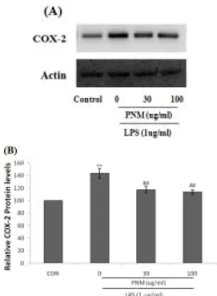
4. 荷梗 메탄올 추출물 (PNM)의 COX-2발현 에 대한 억제 효과
염증반응 또는 발암과정에서 중요한 역할을 하 는 COX-2에 대한 PNM의 억제 효과를 확인하기 위해서 위해 PNM을 30, 100 μg/ml의 농도를 처 리하고 1시간 후 LPS (1 μg/ml)를 처리하였다.
LPS 처리시에는 COX-2 단백질의 발현이 유의하 게 증가되었으나, PNM의 전처치는 COX-2 발현 을 유의하게 억제하였다(Fig. 4).
5. 荷梗 메탄올 추출물 (PNM)의 cytokines에 대한 억제 효과
PNM의 RAW 264.7 세포에서 LPS에 의한 cytokine의 억제 효과를 확인하기 위해 PNM을 18 시간 처리하여 TNF-α의 효과를 ELISA kit를 이 용하여 확인하였다. TNF-α의 경우 LPS처리 군은 control 군에 비해 약 8배 이상의 증가를 확인 할 수 있었지만 PNM의 TNF-α 저해효과는 확인할 수 없었다(Fig. 5A). IL-1β의 경우 LPS처리 군은 control 군에 비해 약 2.4배 이상의 증가를 나타내 었으며. 이러한 증가는 PNM에 의해 농도의존적 으로 억제되었다(Fig. 5B). IL-6는 LPS처리 군은 control 군에 비해 약 12배 이상의 증가하였으며, 이러한 증가는 PNM에 의해 농도의존적으로 감소 되었다(Fig. 5C).
Fig. 4. The inhibitory effects of PNM on COX-2 induction by LPS.
The protein levels of COX-2 was monitored 18 h after treatment with LPS in the presence or absence of PNM 30, 100 μg/ml) (A). The relative levels of COX-2 were measured by scanning densitometry (B).
The actin is used as a loading control. The data represent the mean ± SD of three separate experiments.
The significances of the difference between the treated groups was evaluated using the t-test.
(*: significant compared with the control, **P<0.01, #:
significant compared with the LPS alone, ##P<0.01).
Fig. 5. Inhibition of PNM on LPS-activated cytokines production.
Production of TNF-α (A) IL-1β (B) and IL-6 (C) was measured in the medium of RAW 264.7 cells cultured with LPS (1 μg/ml) in the presence or absence of PNM for 18 h. The data represent the mean ± SD of three separate experiments. The significances of the difference between the treated groups was evaluated using the t-test.
(*: significant compared with the control, **P<0.01, #:
significant compared with the LPS alone, ##P<0.01).
6. 荷梗 메탄올 추출물 (PNM)의 PGE
2에 대한 억제 효과
PNM의 RAW 264.7 세포에서 LPS에 의한 PGE2억제 효과를 확인하기 위해 PNM을 12시간 처리하여 ELISA kit로 평가하였다. PGE2의 경우 LPS처리군은 control 군에 비해 약 6배 이상의 증 가를 나타내었으며, 이러한 증가는 PNM에 의해
농도의존적으로 억제되었다. 특히 PNM 100 μg/ml 의 경우 control군과 차이가 없을 정도의 높은 저 해효과를 나타내었다(Fig. 6).
Fig. 6. Inhibition of PNM on LPS-activated PGE2 production.
RAW 264.7 cells were cultured with LPS (1 μg/ml) in the presence or absence of PNM for 12 h to determine the level of PGE2. The cultured medium was collected and directly assayed for PGE2. The data represent the mean ± SD of three separate experiments. The significances of the difference between the treated groups was evaluated using the t-test.
(*: significant compared with the control, **0.01, #:
significant compared with the LPS alone, #P<0.01).
Ⅳ. 고 찰
荷梗은 수련과 식물 蓮 (Netumbo nucifela Gaeltn)의 잎자루와 꽃자루이다. 6~9월에 채집하 여 햇볕에 말리거나, 신선한 것을 약재로 사용한 다. 건조한 荷梗은 원기둥 모양에 가깝고 지름이 8~15 mm이며 표면은 연한 황갈색이고 높낮이가 일정하지 않는 세로홈과 많은 가시 모양의 돌기가 있다. 꺾은 단면은 연한 백색이고 여러 개의 크기 가 같지 않은 구멍들이 있다. 質은 가볍고 꺾어지 기 쉬우며 꺾어질 때에 가루가 나온다20).
荷梗에는 roemerine, nornuciferine 등의 알칼로 이드, 수지, 탄닌 등이 들어 있으며, 熱을 내리고 暑氣를 제거하며 氣의 순환을 촉진하며 소변이 잘
나오게 하는 효능이 있어, 暑濕胸悶, 水樣性下痢, 痢疾, 淋病, 帶下를 치료한다20).
대식세포는 인체면역계의 중요한 역할을 담당 하고 있는 대표적인 세포이며 탐식세포라고도 한 다21). LPS에 의해 영향을 받는 대식세포의 신호전 달 분자는 serine, threonine kinase로써 세포밖 신 호를 핵으로 전달하게 하는 MAPK (mitogen activated protein kinase)가 있다. LPS에 의해 활 성화되는 MAP Kinase로는 ERK (extracellular signal-regulated kinase)1/2, p38, JNK (c-Jun N-terminal kinase)등이 있고, 대식세포 활성은 TLR4의 발현을 조절한다22). LPS와 TLR4가 결합 하여 활성화되면 세포질 조절 단백질인 MyD88 (myeloid differentiation factor 88)이 모이게 되며 여러 신호전달기전을 통해 NF-κB가 세포질내로 이동하여 활성화된다23). 이러한 NF-κB 전사인자 의 활성화는 염증 매개물질들인 NO, prostaglandins (PGs) pro-inflammatory cytokines등을 조절한다24).
연꽃의 잎자루인 荷梗의 메탄올 추출물 (PNM) 을 이용하여 30과 100 μg/ml에서 세포독성을 일으 키지 않는다는 것을 확인하고 LPS로 유도된 RAW 264.7 세포에서 NO의 저해율을 6, 12, 18, 24시간 간격으로 비교하였다. 그 결과 12, 18, 24 h 에서 30, 100 μg/ml의 농도에서 NO의 유효한 억 제효과를 확인할 수 있었다. 일반적으로 NO는 iNOS에 의하여 다량생성되므로, NO의 억제와 iNOS의 상관성을 확인하기 위해 iNOS의 발현을 평가하였다. PNM은 농도의존적으로 LPS로 증가 된 iNOS의 발현을 유의하게 억제하였다.
LPS의 자극에 의해 활성화된 RAW 264.7 세포 에서 생성되는 cytokine은 자연면역에서 매개 조 절자로서 염증반응을 조절하는 역할을 하기도 하 며, 획득면역에서도 특정항원을 인식하여 T세포에 의해 분비되고 염증반응을 강하게 하거나 특수화 하는데 관여하는 매개 조절자의 기능 등 면역반응 및 염증반응에서 다양한 역할을 수행하는 것으로 알려져 있다25-27).
PNM은 LPS에 의해 유도된 RAW 264.7 세포 에서 IL-1β의 경우 LPS처리 군은 control 군에 비 해 약 2.4배 이상의 증가를 확인 할 수 있었고, PNM은 농도의존적으로 LPS에 의해 증가된 IL-1 β를 억제하였다. IL-6 역시 LPS처리 군은 control 군에 비해 약 12배 이상의 증가를 확인 할 수 있 었고, PNM은 농도의존적으로 억제효과를 나타내 었다. 그러나, TNF-α에서는 LPS처리 군은 control 군에 비해 약 8배 이상의 증가하였지만, PNM처 리군에서는 감소의 경향을 나타내었지만, 유의한 감소를 유도하지 못하였다.
COX-2는 일반 정상조직에서는 그 발현의 정도 가 매우 낮으나 염증성 인자나 세포 성장인자, 발 암원, 종양촉진인자등의 작용에 의하여 빠르고 일 시적으로 발현되는 효소로 알려지고 있어 염증 및 암세포 성장에 주요한 역할을 담당하고 있을 것으 로 여겨지고 있다28). 다수의 염증 억제 약물들의 작용기전은 prostagladin 합성 억제를 나타내며 이 는 COX-2의 생성 및 활성저해에 의한 것이다.
COX-1은 위 및 신장기능의 유지, 혈소판의 형성 에 필요한 prostagladin의 합성에 작용하며, 상대 적으로 COX-2는 동물이나 인간의 염증반응 부위 에서 발현된다29).
본 연구에서 사용된 PNM은 LPS에 의해 발현 이 증가된 COX-2의 발현을 유의하게 억제하였으 며, LPS에 의해 유도된 RAW 264.7 세포에서 PGE2의 생성을 유의성 있게 억제하였다. 이 결과 는 PNM이 COX-2를 억제하며, 이를 통하여 PGE2 생성 억제가 나타나는 것을 의미한다.
결론적으로, PNM은 LPS에 의해 유도된 RAW 264.7 세포에서 iNOS의 발현을 억제하여 NO를 유 의하게 억제하며, COX-2의 발현을 조절하여 PGE2의 생성을 억제하였다.
Ⅴ. 결 론
荷梗추출물 (PNM)의 항염증효능을 평가하기 위하여, RAW 264.7 cell을 LPS로 활성화시킨 후 NO의 생성량, iNOS, COX-2의 발현 및 IL-1β, IL-6, TNF-α, PGE2 등의 염증매개물질에 미치는 영향을 살펴본 바 다음과 같은 결론을 얻었다.
1. LPS는 NO의 생성량을 유의성있게 증가시켰으 며, PNM 30, 100 μg/ml을 처치한 실험군에서 는 증가된 NO를 12-24 h에서 유의하게 억제하 였다. 또한 PNM+LPS처치군은 LPS단독처리군 에 비교하여 유의한 세포독성을 나타내지 않았 다.
2. LPS에 의하여 유의하게 증가된 iNOS 및 COX-2 단백질의 발현은, PNM의 전처치로 발현량이 유의성있게 감소되었다.
3. LPS에 의해 증가된 IL-6와 IL-1β, PGE2는 PNM의 전처치에 의해 유의성 있게 감소되었 다.
감사의 글
본 연구는 지식경제부 지역혁신센터사업(대구 한의대학교 한방생명자원연구센터)의 지원에 의하 여 이루어진 것입니다.
참고문헌
1. Yun HJ, Hoe SK. Anti-inflammatory Effect of Injinho-tang in RAW 264.7 cells. Kor. J.
Herbology. 2008;23:169-78.
2. Posadas I, Terencio MC, Guillén I, Ferrándiz ML, Coloma J, Payá M, Alcaraz MJ. Co- regulation between cyclo-oxygenase-2 and inducible nitric oxide synthase expression in the time-course of murine inflammation. Naunyn Schmiedebergs Arch Pharmacol. 2000;361:98-106.
3. Kim ID, Kang KS, Kwon RH, Yang JO, Lee
JS, Ha BJ. The Effect of Rubus coreanum Miquel Against Lipopolysaccharide-induced Oxidative Stress and Lipid Metabolism. J.Fd Hyg. Safety. 2007;22:213-7.
4. Charles AJ. Immunobiology, 5th. Seoul:Life science Publication co. 2002:76-7.
5. Liu SF, Malik AB. NF-kappa B activation as a pathological mechanism of septic shock and inflammation. Am J Physiol Lung Cell Mol Physiol. 2006;290:L622-45.
6. Brown Z, Robson RL, Westwick J. Regulation and expression of chemokines : potential role in glomerulonephritis. J Leukoc Biol. 1996;59 :75-80.
7. Melcion C, Lachman L, Killen PD, Morel- MarogerL, Striker GE. Mesangial cells, effect of monocyte products on proliferation and matrix synthesis. Transplant Proc. 1982;14 :559-64.
8. Papayianni A. Cytokines, growth factors, and other inflammatory mediators in glomerulonephritis.
Ren Fail 1996;18:725-40.
9. Hibi M, Nakajima K, Hirano T. IL-6 cytokine family and signal transduction: a model of the cytokine system. J Mol Med. 1996;74:1-12.
10. Hirano T, Matsuda T, Nakajima K. Signal transduction through gp130 that is shared among the receptors for the interleukin 6 related cytokine subfamily. Stem Cells. 1994
;12:262-77.
11. Van SJ. Interleukin-6: an overview. Annu Rev Immunol. 1990;8:253-78.
12. Yoon HJ, Moon ME, Park HS, Im SY, Lee JH, Kim YH. Effects of Chitosanoligosaccharide on the C. albicans-induced Inflammatory Effect in Mice and RAW 264.7 Macrophage Cells. J. Chitin Chitosan. 2007;12:15-20.
13. Aggarwal BB, Kohr WJ, Hass PE, Moffat B, Spencer SA, Henzel WJ, Bringman TS, Nedwin GE, Goeddel DV, Harkins RN.
Human tumor necrosis factor. Production, purification, and characterization. J Biol Chem.
1985;260:2345-54.
14. Bacci S, Pieri L, Buccoliero AM, Bonelli A, Taddei G, Romagnoli P. Smooth muscle cells, dendritic cells and mast cells are sources of TNF alpha and nitric oxide in human carotid artery atherosclerosis. Thromb Res. 2008;122 :657-67.
15. Zhu XY, Daghini E, Chade AR, Lavi R, Napoli C, Lerman A, Lerman LO. Disparate effects of simvastatin on angiogenesis during hypoxia and inflammation. Life Sci. 2008;83 :801-9.
16. Vilcek J, Lee TH. Tumor necrosis factor. New insights into the molecular mechanisms of its multiple actions. J Biol Chem. 1991;266:7313-6.
17. Eigler A, Sinha B, Hartmann G, Endres S.
Taming TNF: strategies to restrain this proinflammatory cytokine. Immunol Today.
1997;18:487-92.
18. O'Neill GP, Ford-Hutchinson AW. Expression of mRNA for cyclooxygenase-1 and cyclooxygenase-2 in human tissues. FEBS Lett. 1993;330:156-60.
19. 정보섭, 신민교. 도해향약대사전. 서울:영림사.
2003:516.
20. 김창민, 신민교, 안덕균, 이경순. 중약대사전.
서울:정담. 1998:5932.
21. Park WS. Study on Biological Effect of Multi-Herbal Drug KOCO-P1 on Mouse Macrophage RAW 264.7 Cells. Kor. J.
Herbology 2008;23:151-7.
22. Garrington TP, Johnson GL. Organization and
regulation of mitogen-activated protein kinase signaling pathways. Curr Opin Cell Biol. 1999
;11:211-8.
23. Iontcheva, I., Amar, S., Zawawi, K.H., Kantarci, A. and Van Dyke, T.E. Role for moesin in lipopolysaccharidestimulated signal transduction.
Infect Immun. 2004;72:2312-20.
24. Gomez PF, Pillinger MH, Attur M, Marjanovic N, Dave M, Park J, Bingham CO, Al-Mussawir H, Abramson SB. Resolution of inflammation:
prostaglandin E2 dissociates nuclear trafficking of individual NFkappa B subunits (p65, p50) in stimulated rheumatoid synovial fibroblasts.
J. Immunol. 2005;175:6924-30.
25. Lee BJ, Heo H, Oh SC, Lew JH. Comparison study of Korean and Chinese ginsengs on the regulation of Lymphocyte proliferation and cytokine production. J. Ginseng Res. 2008;32 :250-6.
26. Lee ES, Ju HK, Moon TC, Lee E, Jahng Y,
Lee SH, Son JK, Baek SH, Chang HW.
Inhibition of nitric oxide and tumor necrosis factor-α (TNF-α) production by propenone compound through blockade of nuclear factor (NF)-κB activation in cultured murine macrophage. Biol. Pharm. Bull. 2004;27:617-20.
27. Lazarov S, Balutsov M, lanev E. The role of bacterial endotoxins, receptors and cytokines in the pathogenisis of septic(endotoxin) shock.
Vutr. Boles. 2000;32:33-40.
28. Kim JY, Jung KS, Jeong HG. Suppressive effects of the kahweol and cafestol on cycloocygenase-2 expression in macrophages.
FEBS Letters. 2004;569:321-26.
29. Yoon WJ, Lee JA, Kim JY, Oh DJ, Jung YH, Lee WJ, Park SY. Anti-oxidant Activities and Anti-inflammatory Effects on Artemisia scoparia. Kor. J. Pharmacon. 2006
;37:235-45.