Corresponding Author: Kim, Souk-Boum([email protected])
Received: 2018. 07. 31. Revised: 2018. 08. 01. Accepted: 2018. 08. 22.
대한지역사회작업치료학회지 제8권 제2호
The Journal of Korean Society of Community Based Occupational Therapy Vol. 8 No. 2 (August 2018), 29-37
ISSN 2234-0866 https://doi.org/10.18598/kcbot.2018.8.2.03
A Study of EEG and Melatonin in Plasma According to Exercise Type in Elderly with Sleep Disorder
Kim, Dong-Hyun
*, Ph.D., P.T., Kim, Souk-Boum
**, Ph.D., P.T.
*
Dept. of Occupational Therapy, Gimcheon University
**
Dept. of Occupational Therapy, Cheju Halla University
Abstract
Objective : Sleep disorder caused by stress or disease to elderly currently. We tried to make clear constant exercise according to exercise intensity would have effect on sleep disorder in elderly using EEG and melatonin.
Methods : Thirty subjects were over 65 years old who lived a senior facility in some parts. They were able to communicate and understand the purpose of the study. They also expressed their intension to participate actively in experiment. They should have alert consciousness and orientation about time, place, and people.
Sleep disorder was assessed via below 6 hr total sleep time and Pittsburge Sleep Quality Index, which was satisfied with both of criterion at the same time. Exercises composed of low intensity walking, moderate aerobic exercise, and high intensity resistance strength. We used QEEG 8-System (LAXTHA Inc. KOREA) to check wave type and Polysomnograpy (Compumedics, Australia) to test quality of sleep.
and Histologic features were observed by TTC (triphenyltetrazolium chloride) staining and H & E (Hematoxylin & Eosin) staining.
Results : There was a significant sleep index and change of melatonin after aerobic exercise. There was a significant SOL, S1, S2, and SWS in aerobic exercise, but there was a significant SWS in walking and resistance strength. There was a significant change of delta wave especially in frontal and temporal region between exercises.
Conclusion : They had different effects according to type of exercise, when elderly who have chemical, behavioral change of circadian rhythms did exercise consistently. Aerobic exercise had more effect on sleep disorder than other exercise. Therefore, we may supply proper exercise to elderly and high quality of life.
Key words : EEG, Melatonin, Sleep disorder, Exercise
Ⅰ. Introduction
Population-based studies suggest that around 30%
of the adult population is affected by insomnia to some degree, and 10% report symptoms of chronic insomnia (Fullerton, 2006). Insomnia has a high overall prevalence in the elderly population(Reeder et al, 2007). The factors of sleep disorders in the elderly population may include age-dependent intrinsic lightening of sleep homeostatic processes, chonobiologic changes, higher susceptibility to arousal from sources such as noise and light, awakenings from primary medical conditions (osteoarthritis, gastroesophageal reflux), or specific sleep disorders such as sleep apnea(Bliwise et al, 2009).
The sedentary lifestyle of old people may contribute to the decline in health that often accompanies aging.
The maintenance of high physical function is one of the key factors for successful aging(Rowe & Kahn, 1997).
Elderly persons who practice physical exercise present less sleeping problems. The old physically fit men had shorter sleep onset latencies, less wake time after sleep onset, higher sleep efficiency and more total slow wave sleep than sedentary old men(Edinger et al, 1993).
The macrostructure of sleep includes total sleep time(TST)-actual time spent asleep, time in bed(TIB), sleep efficiency(SE)-the ratio of TST to TIB, sleep latency(SL)-the amount of time required to fall asleep, wake after sleep onset(WASO)-the amount of wake time after initial sleep onset and before final awakening, nREM sleep stages(S1, S2, S3, S4), REM sleep, and REM latency (REM-L)-latency to the first appearance of stage REM sleep. We spend about 80%
of our total sleep time in nREM sleep compared to only 20% time in REM sleep. The specific stages of nREM sleep are beneficial for specific functions of our brain and body(Datta, 2010). SWS or deep sleep occurs during stages three and four of nREM sleep and is thought to be the most restorative of all sleep stages.
In the elderly, polysomnography(PSG) changes consist of increased WASO and S1, and decreased SWS.
Conventional PSG parameters may be strongly affected
also by drugs and by sleep-related disorders(Terzano
& Parrino, 2000).
Adult levels remain relatively unchanged until 35-40 years and a final decline by about 50% then occurs, until very low levels are seen in old age(Kennaway et al, 1996). Melatonin displays high lipid and water solubility which facilitates passage across cell membranes (Pardridge & Mietus, 1980). Circulating melatonin can reach all body tissues including brain and is able to cross the blood-brain barrier to modulate brain activity (Claustrat et al, 2005). The secretion occurs at night, with maximum plasma levels around 03:00-04:00 am, varying with chronotype, whereas diurnal levels are undetectable, or low in rested subjects(Claustrat et al, 2005). The melatonin profile can be simultaneously determined with temperature and sleep recordings and provides and excellent diagnostic element for detecting circadian rhythm sleep disorders(Claustrat et al, 2005).
Therefore, in this study we tried to compare the effect of exercises on old people with sleep disorders by using EEG and melatonin. A limitation of the study was that in spite of many advantages of exercise, stress resulting from exercise may increase interleukin (IL:-6) and tumor necrosis factor(TNF)-α which result in inflammation or cell death. These cause a negative effect to the human body at last(Prabhu, 2004). We didn't investigate these inflammation related factors in the study.
Ⅱ. Method 1. Subject
We studied thirty subjects who were over 65 years
old, who lived a senior facility in K city. They were
able to communicate and understand the purpose of the
study. They also expressed their intension to participate
actively in the experiment. They all had alert
consciousness and orientation about time, place, and
people. For the purpose of this study sleep disorder
was determined by people who slept below 6 hours total(Kozier et al, 2004) and Pittsburge Sleep Quality Index, this satisfied both of criteria at the same time.
2. Exercise Interventions
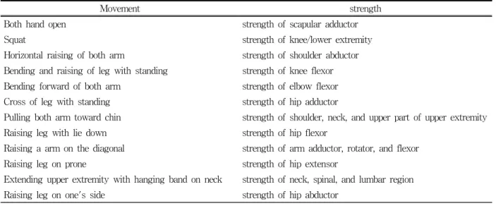
The exercise was composed of 6 upper and lower extremity movements, respectively. The movement was done 10 times per session twice each test day for no longer than 30 min(Table 1).
3. EEG test and analysis
We used QEEG 8-System(LAXTHA Inc. KOREA) machine and Polysomnography(Compumedics, Australia) to record sleep. These machines are designed to minimize error and artifact which produced by participant's movement and to observe EEG waves in real time. The composition of the equipment was EEG measure (QEEG-8) to collect a signal of the body and data collection
and Telescan Program(LAXTHA Inc. KOREA).
International 10/20 system for electrode placement was used(Fz, Cz, Pz, Oz, Fp1, Fp2, F3, F4, F7, F8, C3, C4, T3, T4, T5, T6, P3, P4, O1, O2).
The measured EEG sent signals to a receiver through an A/D converter. The transmitted data was digitized into numbers using a PC program connected to the receiver(Lee, 2009).
Electrode placement and cerebral cortex was as follows, respectively (Table 2).
4. Melatonin test and assay
Plasma melatonin of this study was CPM(counts per minute) from γ-counter equipment and made standard curve using logit-log graph. The CPM of each sample was calculated as melatonin using %B/B0 formula (Mean sample counts - NSB counts/Mean counts of pg/㎖ standard) × 100.
Movement strength
Both hand open strength of scapular adductor
Squat strength of knee/lower extremity
Horizontal raising of both arm strength of shoulder abductor Bending and raising of leg with standing strength of knee flexor Bending forward of both arm strength of elbow flexor Cross of leg with standing strength of hip adductor
Pulling both arm toward chin strength of shoulder, neck, and upper part of upper extremity
Raising leg with lie down strength of hip flexor
Raising a arm on the diagonal strength of arm adductor, rotator, and flexor
Raising leg on prone strength of hip extensor
Extending upper extremity with hanging band on neck strength of neck, spinal, and lumbar region
Raising leg on one's side strength of hip abductor
Table 1. Method of strength exercise using theraband
Cerebral Cortex Electrode Placement
Prefrontal lobe Fp1(left), Fp2(right)
Frontal lobe F3(left), F4(right)
Temporal lobe T3(left), T4(right)
Parietal lobe P3(left), P4(right)
Table 2. Cerebral cortex and EEG electrode placement
5. Data analysis
We conducted the homogeneous of the digitized EEG and melatonin value test first on the groups of people walking, aerobic, and resistance strength exercise before the experiment. We performed test for several independent samples of nonparametric test because there was no normality in sleep scale according to types of exercise after exercises. We performed nonparametric test to compare melatonin after exercises according to types of exercise. In analysis of EEG results, we used parametric test because all cases had normality. We executed one way anova to compare types of exercise after exercises.
We used Duncan test for post verification. Significance level was .05.
Ⅲ. Results
1. Comparison of PSQI score depending on exercise type after exercise
We compared the PSQI score between the 3 groups after walking, aerobic, and resistance strength exercise over the 8 week period. There was a significant difference.
Subjective sleep quality was 1.00±0.47, 0.50±0.53, 1.20±
0.42, respectively. Sleep latency was 1.30±0.48, 0.70±
0.48, 1.40±0.52, respectively. Sleep duration was 1.30±
0.67, 0.30±0.48, 1.60±0.52, respectively. Sleep efficiency was 1.10±0.57, 0.50±0.53, 1.40±0.52, respectively. Sleep disturbance was 1.40±0.52, 0.80±0.42, 1.70±0.48, respectively.
Daytime dysfunction was 0.90±0.32, 1.00±0.00, 1.40±
0.52, respectively. Global PSQI score was 7.00±1.41, 3.80±1.03, 8.70±0.95, respectively(Table 3).
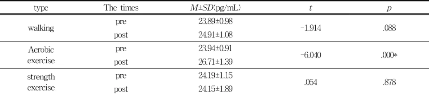
2. Comparison of Melatonin levels depending on exercise type before and after exercise
We compared melatonin levels depending on exercise type before and after the 8 weeks trial period. There was no significant difference before and after walking and strength exercise, but there was a significant difference in aerobic exercise from 23.94±0.91 to 26.71±
1.39(Table 4).
PSQI walking Aerobic exercise strength exercise
psubjective sleep quality 1.00±0.47 0.5±0.53 1.2±0.42 .012*
sleep latency 1.30±0.48 0.7±0.48 1.4±0.52 .014*
sleep duration 1.30±0.67 0.3±0.48 1.6±0.52 .001*
sleep efficiency 1.10±0.57 0.5±0.53 1.4±0.52 .006*
sleep disturbance 1.40±0.52 0.8±0.42 1.7±0.48 .003*
use of sleep medication - - - NS
daytime dysfunction 0.90±0.32 1±0 1.40±0.52 .012*
global PSQI score 7.00±1.41 3.80±1.03 8.70±0.95 .000*
Table 3. Comparison of PSQI score after exercise for 8-weeks
type The times
M±SD(pg/mL) t pwalking pre 23.89±0.98
-1.914 .088
post 24.91±1.08
Aerobic exercise
pre 23.94±0.91
-6.040 .000*
post 26.71±1.39
strength exercise
pre 24.19±1.15
.054 .878
post 24.15±1.89
Table 4. Comparison of melatonin pre and post according to exercise types
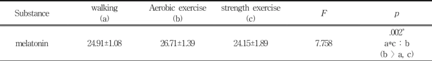
3. Comparison of Melatonin levels between exercise type after exercise
We compared melatonin levels between the 3 groups after exercise. There was a significant difference in aerobic exercise compared to walking and strength exercise in post hoc(Table 5).
4. Comparison of Delta waves after exercise
We compared delta waves of each channel between 3 groups after walking, aerobic, and strength exercise for 8 weeks. There was no significant difference of P3 in the left parietal region of the brain(p=.132). There
was a significant difference of the rest of the channels (p=.000). All aerobic exercises showed higher delta waves than those of walking and strength exercise in post hoc(Table 6).
Ⅳ. Discussion
A sleep disorder is when we don't have normal sleep patterns. We also experience sleep disorders chronically, when we disturb the relationship between the compositions of the biological clock. People don't have a deep sleep when it is induced by circadian time, it is difficult to fall asleep or wake up early morning
Substance walking
(a)
Aerobic exercise (b)
strength exercise
(c)
F pmelatonin 24.91±1.08 26.71±1.39 24.15±1.89 7.758
.002
*a*c : b (b 〉a, c)
Table 5. Comparison of melatonin after exercise for 8-weeks (pg/mL)
Position walking
(a)
Aerobic exercise (b)
strength exercise
(c)
F pFp1 24.29±0.86 26.85±1.69 24.18±0.75 12.872
.000
*a*c : b (b 〉a, c)
Fp2 25.20±1.09 27.41±1.48 24.98±1.41 10.708
.000
*a*c : b (b 〉a, c)
F3 35.01±1.62 40.20±2.27 34.04±1.13 25.873
.000
*a*c : b (b 〉a, c)
F4 33.35±1.80 37.87±2.18 32.97±1.25 18.160
.000
*a*c : b (b 〉a, c)
T3 32.63±2.18 41.12±2.11 36.47±1.65 51.593
.000
*a*c : b (b 〉a, c : c 〉a)
T4 35.04±1.25 40.24±2.31 34.97±1.08 31.915
.000
*a*c : b (b 〉a, c)
P3 24.43±1.78 26.44±3.49 24.22±1.33 2.183 .132
P4 23.89±1.39 26.42±2.77 23.56±0.85 5.545
.000
*a*c : b
(b 〉a, c)
Table 6. Comparison of Delta waves after exercise for 8-weeks
consequentially. These circadian rhythm related sleep disorders lead to decrease in total sleep time (TST) and damage total well-being(Lack & Wright, 2007).
In the study, we tried to know the improvement of sleep disorder in elderly who participated in different exercises.
Physical exercise is a nonphotic signal to entrain the human circadian clock. In addition to its clear health benefits, physical exercise is a preferred method, over pharmaceutical interventions, to synchronize the circadian system. Physical exercise significantly phase delays the human circadian pacemaker, which may help to facilitate circadian adaptation to schedules requiring a delay in the sleep-wake cycle(Eastman et al, 1995).
Therefore, physical exercise could apply as a treatment for circadian rhythm misalignments resulting from jet-lag or shift-work as an example(Germaine et al, 2012). The effects of physical exercise on the circadian system, however, require medium-to-long-term repeated training (Germaine et al, 2012). One of the main disadvantages in assessing the synchronizing effect of exercise on the human circadian system is the inability to directly measure its phase-shifting effects of the central pacemaker. Instead, the levels of one of the main output signals of the clock, melatonin, are commonly used(Germaine et al, 2012).
One neurotransmitter, norepinephrine increases not only cAMP which acts as increasing enzymatic usage in matrix, but also tryptophan.
Melatonin increased with exercise, which caused increasing the metabolic materials because catecholamine is increased considerably when the body active.
Catecholamine begin to increase at 60% maximun VO2(Park, 2010). It is defined high intensity exercise is above 65% maximun VO2, low intensity exercise is below 65% maximun VO2(Davis, 1993). This study also showed there was increasing melatonin in moderate aerobic exercise. Noradrenaline is involved in the control of melatonin synthesis and catecholamine secretions increase markedly during physical exercise(Silva de Lacerda et al, 2007).
The more we increase physical activity while we are awake, the faster we fall asleep and the longer time we spend in SWS stages. But, we can reduce the time in REM, which result in raised sleep efficiency(Kim, 1999).
SWS time depends on individual's physical strength, age, sex, exercise intensity, duration, and exercise type(Shapiro & Driver, 1998; Kim, 1999 requotation). In this study, for these reasons, we took a homogeneity test before polysomnography and then we performed the study after confirmation there was no problem in homogeneity. According to Kim(1999), the ratio of SWS to nonREM in total sleep was increased over two times after exercise. The reason was resulting from additional physical activity while we were awake in response to energy consumption to maintain energy homeostasis of the human body and to prevent more energy consumption during sleep. Therefore, it resulted in an increase of the ratio of SWS which is the lowest energy consumption of sleep stages. In this study, there was a significant increase in SWS after walking, aerobic exercise, and resistance strength, respectively.
There was the most significant increase in SWS after aerobic exercise above all. There was a significant δ wave which is used as an indicator of SWS after all three kinds of exercise, respectively. The increase of δ wave was marked in the frontal and temporal region of the brain.
In relation to exercise and sleep efficiency, exercise elicited increases in SWS and decreases in REM (Youngstedt et al, 1997), it also decreases in SOL and WASO and increases TST(Uchida et al, 2012). Brand et al. (2010) divided adolescents into low and high intensity exercise groups. They reported high intensity exercise resulted in increases in SWS and decreases in REM. In this study, aerobic exercise caused decreases significantly in SOL, walking exercise also caused decreases 1.3 % in SOL which is not significant.
Torsvall et al. (1984) reported there was decreases in
REM, increases in stage 2 sleep, and decreases in SWS
after exercise. In this study, there was no decreases in
REM from any exercise, but significant increases in
SWS.
Exercise training has been proposed as a low-cost, easily accessible and non-pharmacologic treatment alternative(Driver & Taylor, 2000). Youngstedt et al.
(1997) reported an average increase of approximately 9.9 min in total sleep time (TST) and an approximate reduction of 2.1 min in wake after sleep onset (WASO) in good sleepers after acute exercise. Passos et al. (2010) assessed the acute effects of three different modalities of physical exercise on sleep pattern of patients with chronic primary insomnia. The polysomnogram data showed reduction in the sleep onset latency (SOL) and in the total wake time (TWT); increase in total sleep time (TST), and in the sleep efficiency in moderate intensity aerobic exercise. A possible explanation for those differences might be the volume and intensity of exercise.
Shapiro et al. (1975) observed an increase in stages 3 and 4 of NREM sleep and a reduction of REM sleep in physically active good sleepers after graded exercise.
Another study reported increase in stage 2 and a decrease in stages 3 and 4 sleep(Driver, 1988). Following resistance exercise, the results showed significant improvements in all subjective sleep quality(Singh et al, 1997). In this study, there was no effect on improvement of sleep to old people with sleep disorder after resistance exercise. Resistance exercise, which is commonly performed at high intensities for shorter durations of time, enhances muscle size by increasing the synthesis of contractile and structural proteins and, as a result, the muscle is often larger and also more powerful.
Aerobic exercise resulted in reinforcing secretion of melatonin and improved sleep disorders in this study.
It is preferable to induce natural synthesis of melatonin in the body by doing exercise.
Ⅴ. Conclusion
In this study, we subjected elderly who suffer from
sleep disorder and who were over 65 years old in K city. We divided into 3 groups which were low intensity walking, moderate aerobic, and high intensity resistance strength exercise and applied different types of exercise to old people for 8 weeks. We observed the improvement of sleep disorder using melatonin and EEG.
In conclusion, they had different effects according to type of exercise, when elderly who have chemical, behavioral change of circadian rhythms did exercise consistently. Aerobic exercise had more effect on sleep disorder than other exercise. Therefore, we may supply proper exercise to elderly and high quality of life.
However, additional research is encouraged to further confirm the effectiveness of exercise because the old may have many variables like disease, general characteristic, and environmental factor.
This study reveals that the author 's doctoral thesis is summarized.
REFERENCES
Bliwise D. L., Foley D. J., Vitiello M. V., Ansari F. P., Ancoli-Israel, S., Walsh, J. K. (2009). Nocturia and disturbed sleep in the elderly. Sleep Medicine, 10, 540-548.
Claustrat, B., Brun, J., Chazot, G. (2005). The basic physiology and pathophysiology of melatonin. Sleep Medicine Reviews, 9, 11-24.
Davis J. M. (1993). Effects of serotonin agonist during prolonged exercise to fatigue in humans. Medicine
& Science in Sports & Exercise, 25, S78.
Edinger J. D., Morey M. C., Sullivan R. J., et al. (1993).
Aerobic fitness, acute exercise and sleep in older men. Sleep, 16(4), 351-359.
Escames, G., Ozturk, G., Bano-Otalora, B., et al. (2012).
Exercise and melatonin in humans: reciprocal benefits. Journal of Pineal Research, 52, 1-11.
Fullerton, D. S. (2006). The economic impact of
insomnia in managed care: a clearer picture
emerges. American Journal of Managed Care, 12(Suppl 8), S246-252.
Kim J. K. (1999). The Effects of Serotonin changes following the Exercise timing on Melatonin secretion and Sleep Quality. Department of Physical Education The Graduate School Yonsei University.
Kozier, B., Erb, G., Berman, A., Synder, S. (2004).
Fundamentals of nursing; Concepts, process, and practice. New Jersey: Prentice-Hall.
Lack, L. C., Wright, H. R. (2007). Chronobiology of sleep in humans. Cellular and Molecular Life Sciences., 64, 1205-1215.
Lee, W. J. (2009). A study on the cognitive behavior therapy For improvement of sleep habits on EEG pattern. Department of Biomedical Laboratory of Science, Graduate School Inje University.
Pardridge, W. M., Mietus, L. J. (1980). Transport of albumin-bound melatonin through the blood-brain barrier. Journal of Neurochemistry, 34, 1761-1763.
Prabhu, S. D. (2004). Cytokine-induced modulation of cardiac function. Circulation Research, 95, 1140- 1153.
Reeder, C. E., Franklin, M., Bramley, T. J. (2007).
Current landscape of insomnia in manated care.
American Journal of Managed Care, 13(Suppl 5), S112-116.
Rowe, J. W., Kahn, R. L. (1997). Successful aging.
Gerontologist, 37(4), 433-440.
Shapiro, C. M., Driver H. S. (1998). Stress and sleep.
In: Roussel B, Jouvet M(eds): Preceedings of the 27th Defence Research Group NATO colloquium:
sleep and its implications for the millitary. Lyon, France: Plenum Press., 133-146.
Silva de Lacerda, A. F., Janjoppi, L., Scorza, F. A., et al. (2007). Physical exercise program reverts the effects of pinealectomy on the amygdala kindling development. Brain Research Bulletin, 74, 216-220.
Singh, N. A., Clements, K. M., Fiatarone, M. A. (1997).
A randomized controlled trial of the effect of exercise on sleep. Sleep, 20(2), 95-101.
Terzano, M. G., Parrino, L. (2000). Origin and Significance of the Cyclic Alternating Pattern(CAP). Sleep Medicine Reviews, 4(1), 101-123.
Torsvall, L., Akerstedt, T., Lindbeck, G. (1984). Effects on sleep stages and EEG power density of different degrees of exercise in fit subjects. Electroencephalography and Clinical Neurophysiology, 57(4), 347-353.
Youngstedt, S. D., O'Connor, P. J., Dishman, R. K.
(1997). The effects of acute exercise on sleep: a
quantitative synthesis. Sleep, 20, 203-214.
— 국문초록 —
수면장애 노인의 운동유형별 뇌파와 혈 중 멜라토닌 농도 비교
김동현
*, 김석범
***김천대학교 작업치료학과
**제주한라대학교 작업치료학과
목적 : 수면장애는 노인에게 있어 다양한 이유로 빈발하고 있다. 이러한 노인의 수면장애에서 운동 강도에 따른 지속적 인 운동습관이 수면장애의 치료적 효과를 줄 수 있는지를 밝히기 위해서 뇌파와 멜라토닌의 농도를 이용한 과학적인 방법으로 접근하는 것이 본 연구의 목적이다.
연구방법 : 일부지역의 노인시설에 거주하는 65세 이상의 노인 30명을 대상으로 하였다. 수면장애의 판단은 수면시간 6시간 이하의 경우(Kozier et al, 2004)와 PSQI(Pittsburgh Sleep Quality Index)의 점수를 동시에 만족하는 경우로 정하였다. 운동유형별 적용은 저강도의 걷기운동, 중강도의 유산소 운동, 고강도의 저항성 근력운동을 적용하였으며 측정도구는 뇌파에서 파형을 체크하기 위하여 QEEG 8-System(LAXTHA Inc. KOREA) 기기와 수면의 질을 검사 하기 위해서 수면다원검사에 사용하는 Polysomnograpy (Compumedics, Australia) 기기를 사용하였다. 그리고 TTC(triphenyltetrazolium chloride) 염색과 H & E(Hematoxylin & Eosin) 염색을 통해 조직학적 양상을 관찰하였다.
결과 : 중강도의 유산소 운동 후에 멜라토닌의 농도와 뇌파로 측정한 수면지수에는 긍정적인 유의미한 변화가 있었다.
중강도의 유산소 운동에서는 SWS가 저강도, 고강도 운동보다 유의한 효과가 있었고 SWS의 관찰지표인 델타파의 유의한 변화가 있었다.
결론 : 인체의 일주기성 리듬의 생화학적, 행동학적 기능에 변화를 가져오는 노인에게서 수면장애에 대한 지속적인 운 동은 운동의 유형에 따라 효과가 다름을 알 수 있었고 특히 유산소 운동이 다른 운동보다 노인 수면장애에 효과가 크다는 것을 멜라토닌의 농도와 뇌파를 통하여 알 수 있었다. 노인에게 맞춤형 운동을 제공하여 노인들의 삶의 질에 유용하고 건강한 말년을 제공할 수 있을 것이다.
주제어 : 뇌파, 멜라토닌, 수면장애, 운동