• 검색 결과가 없습니다.

Risk factors for respiratory distress syndrome in full-term neonates

N/A
N/A
Protected

Academic year: 2021

Share "Risk factors for respiratory distress syndrome in full-term neonates"

Copied!
5
0
0

로드 중.... (전체 텍스트 보기)

전체 글

(1)

Yeungnam Univ J Med 2018;35(2):187-191

Risk factors for respiratory distress syndrome in full-term neonates

Jin Hyeon Kim, Sang Min Lee, Young Hwan Lee

Department of Pediatrics, Yeungnam University College of Medicine, Daegu, Korea

Background: Respiratory distress syndrome (RDS) is a one of the most common cause of respiratory morbi- dity and mortality in neonates. This study was conducted to investigate the risk factors for RDS in full-term neonates.

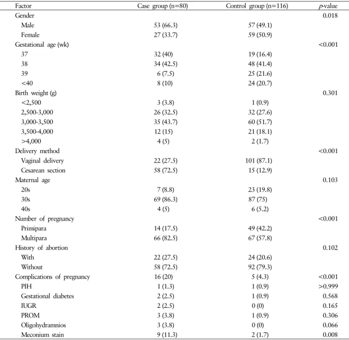
Methods: We conducted this retrospective study using medical records. The study group included 80 full-term neonates diagnosed with RDS and hospitalized in the neonatal intensive care unit between January 2012 and December 2016, at Yeungnam University Hospital. We analyzed sex, gestational age, birth weight, de- livery method, maternal age, number of pregnancy, history of abortion, and complication of pregnancy. The control group included 116 full-time neonates who were hospitalized with jaundice during the same period.

Results: The incidence of full-term RDS was more common in males (odds ratio [OR], 3.288; 95% confidence interval [CI], 1.446-7.479), cesarean section (OR, 15.03; 95% CI, 6.381-35.423), multiparity (OR, 4.216; 95%

CI, 1.568-11.335). The other factors rendered no significant results.

Conclusion: The risk factors for RDS in full-tern neonates were identified as male sex, cesarean section, and multiparity. Further studies involving more institutions are needed to clarify the risk factors for RDS in full- term infants.

Keywords: Full-term; Neonatal respiratory distress syndrome; Risk factor

Copyright ©2018 Yeungnam University College of Medicine

This is an Open Access article distributed under the terms of the Creative Commons Attribution Non-Commercial License (http://creative- commons.org/licenses/by-nc/4.0/) which permits unrestricted non-commercial use, distribution, and reproduction in any medium, provided the original work is properly cited.

Received: October 18, 2018, Revised: October 29, 2018 Accepted: October 30, 2018

Corresponding Author: Young Hwan Lee, Department of Pediatrics, Yeungnam University College of Medicine, 170, Hyeonchung-ro, Nam-gu, Daegu 42415, Korea Tel: +82-53-640-6999, Fax: +82-53-629-2252 E-mail: yhlee@med.yu.ac.kr

INTRODUCTION

Neonatal respiratory distress syndrome (RDS) is one of the major causes of respiratory failure and neonatal death in pre- mature infants and is caused by a lack of pulmonary surfactant due to fetal lung immaturity. Treatment is the administration of a synthesized pulmonary surfactant, following which the course of the disease markedly improves. According to previ- ous reports, the most important risk factors for RDS are pre- maturity and male sex [1,2]. In fact, a study comparing the

incidence of RDS in premature infants showed a 45% inci- dence in premature infants between 23 to 33 weeks; how- ever, the incidence was markedly decreased to 4% and less than 1% in 34-36 weeks and over 37 weeks, respectively [1].

Additionally, other studies have reported an incidence of approximately 10.5% at 34 weeks, 1% at 37 weeks, and 0.3%

at 38 weeks [3]. However, in practice, even if a patient is born

at full-term, RDS frequently develops, and patients are hos-

pitalized in the neonatal intensive care unit (NICU) for mecha-

nical ventilator and pulmonary surfactant treatment. Condò

et al. [4] studied the risk factors of RDS in full-term infants

and compared it with those in preterm infants. The report

mentioned that risk factors such as male sex, low birth weight,

and cesarean section increase the risk of RDS in both preterm

and full-term infants, and gestational diabetes and pregnancy-

induced hypertension are the risk factors for RDS in late pre-

term and full-term neonates. However, as far as the author

(2)

knows, there have been no studies regarding this in Korea.

Therefore, this retrospective study aims to investigate the risk factors for RDS in full-term infants and to help diagnose and treat RDS.

MATERIALS AND METHODS

1. Patients

This retrospective study was approved by the Clinical Re- search Ethics Committee of Yeungnam University Hospital (YUMC-2018-10-032). The subjects were divided into an ex- perimental group (full-term infants diagnosed with RDS) and a control group (full-term infants with neonatal jaundice).

Risk factors were assessed on the basis of history of hospi- talization at the time of admission; we investigated gestatio- nal age, sex, birth weight, delivery method, Apgar score, ma- ternal age at delivery, pregnancy frequency, abortion history, pregnancy-induced hypertension, gestational diabetes, pre- eclampsia, intrauterine growth restriction, premature rupture of membranes, oligohydramnios, polyhydramnios, history of meconium aspiration, placenta previa, placenta abruption.

The study group consisted of 80 full-term infants who were diagnosed with RDS and hospitalized in the NICU of Yeungnam University Hospital, between January 2012 and December 2016. The diagnostic criteria for RDS in full-term infants were as follows [5-8]: (1) full-term infant with gesta- tional age more than 37 weeks; (2) characteristic clinical mani- festations (tachypnea, nasal flaring, progressive dyspnea, grun- ting, cyanosis, chest retraction, and severe respiratory distress requiring positive pressure ventilation); (3) a characteristic finding in the chest radiograph (ground glass appearance, air bronchogram, total white out); (4) decreased oxygen demand after treatment with pulmonary surfactant.

The control group included 116 full-term infants who were admitted with neonatal jaundice during the same period, ex- cept for jaundice caused by sepsis or bacterial infection, or hemolytic anemia.

Among them, a history of meconium aspiration was defi- ned as a meconium stained amniotic fluid at the time of birth, or patients with meconium stained umbilical cord or meco- nium under the fingernail found on physical examination at the time of admission. Intrauterine growth restriction can

result in a baby being small for gestational age (SGA), which is defined as a weight below the 10th percentile for the gesta- tional age. The exclusion criteria were; patients who received pulmonary surfactant therapy before admission to this hospi- tal, and patients with congenital heart disease.

The gestational age was divided into four compartments of 37 weeks, 38 weeks, 39 weeks, 40 weeks, or more, and body weight was divided into five compartments of from 2,000 g to less than 2,500 g, from 2,500 g to less than 3,000 g, from 3,000g to less than 3,500g, from 3,500g to less than 4,000g and more than 4,000 g. Maternal age was divided into three compartments of 20s, 30s, 40s, and over.

2. Statistical analyses

Data were analyzed using SPSS for Windows ver.10.0 (SPSS Inc., Chicago, IL, USA). Chi-square test was used to check for any significant differences between the two groups, and correlation was analyzed by logistic regression analysis. Statis- tical significance was considered in case of p <0.05.

RESULTS

The results are as follows (Table 1). (1) RDS occurred more frequently in males (male, 53 [66.3%]; female, 27 [33.7%];

p =0.018), the incidence of RDS was 3.288 times higher than that in females ( p =0.005). (2) In case of the delivery method, cesarean section was more frequent associated with RDS than vaginal delivery (cesarean section, 58 [72.5%]; vaginal deli- very, 22 [27.5%]; p <0.001), the incidence of RDS was 15.034 times higher with cesarean section than with vaginal delivery ( p <0.001). (3) The mean birth weight was 3,198 g. The fre- quency of each birth weight was 2,000 g to less than 2,500 g in 3 cases, 2,500 g to less than 3,000 g in 26 cases, 3,000 g to less than 3,500 g in 35 cases, 3,500 g to less than 4,000 g in 12 cases, and 4,000 g or more in 4 cases. (4) The mean age of the maternal age at the delivery was 34.0 years (34.01

±3.53). Frequencies in the maternal age categories were fol-

lows: 7 in the 20s, 69 in the 30s, and 4 in the 40s. (5) Frequen-

cies in the gestational age categories were as follows: 32 in

37 weeks to 37 weeks 6 days, 34 in 38 to 38 weeks 6 days, 6

in 39 weeks to 39 weeks 6 days, and 8 in over 40 weeks. The

highest frequency was at 38 weeks ( p <0.001). However,

(3)

Table 1. Patient characteristics and analysis of risk factors in two groups (%)

Factor Case group (n=80) Control group (n=116) p -value

Gender 0.018

Male 53 (66.3) 57 (49.1)

Female 27 (33.7) 59 (50.9)

Gestational age (wk) <0.001

37 32 (40) 19 (16.4)

38 34 (42.5) 48 (41.4)

39 6 (7.5) 25 (21.6)

<40 8 (10) 24 (20.7)

Birth weight (g) 0.301

<2,500 3 (3.8) 1 (0.9)

2,500-3,000 26 (32.5) 32 (27.6)

3,000-3,500 35 (43.7) 60 (51.7)

3,500-4,000 12 (15) 21 (18.1)

>4,000 4 (5) 2 (1.7)

Delivery method <0.001

Vaginal delivery 22 (27.5) 101 (87.1)

Cesarean section 58 (72.5) 15 (12.9)

Maternal age 0.103

20s 7 (8.8) 23 (19.8)

30s 69 (86.3) 87 (75)

40s 4 (5) 6 (5.2)

Number of pregnancy <0.001

Primipara 14 (17.5) 49 (42.2)

Multipara 66 (82.5) 67 (57.8)

History of abortion 0.102

With 22 (27.5) 24 (20.6)

Without 58 (72.5) 92 (79.3)

Complications of pregnancy 16 (20) 5 (4.3) <0.001

PIH 1 (1.3) 1 (0.9) >0.999

Gestational diabetes 2 (2.5) 1 (0.9) 0.568

IUGR 2 (2.5) 0 (0) 0.165

PROM 3 (3.8) 1 (0.9) 0.306

Oligohydramnios 3 (3.8) 0 (0) 0.066

Meconium stain 9 (11.3) 2 (1.7) 0.008

Values are presented as number (%).

PIH, pregnancy induced hypertension; IUGR, intrauterine growth restriction; PROM, premature rupture of membranes.

when logistic regression analysis was performed based on gestational age over 40 weeks, it showed that 37 weeks ( p = 0.483), 38 weeks ( p =0.504), and 39 weeks ( p =0.739) were not significant. (6) Depending on which pregnancy was as- sociated with RDS, the frequencies were as follows: 14 (17.5%) in first pregnancy, followed by 29, 25, 9, and 3 in second, third, fourth, and fifth pregnancies, respectively (36.3%, 31.3%, 11.3%, and 3.8%). The incidence of RDS

was markedly low in the primiparous group ( p< 0.001), and the incidence was increased by 4.216 times ( p =0.004) in pregnancies beyond the second. (7) Based on abortion history:

58 (72.5%) women did not have an abortion history, and 22 (22.5%) women did. This was similar to results of the num- ber of pregnancies, but not statistically significant ( p =0.102).

There was one case where the mother was diagnosed with

pregnancy-induced hypertension. There were 2 cases (2.5%)

(4)

each, born to mothers with gestational diabetes and intraute- rine growth restriction, respectively. Nine cases (11.3%) of suspected meconium aspiration were noted.

DISCUSSION

In this study, it was found that male sex, cesarean section, and multiparity had a statistically significant association with an increased incidence of RDS.

Prematurity is a typical risk factor for RDS in neonates [1]. However, it is possible for RDS to occur even in full- term infants. In a study by Dani et al. [9], 59,990 infants of 63,537 total newborns were reported to be full-term (at 37 weeks or more) and 53 (0.1%) of the 59,990 full-term infants were diagnosed with RDS. In this study, we did not investig ate the incidence of RDS in full-term infants, but we wanted to assess the risk factors.

We confirmed that male sex was one of the risk factors for RDS in full-term infants and this was corroborated by previous studies. The reason is summarized as follows. First, androgen (male sex hormone) regulates the signal transduc- tion pathways of epidermal growth factor and transforming growth factor-beta, delaying the fetal lung maturation. It also delays the secretion of fibroblast-pneumocyte factor, delaying the development of type II alveolar cells, thereby reducing the secretion of the pulmonary surfactant. On the other hand, estrogen (female sex hormone) promotes the synthesis of pul- monary surfactants such as phospholipid, lecithin, surfactant protein A or B, and increases the number of type II alveolar cells by increasing the production of lamellar bodies [10-13].

This study showed that the incidence of RDS was signi- ficantly increased with relatively lower gestational age, al- though it was a study on full-term infants. These results sug- gest that lower gestational age makes maturation of the fetal lungs slower. However, this finding lacks credibility because the subjects were all 37 weeks or older in this study. Addi- tionally, logistic regression analysis of gestational age at 40 weeks showed that it had no significant effect on the develop- ment of RDS. Further research on this subject is warranted in the future.

Cesarean section is known to be an important factor affec- ting the respiratory difficulties in newborns [14]. In this study, only 12.9% of the control group and 72.5% of the patients

with RDS who were hospitalized, were delivered through a cesarean section; these findings were similar to those of a previous study. Cesarean section is a risk factor for RDS be- cause the fetus absorbs about one-third of the fetal lung fluid during vaginal delivery, whereas proper absorption of fetal lung fluid is not achieved in infants delivered through a cesar- ean section. In addition, the labor process itself matures the system that makes the surfactant, and this process is omitted in a cesarean section. Hence, the risk of RDS is increased in the infants delivered through a cesarean section [15].

In this study, it was confirmed that more infants born to multiparous women had RDS, thereby confirming multiparity to be a risk factor for the same. This is similar to the results of studies performed by Melamed et al. [16] and Altman et al. [17] published in 2009 and 2013, respectively. However, while those studies were conducted on preterm infants, this study was performed on full-term infants. Despite this differ- ence, both studies have shown that multiparity increases the risk of RDS in neonates. The mechanism is still unclear and further research is needed in this area.

Factors, such as birth weight, maternal age at birth, history of abortion, pregnancy-induced hypertension, gestational dia- betes, oligohydramnios, premature rupture of membranes, and intrauterine growth restriction were not found to be directly related to the development of RDS in this study. In a study by Liu et al. [18], premature rupture of membrane and gesta- tional diabetes were found to be important risk factors for RDS in full-term infants; however, this study did not show any significant results. This may be because of a smaller num- ber of patients included in the study and unavailability of a detailed history because most of the experimental newborns in this study were born in other hospitals.

The limitations of this study were as follows. First, high possibility that there was no detailed history available of ma- ternal diseases, medications, premature rupture of memb- ranes, etc. Because most of the hospitalized newborns were born in other hospitals. Second, neonates with severe respira- tory distress at the time of hospitalization, who were unable to undergo arterial blood gas analysis at the time of first appearance of symptoms because of early application of arti- ficial respiration including tracheal intubation and rapid use of pulmonary surfactant, could not be included in the study.

Third, the representation is low because the study was con-

ducted on patients who were admitted to a single university

(5)

hospital. Nine newborns with a history of meconium aspira- tion were included in the study group and the results were significant in declaring this as a risk factor when compared with the control group ( p =0.008). However, if meconium stained amniotic fluid at the time of birth, meconium stained umbilical cord, or meconium under the fingernail, is accom- panied by dyspnea and the patient has to undergo positive pressure ventilation and pulmonary surfactant treatment, a controversy is likely to arise as to whether the diagnosis should be RDS or meconium aspiration syndrome [19]. This is anoth- er limitation of this study as it is difficult to know the precise state of the patient at the time of admission because of its retrospective nature.

In conclusion, the risk factors for RDS in full-term infants were male sex, cesarean section, and multiparity. However, prospective studies involving more hospitals are needed.

CONFLICT OF INTEREST

No potential conflict of interest relevant to this article was reported.

ORCID

Jin Hyeon Kim, https://orcid.org/0000-0002-2770-1507 Sang Min Lee, https://orcid.org/0000-0002-6169-3504 Young Hwan Lee, https://orcid.org/0000-0001-8377-5802

REFERENCES

1. Koivisto M, Marttila R, Kurkinen-Räty M, Saarela T, Pokela ML, Jouppila P, et al. Changing incidence and outcome of infants with respiratory distress syndrome in the 1990s: a population-based survey. Acta Paediatr 2004;93:177-84.

2. Perelman RH, Palta M, Kirby R, Farrell PM. Discordance between male and female deaths due to the respiratory dis- tress syndrome. Pediatrics 1986;78:238-44.

3. Hibbard JU, Wilkins I, Sun L, Gregory K, Haberman S, Hoff- man M, et al. Respiratory morbidity in late preterm births.

JAMA 2010;304:419-25.

4. Condò V, Cipriani S, Colnaghi M, Bellù R, Zanini R, Bulfoni C, et al. Neonatal respiratory distress syndrome: are risk fac-

tors the same in preterm and term infants? J Matern Fetal Neonatal Med 2017;30:1267-72.

5. Agrawal V, David RJ, Harris VJ. Classification of acute respi- ratory disorders of all newborns in a tertiary care center. J Natl Med Assoc 2003;95:585-95.

6. Edwards MO, Kotecha SJ, Kotecha S. Respiratory distress of the term newborn infant. Paediatr Respir Rev 2013;14:29- 36.

7. Lee JM, Kim DK, Lee SJ. Probable prognostic factors among the revealing clinical manifestations at admission in neonates with tachypnea. J Korean Soc Neonatol 2006;13:32-9. Korean.

8. Liu J, Shi Y, Dong JY, Zheng T, Li JY, Lu LL, et al. Clinical characteristics, diagnosis and management of respiratory dis- tress syndrome in full-term neonates. Chin Med J (Engl) 2010;

123:2640-4.

9. Dani C, Reali MF, Bertini G, Wiechmann L, Spagnolo A, Tan- gucci M, et al. Risk factors for the development of respiratory distress syndrome and transient tachypnoea in newborn in- fants. Eur Respir J 1999;14:155-9.

10. Bresson E, Seaborn T, Côté M, Cormier G, Provost PR, Pied- boeuf B, et al. Gene expression profile of androgen modu- lated genes in the murine fetal developing lung. Reprod Biol Endocrinol 2010;8:2.

11. Nielsen HC, Torday JS. Sex differences in avian embryo pul- monary surfactant production: evidence for sex chromosome involvement. Endocrinology 1985;117:31-7.

12. Seaborn T, Simard M, Provost PR, Piedboeuf B, Tremblay Y. Sex hormone metabolism in lung development and matu- ration. Trends Endocrinol Metab 2010;21:729-38.

13. Nielsen HC. Androgen receptors influence the production of pulmonary surfactant in the testicular feminization mouse fetus. J Clin Invest 1985;76:177-81.

14. Hansen AK, Wisborg K, Uldbjerg N, Henriksen TB. Risk of respiratory morbidity in term infants delivered by elective cae- sarean section: cohort study. BMJ 2008;336:85-7.

15. Roth-Kleiner M, Wagner BP, Bachmann D, Pfenninger J. Res- piratory distress syndrome in near-term babies after caesarean section. Swiss Med Wkly 2003;133:283-8.

16. Melamed N, Klinger G, Tenenbaum-Gavish K, Herscovici T, Linder N, Hod M, et al. Short-term neonatal outcome in low- risk, spontaneous, singleton, late preterm deliveries. Obstet Gynecol 2009;114:253-60.

17. Altman M, Vanpée M, Cnattingius S, Norman M. Risk factors for acute respiratory morbidity in moderately preterm infants.

Paediatr Perinat Epidemiol 2013;27:172-81.

18. Liu J, Yang N, Liu Y. High-risk factors of respiratory distress syndrome in term neonates: a retrospective case-control study.

Balkan Med J 2014;31:64-8.

19. Wiswell TE, Tuggle JM, Turner BS. Meconium aspiration syndrome: have we made a difference? Pediatrics 1990;85:

715-21.

수치

Table 1. Patient characteristics and analysis of risk factors in two groups (%)

참조

관련 문서

Serum uric acid levels and risk for vascular disease in patients with metabolic syndrome... Prevalence if the metabolic syndrome in a Turkish

In this study, it can be seen that sex and metabolic syndrome are important for biological factors, and occupation, smoking, and exercise are important

This study was conducted to analyze the effects of obesity on the core stability and metabolic syndrome risk factors of middle - aged women.. The effect

 The final results of risk calculation and presentation are individual risk measures, societal risk F-N plots or

The risk of complicated acute appendicitis should be evaluated in terms of medical factors, including psychi- atric disease entity, psychotropic prescription, and

Age at diagnosis after 30 years, baseline serum creatinine levels, and cyst infection are the independent risk factors for kidney failure, as found in this

Further research on other intraoperative risk factors, such as flap thickness and length of incision, in addition to the incision type, may help in improving the postoperative

Prevalence and Factors of Osteoporosis and High Risk of Osteoporotic Fracture in Patients with Ankylosing Spondylitis: A Multicenter Comparative Study of Bone Mineral Density