Received: December 17, 2018 Revised: June 23, 2019 Accepted: June 25, 2019 Journal of
Trauma and InJury
CASE REPORT
J Trauma Inj 2019;32(2):122-125 https://doi.org/10.20408/jti.2018.055
Correspondence to Dae Sung Ma, M.D.
Department of Trauma Surgery, Gil Med- ical Center, Gachon University College of Medicine, 21 Namdong-daero 774beon- gil, Namdong-gu, Incheon 21565, Korea Tel: +82-32-460-3010
Fax: +82-32-460-2372 E-mail: [email protected]
http://www.jtraumainj.org eISSN 2287-1683
pISSN 1738-8767
Copyright © 2019 The Korean Society of Trauma
This is an Open Access article distributed under the terms of the Creative Commons Attribution Non-Commercial License (http://creativecommons.org/licenses/by-nc/4.0/) which permits unrestricted noncommercial use, distribution, and reproduction in any medium, provided the original work is properly cited.
The Successful removal of a foreign Body in the Spleen via diaphragm laceration Site by Video-assisted Thoracoscopic Surgery
Yang Bin Jeon, M.D., Ph.D., Sung Youl Hyun, M.D., Ph.D., Dae Sung Ma, M.D.
Department of Trauma Surgery, Trauma Center, Gachon University Gil Medical Center, Incheon, Korea
A 73-year-old man, who, in an inebriated state, had slipped in a flowerbed and was wounded on the left flank, was transferred to Trauma Center, Gil Medical Center, Gachon University College of Medicine. Based on the chest and abdominopelvic com- puted tomography, he was diagnosed with multiple rib fractures and hemopneumo- thorax on the left hemithorax and was found to have a bony fragment in the spleen.
He had not presented peritonitis and exsanguinous symptoms during the observation period. Seven days later, computed tomography of the abdomen showed suspected di- aphragmatic injury and a retained foreign body in the spleen. On exploration by video assisted thoracoc surgery (VATS), a herniated omentum through the lacerated site of the diaphragm was observed. After omentectomy using Endo Gia, the foreign body in the spleen was observed through the lacerated site of the diaphragm. Traumatic dia- phragm rupture with a foreign body, in the spleen, was successfully managed by video assisted thoracic surgery via the lacerated site of the diaphragm.
Keywords: Diaphragm; Foreign bodies; VATS
INTRODUCTION
The incidence of penetrating diaphragmatic injuries associated with the thoracoab- dominal area is 11-19% [1]. A retained foreign body in a penetrating abdominal injury is a relative indication of laparotomy. However, delayed diagnosis of traumatic dia- phragmatic rupture is associated with high morbidity and mortality in the long-term.
Thoracoscopic exploration of suspected diaphragm injury without peritonitis sign
123
http://www.jtraumainj.org
Yang Bin Jeon, et al. Removal of the Foreign Body in the Spleen via VATS
The Successful removal of a foreign Body in the Spleen via diaphragm laceration Site by Video-assisted Thoracoscopic Surgery
Yang Bin Jeon, M.D., Ph.D., Sung Youl Hyun, M.D., Ph.D., Dae Sung Ma, M.D.
Department of Trauma Surgery, Trauma Center, Gachon University Gil Medical Center, Incheon, Korea
A 73-year-old man, who, in an inebriated state, had slipped in a flowerbed and was wounded on the left flank, was transferred to Trauma Center, Gil Medical Center, Gachon University College of Medicine. Based on the chest and abdominopelvic com- puted tomography, he was diagnosed with multiple rib fractures and hemopneumo- thorax on the left hemithorax and was found to have a bony fragment in the spleen.
He had not presented peritonitis and exsanguinous symptoms during the observation period. Seven days later, computed tomography of the abdomen showed suspected di- aphragmatic injury and a retained foreign body in the spleen. On exploration by video assisted thoracoc surgery (VATS), a herniated omentum through the lacerated site of the diaphragm was observed. After omentectomy using Endo Gia, the foreign body in the spleen was observed through the lacerated site of the diaphragm. Traumatic dia- phragm rupture with a foreign body, in the spleen, was successfully managed by video assisted thoracic surgery via the lacerated site of the diaphragm.
Keywords: Diaphragm; Foreign bodies; VATS
and other peritoneal organs may be a reliable approach for exploration and management of traumatic diaphragm injury. In the thoracoscopic approach, it has a limitation for exploration of the pertitoneal cavity, but it is possible through the lacerated site in the diaphragm.
CASE REPORT
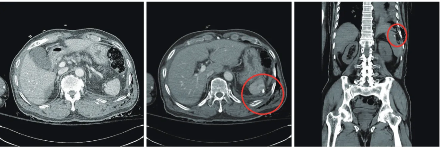
A 73-year-old man, who, in an inebriated state, had slipped down on a flowerbed and was wounded on the left flank, was transferred to Trauma Center, Gil Medical Center, Gachon University College of Medicine. In the previous hospital, computed tomography (CT) of the chest and abdominopelvic (AP) region were evaluated and revealed moderate hemopneumothorax on the left hemithorax with multiple rib fractures from 9th to 12th, fracture of the spinous process of T12 and L1, and spleen laceration due to the presence of a bony fragment (Fig. 1).
The vital signs of the patient were stable without signs of peritonitis. He was admitted to the Trauma Intensive Care Unit after a closed thoracostomy on the left hemithorax and a primary closure on the flank wound (about 5 cm).
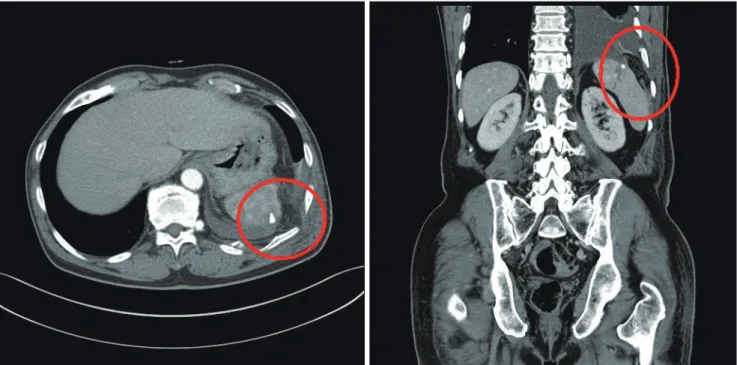
After 1 day, he was transferred to the general ward and was conservatively managed. The chest tube was removed after 5 days. He was stable without signs or symptoms of peritonitis during observation period. After 7 days, he had a follow-up AP CT which revealed improving sub- capsular hematoma of the spleen and retention of foreign
body. However, compared with the previous AP CT, it was observed that there was an increase in the fat density in the thoracic cavity above the spleen and remainder of the hemothorax (Fig. 2). A diaphragmatic injury was suspected. Video-assisted thoracic surgery (VATS) for exploration of the diaphragm was performed. After the evacuation of retained hematoma (300 mL), a herniated omentum was observed through the lacerated site of the diaphragm, which was approximately 4 cm. After resec- tion of omentum using an endoscopic stapler, a foreign body in the spleen was observed directly below the lacer- ated site of the diaphragm. After removal of the foreign body, no exsanguination or complications related to the procedure were observed in the spleen. The diaphragm was repaired by interrupted sutures using 2-0 PROLENE®
Polypropylene Suture (Ethicon Inc; Johnson & Johnson, Somerville, NJ, USA). The foreign body, which was sus- pected to be a bony fragment, was identified as a broken piece of the flowerpot (Fig. 3). He was discharged 14 days post-surgery without complications.
DISCUSSION
The incidence of penetrating diaphragmatic injuries as- sociated with the thoracoabdominal area is 11-19% [1].
Penetrating injuries in the abdomen are generally treated by laparotomy, but in some cases, without hemodynamic instability and signs of peritonitis, conservative manage-
Fig. 1. Chest computed tomography (CT) and abdominopelvic CT finding, red circle indicates a foreign body like a bone fragment in the spleen and
subcapsular hematoma in initially.
124
https://doi.org/10.20408/jti.2018.055Journal of Trauma and Injury Volume 32, Number 2, June 2019
ment is possible. However, delayed diagnosis of traumatic diaphragmatic rupture is associated with high morbidity and mortality in the long-term. It is recommended that thoracoscopy or laparoscopy is used for the diagnosis and repair of a missed diaphragmatic injury [2,3].
In cases with a retained foreign body in the peritoneal cavity, it can sometimes cause complications in the long- term, although it had not resulted in peritonitis or bleed- ing during the relatively short-term period. Some studies reported that a retained foreign body in a penetrating abdominal injury is a relative indication of laparotomy. In these selective patients, without other laparotomy indica-
tions, a more efficient management policy can be chosen [4]. The retained foreign body in the peritoneal cavity which has not been removed in the acute phase might have resulted in unpredicted complication and a long- term follow-up should be required [5-7]. In this case, ini- tial AP CT findings described the foreign body like a bony fragment and there was no peritonitis and other abdomi- nal injuries, so conservative manage was decided.
The diaphragm is the border between the thoracic and the peritoneal cavity. In the acute phase of trauma, if dia- phragmatic injury with concomitant injury of the thorax or the peritoneum is detected, it might be approached
Fig. 3. Intraoperative finding in video-assisted thoracic surgery, arrow indicate a foreign body.Fig. 2. Follow-up abdominopelvic computed tomography after 7 days, fat density increased in the thoracic cavity above spleen and retained a foreign
body in the spleen (red circle).
125
http://www.jtraumainj.org