외상 후 근관내로의 치조골 함입
임예진∙김영진∙김현정∙남순현 경북대학교 치의학전문대학원 소아치과학교실
영구치의 외상성 손상은 전체 외상 환자 중 높은 빈도로 발생하며 사고의 대부분은 치근이 미완성되어 있는 시기에 발생하 여 치수, 치주인대, 치조골, Hertwig 상피 근초(HERS)에 다양한 영향을 주게 된다. 손상 정도에 따라 치수의 완전한 재혈관 화, 근관 석회화, 근관내 치조골 함입 등의 다양한 치유 양상을 나타내며, 치근단의 성장 정지 및 치수 괴사로 인한 염증성 치 근 흡수 등의 합병증을 나타낼 수 있다.
본 증례에서는 미성숙 영구치 치근단을 가진 세 환아에서 외상에 의한 Hertwig 상피 근초의 손상으로 발치와 기저부에 존 재하는 치근막 세포와 골세포의 치수강내로의 증식으로 인해 치근 발육 정지 및 근관내로의 치조골 함입을 나타내어 보고하 고자 한다.
외상 후 Hertwig 상피 근초의 손상에 의한 근관내로의 치조골 함입 치유 양상은 치수는 정상적인 기능을 하는 것으로 생각 되며, 유착 등의 합병증이 동반되지 않는 경우 특별한 치료를 필요로 하지 않으므로 감별 진단이 요구되며, 외상 받은 치아의 치료시 Hertwig 상피 근초에 대한 부가적인 외상을 가하지 않도록 주의해야 한다.
주요어: Hertwig 상피 근초, 미성숙 치근단, 치조골 함입
Ⅰ. 서 론
여러 가지 질환 예방법의 발달에도 불구하고 치아 외상은 우 발적으로 발생하기 때문에 완전히 예방할 수 있는 방법은 없으 며, 특히 활동력이 왕성한 학령 전 및 학령기 아동에서는 구강 조직 및 치아에 손상을 받는 경우가 빈발하고 있다. 치아 외상 의 71~92%가 19세 이전에 발생하며1), 미성숙 영구 전치 치 근단을 가지는 혼합치열기인 8~10세 사이에 가장 많이 발생한 다2,3).
미성숙 영구치에서의 치아 외상의 경우 치아 경조직 및 치수, 치주인대, Hertwig 상피 근초, 치조골, 치은 및 구강 점막 등에 대한 손상이 포함될 수 있으며, 치수 괴사나 근관의 석회화, 치 근 성장의 장애, 치근의 염증성 및 대치성 흡수, 인접 치조골의 소실 등의 다양한 합병증이 발생할 수 있고, 외상받은 정도 및 치아의 발육 상태 등에 따라 치료 술식 및 예후도 달라지므로, 정확한 진단하에 치료하는 것이 중요하다1,4-7).
외상 후 합병증은 치수, 치주인대 등의 손상과 함께 Hertwig 상피 근초 등의 손상에 의해 발생할 수 있으며, 그 발생빈도는 치수와 치아 지지조직이 모두 손상을 받는 치아 탈구성 손상의 경우에 가장 높게 나타난다1).
특히 탈구성 손상에서는 외상 당시나 치료 당시 직접적인 손상 이나 외상에 대한 처치 후 불충분한 영양공급에 의해 Hertwig 상피 근초가 손상을 받게 될 가능성이 많다. Hertwig 상피 근초 가 손상된 경우 치근단 성장의 정지와 함께 발치와 기저부에 존 재하는 치근막 세포와 골세포가 치수강내로 증식 할 수 있다. 치 근막은 치수강 내벽에 백악질을 첨가하면서 치관측으로 증식하 고, 동시에 골조직이 치관측으로 침입 증식하게 되며 이로 인해 내부 치주인대에 의해 근관벽을 분리시키게 된다8,9).
본 증례들은 미성숙 치근단을 가진 영구 전치들의 탈구성 손 상 후 근관내 치조골 함입 양상을 보이며 치수 생활력을 유지하 고 있어 특별한 치료 없이 정기 검진을 시행하고 있어 보고하고 자 한다.
교신저자 : 남 순 현
대구광역시 중구 삼덕 2가 50번지 / 경북대학교 치의학전문대학원 소아치과학교실 / 053-600-7201 / [email protected] 원고접수일: 2011년 07월 30일 / 원고최종수정일: 2011년 10월 06일 / 원고채택일: 2011년 10월 08일
국문초록
Ⅱ. 증례보고 1. 증례 1
만 7세 된 남자 환아가 내원 5일 전 외상에 의해 앞니를 다친 후 잇몸이 찢어진 것을 봉합하고 치아 검사를 위해 내원하였다.
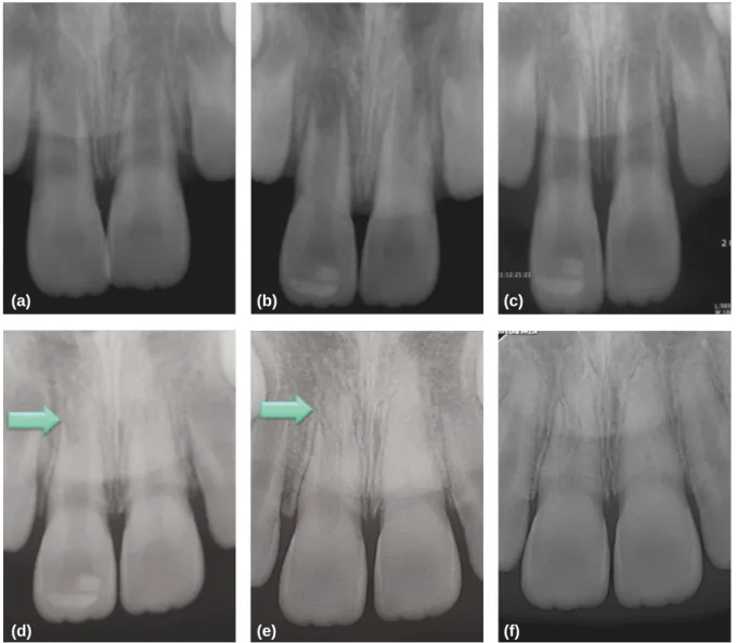
임상, 방사선 검사상 상악 우측 중절치의 정출과 상악 좌측 중 절치의 아탈구 소견을 보여(Fig. 1a), 정출된 치아를 재위치 시 킨 후 선부자 고정술을 시행하였다.
2주 후, 방사선 사진상 상악 우측 중절치 근단부의 방사선 투 과상이 약간 증가된 양상을 보이고(Fig. 1b), 상악 좌, 우측 중 절치 모두 냉검사와 전기 치수검사로 치수 생활력 검사시 반응 을 나타내지 않았으나 외상 초기의 미완성 치근단이고 치아의
변색이 관찰되지 않아 정기 검진을 시행하기로 하였다.
정기 검진시 지속적으로 치수 생활력 검사에 반응을 나타내 지 않다가, 4주후 방사선 사진상 상악 좌, 우측 중절치 근단부 는 비슷한 방사선 투과도를 보이며(Fig. 1c), 상악 우측 중절치 는 치수 생활력을 회복하였으나 상악 좌측 중절치는 여전히 반 응이 없었다.
6개월 후 방사선 사진상 상악 좌측 중절치의 근관벽이 많이 두꺼워진 것을 관찰 할 수 있고 냉검사에만 미약하게 반응을 하 였다. 이 시기에 상악 우측 중절치 치근의 원심측 치근단 1/3 위치에서 치근의 연속성이 끊어지는 듯이 보였는데(Fig. 1d), 1년 후 내원시 근관내 방사선 불투과성이 증가되어있으며, 선 형의 방사선 투과상이 함께 보여(Fig. 1e) 치조골과 치주인대 로 치유된 것으로 진단하였다.
(a) (b) (c)
(d) (e) (f)
Fig. 1. Periapical views (a) initial, apical PDL widening of upper right central incisor was seen. (b) after 2 weeks, apical radio-lucency of upper right central incisor was observed. (c) after 4 weeks, normal radio-graphic findings of upper central incisors was seen. (d) after 6 months, thickening of dentinal wall of upper left central incisor and root discontinuity of upper right central incisor were observed. (e, f) after 1,3 years, canal obliteration of upper left central incisor and increased radio-opacity in canal and inner PDL of upper right central incisor(invasion of alveolar bone into root canal) were seen.
3년 후의 방사선 사진상(Fig. 1f) 상악 우측 중절치의 치조골 의 함입과 상악 좌측 중절치의 근관 석회화로 진단하였으며 냉 검사와 전기 치수 검사에 모두 양성 반응을 나타내어 치수 생활 력을 유지하고 있었다.
2. 증례 2
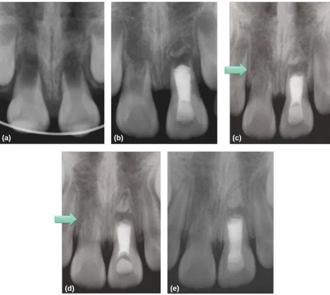
만 6세 된 여아로 외상으로 본원 응급실에 내원하였다. 상악 우측 중절치의 함입과 상악 좌측 중절치가 완전 탈구되었으며 이를 우유에 보관하여 내원하여, 탈구된 치아를 재위치 시킨 후 선부자 고정술을 시행하였다(Fig. 2a). 5주 후 상악 좌측 중절 치의 누공이 발견되어 근관 치료를 시행하였고, 상악 우측 중절 치는 냉검사와 전기 치수 검사에 반응이 없었으나 3개월 후 방
사선 사진상에서 치근단 부위 방사선 불투과상이 증가하는 양 상을 보였고(Fig. 2b) 이 시기에 치수 생활력은 유지되고 있었 다. 6개월 후 치수 생활력은 유지되고 있었고 근관내 치조골의 함입이 의심되었다(Fig. 2c). 1년 후 방사선 사진상 명확한 근 관내 치조골의 함입으로 진단할 수 있었으며(Fig. 2d), 수상 후 2년 후 마지막 검진시까지 치수 생활력은 유지되어 있었고, 초 진 방사선 사진과 비교하여 치근단의 성장은 정지된 양상을 보 이며 근관내 치조골의 함입을 확인할 수 있었다(Fig. 2e).
또한 완전 탈구된 상악 좌측 중절치의 치근단 부위에서 분리 된 형태의 치근의 성장을 관찰할 수 있었는데 이것은 외상당시 분리되어 남아있는 Hertwig 상피 근초가 정상적으로 기능을 하여 형성된 환상치근(phantom root)으로 보인다(Fig. 2b-f).
(a) (b) (c)
(d) (e)
Fig. 2. Periapical views (a) short root length and wide open apex of upper central incisors were seen. (b) after 3 months, increased radio-opacity in canal of upper right central incisor and phantom root formation of upper left central incisor were observed. (c) after 6 months, radio-opacity in canal and inner PDL upper right central incisor was seen. (d) after 1 years, invasion of alveolar bone into root canal upper right central incisor and growth of phantom root with apical barrier of upper left central incisor were seen. (e) after 2 years, root growth suspension with invasion of alveolar bone into root canal upper right cen- tral incisor was seen.
3. 증례 3
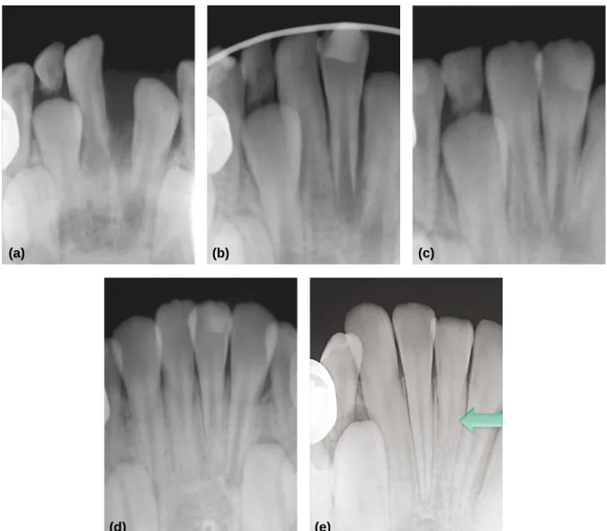
만 7세 된 남자 환아가 외상으로 하악 좌측 중절치의 완전 탈 구(Fig. 3a)를 주소로 응급실에 내원하여 탈구된지 4시간만에 재식 후 선부자 고정술을 시행였다. 치수괴사의 가능성이 높아 보였으나 외상 후 1개월(Fig. 3b), 3개월(Fig. 3c) 정기 검진 시에 치아는 생활력을 가지고 있었고 특별한 방사선 소견을 보 이지 않다가, 1년 후 방사선 사진상 인접한 하악 우측 중절치의 치근 길이에 비해 다소 짧은 치근 길이를 보였으며(Fig. 3d) 치 수 생활력은 유지하고 있었다. 2년 후 방사선 사진상 하악 좌측 중절치의 방사선 사진상 근단 1/3 부위에서 근관 내 치조골과 치주인대의 치유 양상을 보여(Fig. 3e) 정기 검진을 시행하고 있으며, 유착의 가능성이 있어 장기간의 검진을 필요로 한다.
Ⅲ. 총괄 및 고찰
치아의 형성과정에서 치관부의 법랑질과 상아질이 거의 완성 되면 치경 변곡부에서 내법랑질상피와 외법랑질상피가 거의 접 한 상태로 치근단부를 향하여 성장한다. 이렇게 서로 접한 두 층의 법랑질 상피를 Hertwig 상피 근초라고 하며 입방세포의 안쪽층과 좀더 평편한 세포의 바깥쪽으로 구성되어 있고, 때때 로 늘어난 세포의 중간층이 나타나기도 한다8,10,11).
치근 형성은 Hertwig 상피 근초의 활성에 의해 결정되며1,12-15), 치근 발육은 상피의 연속적인 증식에 의해 이루어진다16,17). Hertwig 상피 근초는 치아낭으로부터 치수를 분리하는 연속적 인 상피세포관으로, 치유두의 세포를 상아질 모세포로 유도분 화하여 치근부의 상아질을 형성하고 완성하도록 하며, 치근부
(a) (b) (c)
(d) (e)
Fig. 3. Periapical views (a) initial, totally avulsion of lower left central incisor was seen. (b, c) after 1, 3 months, normal radio-graphic findings of trauma- tized teeth were seen. (d) after 1 year, suspensions of root growth and dentinal wall thickening of lower left central incisor were seen. (e) after 2 years, inva- sion of alveolar bone into root canal of lower left central incisor was seen.
상아질이 치근단부로 성장함에 따라 Hertwig 상피 근초는 법 랑질 상피로부터 분리되어 근첨부로 이동한다8).
Hertwig 상피 근초는 외상에 민감한데, 미성숙 치아의 교정 적 함입과 같은 만성 외상은 종종 그것의 분열을 일으켜 근관 내로 들어간 상피 조각은 진성 치수석을 형성할 수 있고18-22), 치 주인대의 열상과 같은 근초에 가까운 외상은 근초의 일시적 과 활성을 일으켜 근첨부의 상아질과 백악질의 급격한 형성을 개 시하게 될 수 있으나, 근초의 활성은 나중에 정상으로 돌아오게 된다1).
유치의 함입 혹은 미성숙 영구치의 강력한 변위와 같은 급성 외상은 Hertwig 상피 근초에 직접적으로 전달되어 근초에 손 상을 일으켜 치근 발육의 부분 혹은 완전 정지를 가져올 수 있 으며1,23-27,28)
, 손상받은 Hertwig 상피 근초가 치주인대 기원 세 포의 근관내 이동을 막아주는 기능을 실행하지 못해, 발치와 기 저부의 치주인대 기원의 세포와 골세포가 치수강내로 증식하 며, 치근막은 치수강 내벽에 백악질을 첨가하면서 치관측으로 증식하고, 동시에 골조직이 치관측으로 침입 증식하게 된다. 최 종적으로는 근관내로 치조골이 함입되는 치유 양상을 보이게 된다8,29,30).
Hertwig 상피 근초의 손상에 따른 근관내 치조골 함입은 외 상성 손상 외에도 화학적 손상, 교정적 치료, 치아의 재식 치료 시에도 나타날 수 있는데, Robert 등31)은 함입된 미성숙 영구치 의 교정적 맹출에서 치주인대로 정상 치유되고 치수괴사의 소 견이 보이지 않던 치아에서 Hertwig 상피 근초의 합병증의 일 부로 근관내로 치조골과 치주인대가 성장한 증례를 소개하였으 며, Cveck과 Granatha 등26,27)의 실험에서 미성숙 치수내로의 치조골 함입이 상피 근초가 잘릴 때 혹은 포름 알데하이드를 적 용하여, 실활시키는 화학적 손상을 입을 때 발견되었다. 또한 Andreasen32)은 자가 이식 후 미완성 치근의 경우 76~94%, 완성치근의 경우 0~22% 정도에서 치수 생활력을 보이며, 재 식한 치아의 대부분의 경우가 치수의 재혈관화가 일어나지 못 하고 치근 발육이 부분적으로 혹은 완전히 정지하게 되며 치수 강은 폐쇄되거나 골조직으로 채워지게 된다고 하였다1).
건강한 치수와 Hertwig 상피 근초는 일반적으로 성장중인 치아의 적절한 치근 형성에 필수적인 것으로 간주되지만33) Andreasen34) 등은 재식된 치아의 치수 일부가 괴사된 상태에 서도 근관 치료 시행여부와 관계없이 치근 발육이 계속된 경우 를 보고하면서 Hertwig 상피 근초가 특정 환경에서는 약간의 염증 상태나 감염에 저항을 하면서 치근을 형성하거나 성장하 는 능력을 지속한다고 하였다. Hertwig 상피 근초가 파괴되지 않는다면 근단의 세포들을 구성하여 치수가 괴사된 후에도 지 속적인 치근 성장을 할 수 있도록 해주는데, 이것은 손상되지 않고 생활력을 유지하며 남아있는 Hertwig 상피 근초를 포함 하는 치수의 근단 부위에 존재하는 잔존하는 치성 세포(odon- togenic cell)에 의해서 가능하다35).
미성숙 영구치의 근관치료에서 reaming, filing 동작은 치근 단 조직에 손상을 가하여 잔존한 치성세포(odontogenic cell)에 손상을 가하여 정상적인 치근의 성장을 멈출 수도 있으므로36)
치수 괴사된 환아에서 근관 치료시 Hertwig 상피 근초를 손상 시키지 않도록 주의해야 하며, 또한 부분적으로 손상받은 근초 는 인접 근초의 성장에 따라 정상 기능을 회복할 수 있으므로30) 탈구성 손상을 받은 외상 환아에서 치아를 정복하는데 있어 부 가적으로 손상을 가하는 것을 최소화하도록 부드러운 조작이 필요하다.
근관내 치조골에 의한 치유에서 치수는 정상적인 기능을 하 며, 치료의 대상이 아니므로8)감별진단이 필요하다. 치수의 반 응으로 근관내 경조직의 침착이 특징적으로 나타나는 근관 석 회화와의 감별 진단이 필요한데 대부분 증상이 없고, 두가지 치 유 양상 모두 근관 치료를 필요로 하지 않는다.
근관내 치조골의 형성은 대부분이 치근 성장 정지와 함께 나 타나며, 근관내 방사선 불투과성이 증가하지만 상아질의 두께 증가는 없으며 선형태의 방사선 투과상 즉, 치주인대를 함께 보 인다. 반면 근관 석회화는 방사선학적 징후로 상아질의 두께 증 가로 인해 치관부 치수강의 크기가 작아지고 이어서 전반적인 근관이 좁아지는데 때때로 이는 부분 혹은 완전한 폐쇄를 보이 고 치근단 부위는 정상이다1,37). 또한 이환된 치아의 치관은 상 아질의 두께 증가로 인해 투과도가 감소함에 따라 인접한 정상 치아에 비해 어두운 색을 나타내며 노란색이나 회색을 나타낸 다. 일부 연구에서는 만약 재혈관화된 치수강 부위가 치주인대 조직 세포의 이동 없이 골조직으로만 대체된다면 유착이 발생 할 수 있다고도 하였다1,29,38,39). 이번 증례에서는 임상 소견상 유 착은 발생하지 않았으며 치수강 내 치주인대가 함께 함입된 것 을 확인할 수 있었다. 아쉽게도 세 번째 증례는 남아있는 치근 의 크기가 작아 치수강 내 치주인대 형성 유무나 유착 여부를 정확히 판단하기는 어려웠으나 치수강 부위의 방사선 투과성이 감소하는 것으로 보아 치수 부위가 재혈관화 되어 골조직으로 대체되어 가고 있으며, 임상 소견상 생리적 동요도를 보이고 치 아의 침강 현상은 보이지 않아 유착은 일어나지 않는 것으로 보 인다. 하지만 장기간의 관찰이 필요할 것으로 생각된다.
탈구성 손상을 받은 미성숙 영구치에서의 정기 검진을 통해 Hertwig 상피 근초의 손상으로 치근단의 성장 정지 및 근관내 치조골이 함입된 치유 양상을 관찰할 수 있었던 본 증례들을 통 해, Hertwig 상피 근초가 정상적인 치근 발육에 중요한 역할을 시행하고 있는 것을 확인할 수 있었으며 비록 치수가 골조직으 로 대체되었지만 정상적인 기능을 하고 있는 것을 확인할 수 있 었다. 아쉬운 점은 외상으로 인한 조직 반응이 다양하고, 합병 증의 발생 시기 또한 예측할 수 없는데 본 증례의 환아들은 5년 이하의 관찰기간으로만 한정되어 있어 앞으로의 장기간의 관찰 이 필요하다는 점이다.
Ⅳ. 요 약
미성숙 영구치 치근단을 가진 환아에서 탈구성 손상을 받아 응급 치료를 시행 후 정기 검진을 하던 중 치근 발육 정지 및 근 관 내 치조골 함입의 치유 양상을 보였다. 근관내 치조골 함입 에 의한 치유는 근관 석회화와 매우 유사한 방사선적 소견 및
임상양상을 나타내기 때문에 감별진단이 필요하며, 근관내 치 조골 함입의 중요한 방사선적 소견은 치근의 성장 정지, 근관내 치조골 함입 및 치주인대가 관찰되며 상아질의 형성이 중단되 는 등의 특징을 나타낸다. 또한 치수는 생활력을 유지하고 정상 적인 기능을 하고 있어 특별한 치료를 필요로 하지 않으나 유착 등의 합병증이 발생할 수 있으므로 정기적인 검사가 필요하며 본 증례에서 관찰한 바와 같이 Hertwig 상피 근초는 정상적인 치근형성에 매우 중요하므로 외상을 받은 초기 미성숙 영구치 의 더 이상의 부가적인 손상이 가해지지 않도록 주의하여야할 것으로 생각된다.
참고문헌
1. Andreasen JO, Andreasen FM, Andersson L : Textbook and color atlas of traumatic injuries to the tooth 4th edition. Blackwell Munksgaard, Copenhagen, Denmark, 71-72, 151-202, 217-254, 377-385, 302-304, 372-403, 415-416, 444-488, 669-715, 2007.
2. Malgren B : Decoronation - how, why, and when? J Calif Assoc, 28:846-845, 2000.
3. 정주현, 이제호, 김성오, 최병재 : 유치열과 혼합치열기 어 린이의 상악 절치부 외상. 대한소아치과학회지, 31:290- 298, 2004.
4. Andreasen JO : Etiology and pathogenesis of trau- matic dental injuries. A clinical study of 1,298 cases.
Scand J Dent Res, 78:329-342, 1970.
5. Andreasen JO, Ravn JJ : Epidermiology of traumat- ic dental injuries to primary and permanent teeth in a Danish population sample. Int J Oral Surg, 1:235- 239, 1972.
6. Hedegard B, Stalhane I : A study of traumatized permanent teeth in children aged 7-15 years. Part I.
Swed Dent J, 66:431-450, 1973.
7. Ravn JJ : Dental injuries in Copenhagen school chil- dren, school years 1967-1972. Community Dent.
Oral Epidermiol, 2:231-235, 1974.
8. 김도완 역 : 자가치아 이식. 나래출판사(주), 서울, 16, 49-50, 2000.
9. Andersson AW, Massler M : Periapical tissue reac- tions following root amputation in immediate tooth replants. Isr J Dent Med, 19:1-8, 1970.
10. Noble HW, Carmichael AF, Rankine DM : Electron microscopy of human developing dentine. Arch Oral Biol, 7:395-399, 1962.
11. OoE T : Human tooth and dental arch development.
Tokyo Ishiyaku Publishers, 86-94, 1981.
12. Diamond M, Applebaum E : The epithelial sheath :
histogenesis and fuction. J Dent Res, 21:403-411, 1942.
13. Grant D, Bernick S : Morphodifferentiation and structure of Hertwig’s root sheath in the cat. J Dent Res, 50:1580-1588, 1971.
14. Owens PDA : A light microscopic study of the devel- opment of the roots of premolar teeth in dogs. Arch Oral Biol, 20:525-538, 1974.
15. Owens PDA : Ultrastructure of Hertwig’s epithelial root sheath during early root development in premo- lar teeth in dogs. Arch Oral Biol, 23:91-104, 1978.
16. Diab MA, Stallard RE : A study of the relationship between epithelial root sheath and root develop- ment. Periodontics, 3:10-17, 1965.
17. Shibata F, Stern IB : Hertwig’s sheath in the rat incisors. II. Autoradiographic study. J Periodont Res, 2:111-120, 1968.
18. Stenvik A : Pulp and dentine reactions to experi- mental tooth intrusion(a histological study - long term effects). Trans Eur Orthod Soc, 45:449-464, 1970.
19. Stenvik A : The effect of extrusive orthodontic forces on human pulp and dentin. Scand J Dent Res, 79:430-435, 1971.
20. Stenvik A, Mjor IA : Epithelial remnants and denti- cle formation in the human dental pulp. Acta Odontol Scand, 28:721-728, 1970.
21. Stenvik A, Mjor IA : Pulp and dentine reactions to experimental tooth intrusion. A histological study of the initial change. Am J Orthod, 57:370-385, 1970.
22. Stenvik A, Mjor IA : The effect of experimental tooth intrusion on pulp and dentine. Oral Surg Oral Med Oral Pathol, 32:639-48, 1971.
23. Andreasen JO, Sundstrom B, Ravn JJ : The effect of traumatic injuries to primary teeth on their perma- nent successors. I. A clinical and histological study of 117 injured permanent teeth. Scand J Dent Res, 79:219-283, 1971.
24. Andreasen JO, Ravn JJ : The effect of traumatic injuries to primary teeth on their permanent succes- sors. II. A clinical and radiographic follow up study of 213 teeth. Scand J Dent Res, 79:284-294, 1971.
25. Andreasen JO : The influence of traumatic intrusion of primary teeth on their permanent successors. A radiographic and histological study in monkeys. Int J Oral Surg, 5:207-219, 1976.
26. Cvek M, Rudhmer M, Granath L-E, et al. : First permanent molars subjected to mortal amputation
before maturation of roots. I. Roentgenographic peri- radicular changes and peripheral root resorption after four years' observation. Odont Rev J, 20:119- 122, 1969.
27. Granath L-E, Hollender L, Cvek M, et al. : First permenent molars subjected to mortal amputation before maturation of roots. II. Roentgenologic and histologic study of ingrowth of vital tissue into devi- talized, fixed pulp tissue. Odont Rev J, 21:319-330, 1970.
28. Skoglund A, Tronstad L : Pulp changes in replanted and autotransplanted immature teeth of dogs. J Endod, 7:309-319, 1981.
29. Anderson AW, Massler M : Periapical tissue reac- tions following root amputation in immediate tooth replants. Isr J Dent Med, 19:1-8, 1970.
30. Andreasen JO, Kristerson L, Andreasen FM : Damage to the Hertwig’s epithelial root sheath : effect upon root growth after autotransplantation of teeth in monkeys. Endod Dent Traumatol, 4:145- 151. 1988.
31. Roberts J, Chrostopher O, Harold M : Conservative management of an intruded immature maxillary permanent central incisor with healing complication of pulp bone. Aust Enod J, 27;29-32, 2001.
32. Andreasen JO : Atlas of replatation and transplan- tation of teeth. Saunders, St. Louis, 16-57, 1992.
33. Torneck CD, Smith J : Biologic effects of endodontic
procedures on developing incisor teeth. I. Effect of partial or total pulp removal. Oral Sur Oral Med Oral Pathol, 30;258-66, 1970.
34. Andreasen JO, Borum MK, Andreasen FM : Replantation of 400 avulsed permanent incisors. 3.
Factors related to root growth. Endod Dent Traumatol, 11:69-75, 1995.
35. Safi L, Ravanshad S : Continued root formation of a pulpless permanent incisor following root canal treatment : a case report. Int Endod J, 38:489-493, 2005.
36. Torneck CD, Smith JS, Grindahl P : Biologic effects of endodontic procedures on developing incisor teeth.
II. Effect of debridement and disinfection procedures in the treatment of experimentally induced pulp and periapical disease. Oral Sur Oral Med Oral Pathol, 34: 532-540, 1973.
37. Patterson S, Mitchell D : Calcific metamorphosis of the dental pulp. Oral Surg Oral Med Oral Pathol, 20:94-101, 1965.
38. Andreasen JO, Borum MK, Jacobsen HL : Replantation of 400 avulsed permanent incisors. 2.
Factors related to pulpal healing Endod Dent Traumatol, 11:59-68, 1995.
39. 강유진, 김영진, 김현정, 남순현 : 외상으로 유착된 영구 전 치에서의 치관 절제술. 대한소아치과학회지, 37:252-259, 2010.
Abstract
INVASION OF ALVEOLAR BONE INTO ROOT CANAL AFTER TRAUMATIC INJURY
Ye-Jin Im, Young-Jin Kim, Hyun-Jung Kim, Soon-Hyeun Nam
Department of Pediatric Dentistry, School of Dentistry, Kyungpook National University
Traumatic injury on tooth occurs frequently among trauma patients, and mainly occurs on tooth with prema- ture roots which influences pulp tissue, periodontal ligament, alveolar bone, and Hertwig’s epithelial root sheath. According to the degree of trauma, a number of kinds of healing process can be observed, such as com- plete re-vascularization of pulp, root canal obliteration, growth suspension of root apex, and invasion of alveolar bone into root canal, and there can be some complications such as necrotic change of inflammatory root resorp- tion and partial pulp necrosis due to pulp necrosis toward complete necrosis.
In this clinical case, 3 patients who had traumatic injury showed root growth suspension and alveolar bone invasion into root canal due to proliferation of periodontal ligament cell and osteocyte at the base of extraction socket into pulp chamber because of the injury on Hertwig’s epithelial root sheath.
If intrusion of alveolar bone into root canal due to injury on Hertwig’s epithelial root sheath after having trau- matic injury doesn’t show any complication, the pulp may be considered to have normal vitality and doesn’t need any further treatment, therefore differential diagnosis is very necessary. However, it may be accompanied with suspension of root growth, therefore, additional trauma during the treatment of injured tooth should not be ap- plied.
Key words : Hertwig’s epithelial root sheath, Premature root, Intrusion of alveolar bone