Ann Hepatobiliary Pancreat Surg 2017;21:243-246
https://doi.org/10.14701/ahbps.2017.21.4.243
Case Report
Xanthogranulomatous pancreatitis mimicking potentially malignant pancreatic neoplasm: report of a case
Hyung Jun Kwon
Department of Surgery, Kyungpook National University Chilgok Hospital, Kyungpook National University School of Medicine, Daegu, Korea
Xanthogranulomatous pancreatitis (XGP) is a rare benign disease that may mimic or accompany other pancreatic diseases. Here we report a case of XGP initially suspected as malignant cystic neoplasm of the pancreas. A 64-year-old man had been incidentally found to have hypodense lesion at the body of pancreas during a lung cancer workup.
All laboratory tests were within normal limits except that carcinoembryonic antigen was elevated to 31.3 ng/ml. Imaging study showed 1.8 cm sized, well demarcated, and low-attenuated mass on computed tomography (CT) with heteroge- neously high intensity on T2-weighted images of magnetic resonance imaging (MRI). Under the impression of pancreas cystic neoplasm as a rare case of male solid-pseudopapillary tumor or pancreatic metastasis of lung cancer, laparo- scopic distal pancreatectomy was performed. Microscopically, the mass had many foamy histiocytes with cholesterol clefts, consistent with xanthogranulomatous inflammation. Therefore, it is important to consider XGP in the differential diagnosis of pancreatic diseases. (Ann Hepatobiliary Pancreat Surg 2017;21:243-246)
Key Words: Xanthogranulomatous; Inflammation; Pancreatitis; Neoplasms
Received: July 21, 2017; Accepted: August 13, 2017 Corresponding author: Hyung Jun Kwon
Department of Surgery, Kyungpook National University Chilgok Hospital, 807 Hoguk-ro, Buk-gu, Daegu 41404, Korea Tel: +82-53-200-2166, Fax: +82-53-200-2027, E-mail: [email protected]
Copyright Ⓒ 2017 by The Korean Association of Hepato-Biliary-Pancreatic Surgery
This is an Open Access article distributed under the terms of the Creative Commons Attribution Non-Commercial License (http://creativecommons.org/
licenses/by-nc/4.0) which permits unrestricted non-commercial use, distribution, and reproduction in any medium, provided the original work is properly cited.
Annals of Hepato-Biliary-Pancreatic Surgery ∙ pISSN: 2508-5778ㆍeISSN: 2508-5859
INTRODUCTION
Xanthogranulomatous inflammation is a rare idiopathic condition characterized by the aggregation of foamy his- tiocytes at various locations of the body along with chron- ic inflammation. These changes are found relatively com- mon in the gallbladder and kidney. However, they are ex- tremely rare in the pancreas.1-3 Including the present case, only 16 cases of xanthogranulomatous pancreatitis (XGP) have been reported previously in literatures written in English. Here we report a case of XGP initially suspected as malignant cystic neoplasm of the pancreas.
CASE
A 64-year-old man was admitted to the Surgical Department for further examination of a 1.8-cm sized hy- podense lesion incidentally found at the body of pancreas.
He was under examination for a 3.6-cm sized lung mass at the left upper lobe which was diagnosed as moderately
differentiated adenocarcinoma after left upper lobe lobectomy. The patient was a 44 pack-year smoker with diabetes requiring hyperglycemic agents. He had no re- ported history of alcohol intake, gallstones, or abdominal trauma.
His carcinoembryonic antigen (CEA) was elevated to 31.3 ng/ml. Liver function tests were normal. His serum levels were: AST, 29 IU/L; ALT, 24 IU/L; alkaline phos- phatase, 71 IU/L; total bilirubin, 0.81 mg/dl;. amylase, 49 IU/L; C-reactive protein (CRP), 2.78 mg/dl; and hemoglo- bin A1c level, 6.9%. CT scan showed a 1.8-cm sized and low-attenuated mass at the body of pancreas with upstream pancreatic duct dilatation (Fig. 1A). MRI demonstrated heterogeneously high intensity on T2-weighted images and low intensity on T1-weighted images (Figs. 1B and C).
The mass was well demarcated from the surrounding pan- creas tissue. There was no apparent peripancreatic fat tis- sue involvement or lymph node enlargement. Positron emission tomography-computed tomography (PET-CT) scan showed hypermetabolic mass at the upper lobe of the
244 Ann Hepatobiliary Pancreat Surg Vol. 21, No. 4, November 2017
Fig. 1. (A) Axial section of CT scan showing a round 1.8-cm sized cystic mass with upstream pancreatic duct dilation (arrow).
(B) MRI showing a lesion with heterogeneous low intensity on T1-weighted images (arrow). (C) MRI showing a heterogeneous high intensity on T2-weighted images (arrow). (D) PET-CT showing no significant FDG uptake.
left lung, the known lung cancer lesion site. However, no significant 2-[18F]-fluoro-2-deoxy-d-glucose (FDG) uptake was found at the pancreas lesion (Fig. 1D). Endoscopic ultrasound-guided fine needle aspiration was performed for pathological diagnosis. However, it failed due to in- adequately retrieved specimen for determination. Under the impression of pancreas cystic neoplasm as a rare case of male solid-pseudopapillary tumor or pancreatic meta- stasis of lung cancer, laparoscopic distal pancreatectomy with splenectomy was performen on June 13, 2016.
Gross examination of resected specimen revealed a 1.6 cm×1.5 cm sized yellowish mass in the pancreatic body (Fig. 2A). Microscopically, the mass had many foamy his- tiocytes with cholesterol clefts (Figs. 2B and C), con- sistent with xanthogranulomatous inflammation. Thus, the cystic lesion was diagnosed as XGP. The patient recov-
ered fully without any complication at two weeks after surgery. There were no signs of recurrence during seven months of follow up.
DISCUSSION
Including the present case, a total of 16 cases of XGP have been reported world-wide in literatures written in English. Among them, males were predominant (12 males and 4 females). The mean age of these patients was 59.8 year (range, 30-82). These lesions were evenly distributed throughout the pancreas in the head (4/16), body (7/16), and tail (5/16). A total of 11 cases underwent surgery with clinically suspected malignant pancreas lesion.1-9 Among them, three cases2,5,7 were postoperatively confirmed to be combined intraductal papillary mucinous neoplasm. One
Hyung Jun Kwon. Xanthogranulomatous pancreatitis mimicking malignant pancreatic neoplasm 245
Fig. 2. (A) Gross examination of the resected specimen showing a 1.6 cm×1.5 cm sized yellowish mass. (B, C) Microscopic examination of the specimen by H&E staining showing many cholesterol clefts with foamy histiocytes (B: ×20, C: ×200).
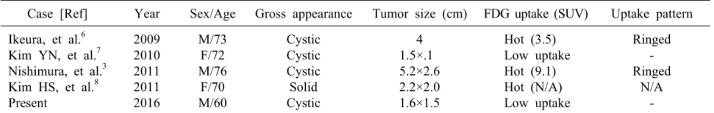
Table 1. FDG uptake pattern of xanthogranulomatous pancreatitis lesion
Case [Ref] Year Sex/Age Gross appearance Tumor size (cm) FDG uptake (SUV) Uptake pattern Ikeura, et al.6
Kim YN, et al.7 Nishimura, et al.3 Kim HS, et al.8 Present
2009 2010 2011 2011 2016
M/73 F/72 M/76 F/70 M/60
Cystic Cystic Cystic Solid Cystic
4 1.5×.1 5.2×2.6 2.2×2.0 1.6×1.5
Hot (3.5) Low uptake Hot (9.1) Hot (N/A) Low uptake
Ringed - Ringed
N/A - FDG, 2-[18F]-fluoro-2-deoxy-d-glucose; SUV, standardized uptake value; N/A, not available
case9 was combined with mucinous cystic neoplasm.
Xantogranulomatous cholecystitis (XGC) is well-recog- nized entity characterized by aggregation of foamy histio- cytes and inflammatory cells.4 The pathogenesis of XGC is currently unclear, although many hypotheses have been proposed. The generally accepted pathogenesis of XGC involves a series of events similar to xanthogranulomatous pyelonephritis. As described by Roberts and Parsons,10 es- sential components of XGC are inflammation associated with infection, obstruction of the biliary outflow from the gall bladder due to calculi, and a source of lipid such as biliary cholesterol. Gall bladder with acute or chronic cholecystitis and partial or total obstruction of bile out- flow can results in rupture of Rokitansky-Aschoff sinuses or mucosal ulceration, causing extravasation of bile into the gall bladder wall. Histiocytes accumulation in attempt to phagocytose the biliary cholesterol might result in for- mation of xanthoma cells. Similarly, it has been postu- lated that XGP might develop in the pancreas following extravasation of mucin due to obstructive changes in the pancreatic duct system.2,5,9 With extravasation of mucin to adjacent pancreas parenchyma, inflammatory reaction might have developed. As histiocytes accumulate to phag- ocytose destroyed tissues, consumed adipocytes may re- sult in xanthogranulomatous changes. In our case, the pa-
tient first developed 1.2-cm sized cyst that was increased to 1.8-cm over six months. Upstream pancreatic duct dila- tation was prominent next to the mass at the body of pancreas. Grossly, it appeared as a 1.6-cm sized well-de- marcated mass filled with yellowish material as if cystic lesion first developed and xanthogranulomatous material filled up the cavity later on. We believe that cystic lesion with necrotized parenchyma might have first developed through unknown reason. Over a long period of time, in- flammatory change of the lesion might have caused xan- thogranulomatous changes. However, the pathogenesis of XGP needs to be studied further, considering the fact that XGP is extremely rare condition compared to numerous diseases. In addition, the conditions that can cause ductal obstruction with inflammation should be considered.
XGP clinically and grossly often mimics other mass forming pancreatic lesions.9 It is extremely difficult to dis- tinguish XGP from pancreatic neoplasm through imaging studies.5 Thus, radiologically it is often confused with car- cinoma of the pancreas. There are no definite character- istics that can differentiate XGP from other pancreatic lesions. In our case, the lesion appeared as cystic lesion with upstream pancreatic duct dilatation. The increase in size in six months led to an impression of possible malig- nant cystic neoplasm. MRI showed heterogeneous high in-
246 Ann Hepatobiliary Pancreat Surg Vol. 21, No. 4, November 2017
tensity on T2-weighted images and heterogeneous low signal intensity on T1-weighted images, leading to the im- pression of solid pseudopapillary tumor. Nishimura et al.3 have suggested that ringed uptake pattern of FDG-PET may indicate a diagnostic feature of cystic-type XGP.
After reviewing six cases of XGP with FDG-PET, four cases showed hot uptake (Table 1). Among these four cystic type cases, only two cases had ringed uptake pattern. The other two cases did not show significant up- take, including the present case. They were relatively small in gross size (1.5 cm and 1.6 cm, respectively) com- pared to those with ringed uptake (4 cm and 5.2 cm, re- spectively). In comparison, two solid type cases showed hot uptake, although they were relatively small in size (2.2 cm, both). Since XGP is associated with in- flammatory reaction, glucose metabolism of the lesion might be increased. Therefore, false-positive FDG uptake may result in XGP. Furthermore, ringed uptake pattern may indicate cystic type XGP. However, such findings alone are insufficient for differential diagnosis of XGP from other pancreatic diseases.
Xanthogranulomatous pancreatitis is a rare benign dis- ease that may mimic or accompany other pancreatic dis- eases in imaging studies. Therefore, it is important to con- sider it in the differential diagnosis of pancreatic diseases.
However, more cases need to be reviewed to identify unique characteristics of XGP for differential diagnosis.
REFERENCES
1. Iyer VK, Aggarwal S, Mathur M. Xanthogranulomatous pan- creatitis: mass lesion of the pancreas simulating pancreatic carci- noma--a report of two cases. Indian J Pathol Microbiol 2004;
47:36-38.
2. Kamitani T, Nishimiya M, Takahashi N, Shida Y, Hasuo K, Koizuka H. Xanthogranulomatous pancreatitis associated with in- traductal papillary mucinous tumor. AJR Am J Roentgenol 2005;185:704-707.
3. Nishimura M, Nishihira T, Hirose T, Ishikawa Y, Yamaoka R, Inoue H, et al. Xanthogranulomatous pancreatitis mimicking a malignant cystic tumor of the pancreas: report of a case. Surg Today 2011;41:1310-1313.
4. Shima Y, Saisaka Y, Furukita Y, Nishimura T, Horimi T, Nakamura T, et al. Resected xanthogranulomatous pancreatitis.
J Hepatobiliary Pancreat Surg 2008;15:240-242.
5. Iso Y, Tagaya N, Kita J, Sawada T, Kubota K.
Xanthogranulomatous lesion of the pancreas mimicking pancre- atic cancer. Med Sci Monit 2008;14:CS130-CS133.
6. Ikeura T, Takaoka M, Shimatani M, Koyabu M, Kusuda T, Suzuki R, et al. Xanthogranulomatous inflammation of the peri- pancreatic region mimicking pancreatic cystic neoplasm. Intern Med 2009;48:1881-1884.
7. Kim YN, Park SY, Kim YK, Moon WS. Xanthogranulomatous pancreatitis combined with intraductal papillary mucinous carci- noma in situ. J Korean Med Sci 2010;25:1814-1817.
8. Kim HS, Joo M, Chang SH, Song HY, Song TJ, Seo JW, et al. Xanthogranulomatous pancreatitis presents as a solid tumor mass: a case report. J Korean Med Sci 2011;26:583-586.
9. Hanna T, Abdul-Rahman Z, Greenhalf W, Farooq A, Neoptolemos JP. Xanthogranulomatous pancreatitis associated with a mucinous cystic neoplam. Pathol Int 2016;66:174-176.
10. Roberts KM, Parsons MA. Xanthogranulomatous cholecystitis: clin- icopathological study of 13 cases. J Clin Pathol 1987;40:412-417.