ISSN 2234-3806 • eISSN 2234-3814
http://dx.doi.org/10.3343/alm.2013.33.1.52
Elevated Levels of T Helper 17 Cells Are Associated with Disease Activity in Patients with Rheumatoid Arthritis
Jimyung Kim, M.D.1, Seongwook Kang, M.D.2, Jinhyun Kim, M.D.2, Gyechul Kwon, M.D.1, and Sunhoe Koo, M.D.1
Departments of Laboratory Medicine1, Rheumatology2, Chungnam National University College of Medicine, Daejeon, Korea
Background: Interleukin-17 (IL-17)-producing T helper (Th) 17 cells are considered as a new subset of cells critical to the development of rheumatoid arthritis (RA). We aimed to investigate the distribution of Th1 and Th17 cells and their association with disease activi- ty, and determine the Th17-related cytokine levels in the peripheral blood of RA patients.
Methods: Peripheral blood mononuclear cells from 55 RA and 20 osteoarthritis (OA) pa- tients were stimulated with mitogen, and the distributions of CD4+Interferon (INF)+IL-17- (Th1 cells) and CD4+INF-IL-17+ (Th17 cells) were examined by flow cytometry. Serum lev- els of IL-6, IL-17, IL-21, IL-23, and tumor necrosis factor (TNF)-α were measured by ELI- SA. Erythrocyte sedimentation rate (ESR) and C-reactive protein (CRP) were recorded.
The 28-joint disease activity score (DAS28) was also assessed.
Results: The median percentage of Th17 cells was higher in RA patients than in OA pa- tients (P =0.04), and in active than in inactive RA (P =0.03), whereas that of Th1 cells was similar in both groups. Similarly, the levels of IL-17, IL-21, and IL-23 were detected in a significantly higher proportion of RA patients than OA patients and the frequencies of de- tectable IL-6, IL-17, and IL-21 were higher in active RA than in inactive RA group. The percentage of Th17 cells positively correlated with the DAS28, ESR, and CRP levels.
Conclusions: These observations suggest that Th17 cells and Th17-related cytokines play an important role in RA pathogenesis and that the level of Th17 cells in peripheral blood is associated with disease activity in RA.
Key Words: Rheumatoid arthritis, T helper 1 cell, T helper 17 cell, Cytokines
Received: June 5, 2012 Revision received: August 8, 2012 Accepted: November 6, 2012 Corresponding author: Sunhoe Koo Department of Laboratory Medicine, Chungnam National University College of Medicine, 282 Munhwa-ro, Jung-gu, Daejeon 301-721, Korea
Tel: +82-42-280-7798 Fax: +82-42-257-5365 E-mail: [email protected]
© The Korean Society for Laboratory Medicine.
This is an Open Access article distributed under the terms of the Creative Commons Attribution Non-Commercial License (http://creativecom- mons.org/licenses/by-nc/3.0) which permits unrestricted non-commercial use, distribution, and reproduction in any medium, provided the original work is properly cited.
INTRODUCTION
Rheumatoid arthritis (RA) is a systemic autoimmune disorder characterized by articular inflammation eventually leading to joint destruction. Although the mechanism of RA pathogenesis is not fully understood, humoral and cellular immunity are known to be involved. In particular, CD4+ T lymphocytes and their cytokines have been reported to play a major role in the initiation of inflam- mation in this disease [1, 2].
CD4+ T helper (Th) cells are activated by the antigenic stimu-
lation of T-cell receptors and differentiated into different subsets of effector Th cells. Among these cells, interferon-γ (INF-γ)-pro- ducing Th1 cells are predominant in RA [3]. Recent reports have suggested that interleukin-17 (IL-17)-producing Th17 cells are a new subset of cells critical to the pathogenesis of RA [2, 4]. IL- 17 induces the production of inflammatory cytokines such as IL-1, IL-6, IL-8, and tumor necrosis factor-α (TNF-α) [5, 6], and it has been detected in the serum, synovial fluid (SF), and syno- vium of patients with RA [4, 7].
However, very few studies have focused on the role of Th17
cells in arthritis, and the published results are controversial. A previous report has suggested that Th17 cells are proinflamma- tory and play an important role in various autoimmune diseases, including RA [8]. Subsequently, Zhang et al. reported that the percentage of pure Th17 cells (CD4+INFγ-IL-22-IL17+) was signif- icantly higher in RA patients than in osteoarthritis (OA) patients and healthy controls [9]. In contrast, Yamada et al. [10] reported that the frequency of Th17 cells was neither increased in the SF or peripheral blood (PB) of RA patients nor correlated with dis- ease activity. Another study showed that the percentage of Th17 cells in the SF correlated with inflammatory activity in arthritis, irrespective of the type of disease [11]. To date, a few studies have quantified the prevalence of Th17 cells in RA, and their re- sults have been discrepant [9-12]. Therefore, it is necessary to analyze the prevalence of Th17 cells in RA.
Recently, the Th17 cell-associated cytokines have been pro- posed. IL-6 and TNF-α induce the differentiation of Th17 cells, whereas IL-21 and IL-23 promote the proliferation of Th17 cells [8, 13, 14]. Recent studies [12, 15] have shown that the plasma levels of IL-21 and IL-23 are increased in RA and correlated with disease activity. In addition, a previous study reported that IL-23 levels are correlated with the SF and serum levels of IL-17 and TNF-α [16]. However, only a few previous studies have simulta- neously examined the levels of major cytokines in RA.
In this study, we investigated the distribution of Th1 and Th17 cells in the PB and SF of RA and OA patients and measured the levels of related cytokines. We also evaluated the frequencies of Th1 and Th17 cells with regard to disease activity of RA and the relationship between their frequencies and inflammatory markers.
METHODS 1. Patients
In this study, we prospectively enrolled 55 RA patients (age, 55.2
±14.1 yr; 14 men and 41 women) and 20 OA patients (age, 61.1
±13.3 yr; 4 men and 16 women) referred to the outpatient clinic of the Rheumatology Department at Chungnam National Uni- versity Hospital between September and November 2011. Among the 55 RA patients, 46 had been receiving disease-modifying antirheumatic drugs (DMARD). Corticosteroids ( ≤10 mg/day) were given to 39 patients, whereas the remaining 9 were newly diagnosed patients naïve to steroids and DMARD. Disease activ- ity was assessed using the 28-joint disease activity score (DAS28) [17]. The study protocol was approved by the Institutional Re- view Boards of Chungnam National University Hospital and writ- ten informed consent was obtained from all participants.
The erythrocyte sedimentation rate (ESR) level and serum levels of C-reactive protein (CRP), rheumatic factor (RF), and anti-cyclic citrullinated peptide (CCP) were also measured. PB and SF samples were collected for mononuclear cell separation.
SF was aspirated only in patients presenting with symptomatic effusion. Serum samples were collected and stored at 80˚C until assayed.
2. Th1 and Th17 analyses
1) Lymphocyte stimulationPB and SF were diluted with phosphate-buffered saline (PBS;
1:1 and 1:2, respectively) and then layered on Ficoll-Paque sep- aration media (GE Healthcare Bio-Science AB, Uppsala, Swe- den). Mononuclear cells were separated by centrifugation and stimulated in 48-well plates at 1×10⁶ cells/well with 50 ng/mL of phorbol myristate acetate (PMA) (Sigma Aldrich, St. Louis, MO, USA) and 1 μg/mL ionomycin in the presence of 10 μg/mL Brefeldin A for 6 hr at 37˚C in a 5% CO2 incubator.
2) Flow cytometric analysis
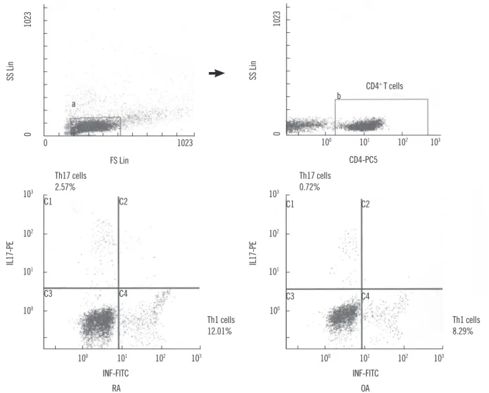
After stimulation, the cells were collected and washed with PBS (1,500 rpm for 10 min); thereafter, 5×10⁵ cells were resus- pended in 100 μL of PBS and incubated in the dark at room temperature for 30 min with 10 μL of anti-CD4-phycoerythrin- cyanin (PC) 5 (Beckman Coulter, Marseille, France). The cells were then fixed and permeabilized with IntraPrep (Beckman Coulter) according to the manufacturer’s instructions. Subse- quently, the cells were incubated in the dark at room tempera- ture for 30 min with 20 μL anti-INF-γ-fluorescein-isothiocyanate (FITC) (Beckman Coulter) and 5 μL anti-IL-17-phycoerythrin (PE) (eBioscience, San Diego, CA, USA). The samples were an- alyzed using a Cytomics FC 500 flow cytometer (Beckman Coul- ter). In the flow cytometric analysis, lymphocytes were first gated in a forward scatter and side scatter, and then, at least 10,000 CD4-positive cells were analyzed. The CD4-positive cells were further classified as CD4+INFγ+IL17- cells (Th1), CD4+INFγ-IL17+ cells (Th17), and CD4+INFγ+IL17+ cells (Fig. 1). The isotype con- trols IgG1-FITC and IgG1-PE (Beckman Coulter) were used to correct compensation and detect nonspecific staining.
3. Determination of IL-6, IL-17, IL-21, IL-23, and TNF-α
The serum levels of IL-6, IL-17, IL-21, IL-23, and TNF-α were quantified by ELISA according to the manufacturer’s instruc- tions (eBioscience). All stored samples were diluted 1 in 2 with the sample diluent provided in the kit and analyzed in duplicate.The concentration was calculated using the average optical den-
sity (OD) value. Serum samples with a concentration exceeding the highest standard were rediluted. The minimum detection limits for IL-6, IL-17, IL-21, IL-23, and TNF-α were 0.92, 0.5, 20.0, 4.0, and 2.3 pg/mL, respectively.
4. Statistics
All statistical analyses were performed using the MedCalc statis- tical software 11.3.0.0 (MedCalc Software, Mariakerke, Belgium).
Continuous variables are presented as the mean±SD or median (95% confidence interval [CI]). Intergroup comparisons were made using the Student’s t-test, Mann-Whitney U test, or Krus- kal-Wallis test, where applicable. To assess the correlation be- tween 2 parameters, linear regressions were calculated. Detec- tion frequencies of cytokines were compared between groups
Fig. 1. The percentage of Th1 cells and Th17 cells in representative rheumatoid arthritis (RA) and osteoarthritis (OA) patients. Lympho- cytes (a) were gated in a forward scatter (FS)/side scatter (SS) dot plot, and then CD4+ T cells (b) within indicated lymphocytes were isolat- ed. CD4+ T cells were divided into CD4+INFγ+IL17- (Th1 cells), CD4+INFγ-IL17+ (Th17 cells) and CD4+INFγ+IL17+ cells.
Abbreviations: Th1, T helper 1; Th17, T helper 17; RA, rheumatoid arthritis; OA, osteoarthritis; INF, interferon; IL, interleukin.
SS Lin
FS Lin a
0 0
1023
1023
IL17-PE
100 101 102 103
C1
C3
C2
C4
INF-FITC RA Th17 cells 2.57%
Th1 cells 12.01%
100 101 102 103
IL17-PE
C1
C3
C2
C4
OA Th17 cells
0.72%
Th1 cells 8.29%
INF-FITC
100 101 102 103 100
101 102 103
SS Lin
CD4-PC5 CD4+ T cells b
0 100 101 102 103
1023
using the chi-square test or Fisher’s exact test when appropri- ate. A P value of less than 0.05 was considered statistically sig- nificant.
RESULTS
1. Patient characteristics
As illustrated in Table 1, no significant differences were observed between the RA and OA patients with regard to age and sex dis- tribution. Furthermore, hemoglobin concentration, leukocyte count, and platelet count in RA patients were similar to those in OA patients. In addition, the 2 patient groups showed similar se- rum levels of CRP and ESR.
2. Frequency of Th1 and Th17 cells in RA and OA
PB samples were obtained from all 75 patients, and SF samples were collected from 3 RA patients and 5 OA patients. We identi- fied Th1 and Th17 cells in PB and/or SF mononuclear cells after the samples were treated with PMA and ionomycin. Neither Th1 cells nor Th17 cells were detected in the RA and control speci- mens without stimulation treatment (data not shown).
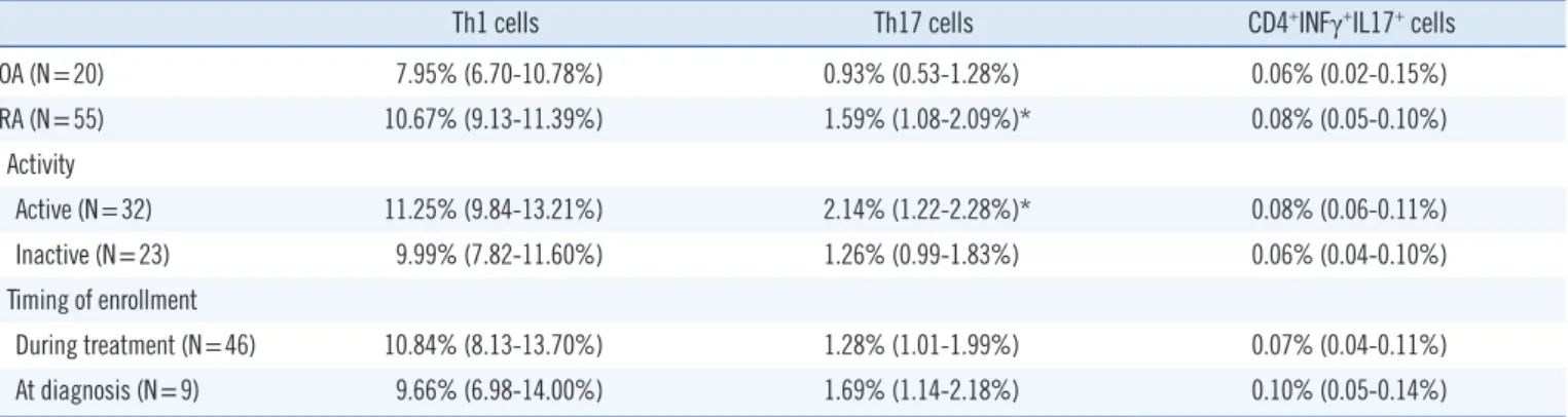
The median percentages of CD4+ T cells were 30.05% (26.62- 33.05%) and 32.42% (15.15-34.28%) in the RA and OA patients, respectively, indicating no significant intergroup difference (P = 0.69). The median percentages of Th1 cells, Th17 cells, and CD4+INFγ+IL17+ cells were 10.67% (9.13-11.39%), 1.59% (1.08- 2.09%), and 0.08% (0.05-0.10%), respectively, in the PB mono- nuclear cells (PBMCs) of RA patients, and 7.95% (6.70-10.78%), 0.93% (0.53-1.28%), and 0.06% (0.02-0.15%), respectively, in PBMCs of OA patients. The frequency of Th17 cells was signifi- cantly higher in RA patients than in OA patients (P =0.04), but the frequency of Th1 cells did not differ significantly between
the 2 groups (P =0.19) (Table 2).
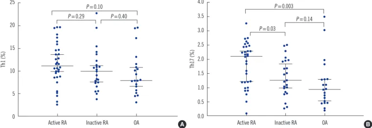
The median percentages of Th1 cells and Th17 cells were 11.25% (9.84-13.21%) and 2.14% (1.22-2.28%) in the 32 active RA patients (DAS≥2.6), and 9.99% (7.82-11.60%) and 1.26% (0.99-1.83%) in the 23 inactive RA patients. The frequency of Th17 cells was significantly higher in active RA patients than in inactive RA patients (P =0.03) or OA patients (P =0.003), but no significant difference in the frequency of Th1 cells was observed between active RA and inactive RA (P =0.29) or between active RA and OA patients (P =0.10) (Fig. 2). The median percentages of Th1 cells and Th17 cells were 9.66% (6.98-14.00%) and 1.69% (1.14-2.18%) in the 9 newly diagnosed RA patients, and 10.84% (8.13-13.70%) and 1.28% (1.01-1.99%) in the 46 RA patients under treatment. The frequencies of Th1 and Th17 cells did not show any significant difference between the 2 groups according to the treatment (P =0.53 and P =0.39, respectively) (Table 2). SF samples of 8 patients were analyzed. For the 3 RA patients, the median percentages of Th1 cells, Th17 cells, and CD4+INFγ+IL17+ cells were 12.91%, 1.92%, and 0.26% of the SF mononuclear cells, respectively, and for the 5 OA patients, the corresponding values were 10.68%, 1.19%, and 0.13%. These 8 patients, whose SF samples were analyzed for determining the frequency of Th1 and Th17 cells, showed higher percentages of both cell types in the SF samples than in PB samples although the number of samples tested was very small.
3. Relationships of the frequency of Th1 and Th17 cells with inflammatory indices or disease activity in RA patients
The percentage of Th1 cells was not significantly correlated with that of Th17 cells in both RA (r=0.26, P =0.07) and OA patients (r=0.31, P =0.12). In the RA patients, a significant positive cor- relation was found between the percentages of Th17 cells in theTable 2. The percentages of Th1 cells, Th17 cells, and CD4+INFγ+ IL17+ cells in the peripheral blood from the subgroups of patients
Th1 cells Th17 cells CD4+INFγ+IL17+ cells
OA (N=20) 7.95% (6.70-10.78%) 0.93% (0.53-1.28%) 0.06% (0.02-0.15%)
RA (N=55) 10.67% (9.13-11.39%) 1.59% (1.08-2.09%)* 0.08% (0.05-0.10%)
Activity
Active (N=32) 11.25% (9.84-13.21%) 2.14% (1.22-2.28%)* 0.08% (0.06-0.11%)
Inactive (N=23) 9.99% (7.82-11.60%) 1.26% (0.99-1.83%) 0.06% (0.04-0.10%)
Timing of enrollment
During treatment (N=46) 10.84% (8.13-13.70%) 1.28% (1.01-1.99%) 0.07% (0.04-0.11%)
At diagnosis (N=9) 9.66% (6.98-14.00%) 1.69% (1.14-2.18%) 0.10% (0.05-0.14%)
Data are presented as median (95% confidence interval).
*P <0.05 vs. OA.
Abbreviations: Th1, T helper 1; Th17, T helper 17; INF, interferon; IL, interleukin; RA, rheumatoid arthritis; OA, osteoarthritis.
Table 1. Clinical characteristics and laboratory findings in RA and OA patients
RA (N=55) OA (N=20) P value
Mean age (yr) 55.2±14.1 61.1±13.3 0.11
Male:Female 14:41 4:16 0.76
Hb (g/L) 125±14 121±18 0.39
Leukocyte (×109/L) 7.4±2.2 6.6±2.3 0.17
Platelet (×109/L) 261±54 265±80 0.81
CRP (mg/L) 12±4 11±4 0.81
ESR (mm) 28.2±3.8 26.0±5.1 0.70
Data are presented as mean±SD or as numbers.
Abbreviations: RA, rheumatoid arthritis; OA, osteoarthritis; CRP, C-reactive protein; ESR, erythrocyte sedimentation rate.
PBMCs and serum CRP (r =0.34, P =0.01) or ESR levels (r = 0.30, P =0.03). However, Th1 cells in the PBMCs were not cor- related with CRP or ESR levels. Furthermore, the percentages of the Th1 and Th17 cells in the PB were not correlated with leu- kocyte count or neutrophil percentage. In addition, Th1 and Th17 cell percentages in the PB were not correlated with the serum RF or anti-CCP levels. However, the frequencies of Th17 cells were positively correlated with the DAS28 (r=0.43, P =0.01), whereas those of Th1 cells were not (r=0.14, P =0.27) (Fig. 3).
A trend of positive correlation between the percentages of Th17 cells and DAS28 was also observed in the 32 active RA patients but was not significant (r=0.30, P =0.13).
4. Serum cytokines in RA and OA
IL-17 and IL-21 levels were detectable in 19 (34.5%) and 25
(45.4%) RA patients, respectively, but in none of the OA patients (Table 3). Detectable serum levels of IL-23 were present in 40 (72.7%) RA patients and 1 (5.0%) OA patient, whereas serum TNF-α levels were detected in the serum of only 2 (3.6%) RA Table 3. Frequencies of positivity (%) of serum cytokines in RA and OA patients
Cytokine RA (N=55) OA (N=20) P value
IL-6 36.4 (20/55) 50.0 (10/20) 0.30
IL-17 34.5 (19/55) 0.0 (0/20) 0.002
IL-21 45.4 (25/55) 0.0 (0/20) <0.0001
IL-23 72.7 (40/55) 5.0 (1/20) <0.0001
TNF-α 3.6 (2/55) 0.0 (0/20) 1.00
Abbreviations: IL, interleukin; TNF, tumor necrosis factor; RA, rheumatoid arthritis; OA, osteoarthritis.
Fig. 3. Correlation between the percentage of Th17 cells and C-reactive protein (A) as well as DAS28 (B) in RA patients.
Abbreviations: Th, T helper; DAS28, 28-joint disease activity score; RA, rheumatoid arthritis.
A B
Percentage of Th17 cells
3.5 3.0 2.5 2.0 1.5 1.0 0.5 0.0
C-reactive protein (mg/dL)
0 2 4 6 8 10 r=0.34, P =0.01
Percentage of Th17 cells
3.5 3.0 2.5 2.0 1.5 1.0 0.5 0.0
DAS28 score
0 1 2 3 4 5 6 r=0.43, P =0.01
Fig. 2. Frequencies of Th1 cells (A) and Th17 cells (B) in patients with active rheumatoid arthritis (RA), patients with inactive RA, and pa- tients with osteoarthritis (OA). Horizontal lines represent median and error bars represent a 95% interval.
Abbreviation: Th, T helper.
A B
Th1 (%)
25
20
15
10
5
0 Active RA
P =0.10 P =0.29 P =0.40
Inactive RA OA
Th17 (%)
4.0 3.5 3.0 2.5 2.0 1.5 1.0 0.5
0.0 Active RA
P =0.003
P =0.03
P =0.14
Inactive RA OA
patients. Serum IL-6 levels were detectable in 20 (36.4%) RA pa- tients and 10 (50.0%) OA patients. The frequency of detectable serum levels of IL-17, IL-21, and IL-23 was significantly higher in RA patients than in OA patients (P =0.002, P <0.0001, and P <
0.0001, respectively), but that of IL-6 and TNF-α did not show any intergroup differences (P =0.30 and P =1.00, respectively).
In active RA patients, IL-6, IL-17, IL-21, IL-23, and TNF-α were detected in 50.0% (16/32), 46.9% (15/32), 62.5% (20/32), 81.3% (26/32), and 6.3% (2/32) patients, respectively. However, inactive RA patients showed detectable cytokines in 17.4% (4/23), 17.4% (4/19), 21.7% (5/23), 60.9% (14/23), and 0.0% (0/23) patients, respectively. The frequency of detectable serum levels of IL-6, IL-17, and IL-21 was significantly higher in active RA patients than in inactive RA patients (P =0.02, P =0.04, and P =0.006, re- spectively), but the frequency of detectable serum levels of IL-23 and TNF-α did not show any significant intergroup differences (P =0.13, and P =0.50, respectively).
For each cytokine, the frequencies of Th1 and Th17 cells showed no significant difference according to the detection of cytokine.
DISCUSSION
Th17 cells are a distinct subset of T helper cells that play a role in the activation of the immune system by stimulating proinflam- matory cytokine production via IL-17 [8]. Generally, INFγ or IL- 17 in mononuclear cells cannot be detected by staining without stimulation. This fact held true in this study, where INFγ- or IL- 17-expressing helper T cells in the PB or SF could be detected only after mitogen stimulation.
The percentage of Th17 cells was found to be relatively smaller than that of Th1 cells. In addition, the number of Th1 or Th17 cells in the SF was higher than that in the PB, although the number of comparable patients was very small. These results are consistent with those of previous studies [11, 18] that ana- lyzed the frequency of peripheral and synovial Th17 cells in RA patients.
We found that the frequency of peripheral Th17 cells was sig- nificantly higher in RA patients than in OA patients, whereas the frequency of peripheral Th1 cells showed no significant inter- group differences. Although a previous study has reported con- tradictory results and has suggested no such difference between RA and healthy controls [10], the findings of more recent stud- ies [9, 15, 19] are consistent with our current results. The higher frequency of Th17 cells in RA patients implies that these cells may potentially play a role in the pathogenesis of RA.
We also observed that the frequencies of peripheral Th17 cells
were higher in patients with active RA than in those with inactive RA and that the frequencies were positively correlated with the DAS28 as well as serum CRP levels in RA patients. However, pe- ripheral Th1 cells were not correlated with inflammatory markers or disease activity. Chen et al. reported that the baseline levels of peripheral Th17 cells were positively correlated with DAS28 be- fore therapy and that a significant decrease in the frequencies of peripheral Th17 cells was observed after anti-TNF-α therapy, in parallel with decreased DAS28 [15]. A recent report also sug- gested that the percentages of synovial Th17 cells show a direct correlation with joint and systematic inflammation markers, whereas percentages of synovial Th1 cells do not show such a correlation [11]. Our findings support the notion that peripheral Th17 cells may influence inflammatory status and that their lev- els may reflect the disease activity in RA.
The present study showed that the serum levels of cytokines, determined by ELISA, were detectable in some RA patients but not in most OA patients. Compared to previous studies [9, 12, 15, 20, 21], the measured levels of cytokines in our study were rather low. We infer that the low cytokine levels in our study were due to variation in cytokine measurements because differ- ences among cytokine levels have been reported in previous studies. Thus, we compared the frequency of detectable cyto- kines between RA and OA instead of the quantitative analysis of cytokine levels to investigate the effects of Th17-related cytokines in the pathogenesis of RA. A previous study has proposed that IL-6 plays a predominant role in directing the differentiation of Th17 cells in animal models of autoimmune disease [22]. An- other recent study has suggested that TNF-α triggers the differ- entiation of T cells into Th17 cells [14]. In this study, the detec- tion frequency of IL-6 and TNF-α in PB was similar in both OA and RA patients, but detectable levels of serum IL-6 were ob- served more frequently in active RA (50.0%) patients than in in- active RA (17.4%). Kang et al. [20] reported that IL-6 levels were higher in active RA patients than in inactive RA patients and that TNF-α levels were not different between the 2 groups. Al- though we could not accurately compare IL-6 levels between the 2 groups because IL-6 was not detected in some RA pa- tients, we assumed that the measurement of serum levels of IL- 6, unlike TNF-α, is related to disease status in RA.
We observed that the serum levels of IL-17, IL-21, and IL-23 were mainly detected in RA patients, and IL-17 and IL-21, in particular, were more frequently detected in active RA patients.
Similarly, the serum levels of IL-21 and IL-23 were previously re- ported to be higher in RA patients than in healthy controls [12, 15, 21]. It is estimated that the serum levels of IL-21 and IL-23,
which are related to the proliferation of Th17 cells, are highly specific for RA, and IL-21 and IL-23 could be candidate inflam- matory biomarkers in RA. However, reports on levels of IL-17 have been inconsistent: some recent studies have shown that plasma IL-17 levels in RA patients did not differ from those in OA patients or healthy controls [9, 12, 23], whereas other stud- ies have reported an elevation of the serum levels of IL-17 in RA patients [15, 24]. Notably, our finding supported a pathogenic role of IL-17 in RA. Inconsistent with our data, the disparate findings concerning IL-17 levels in a few previous reports may because of differences among the RA patients, medication ad- ministered, and the presence of RF [25].
Thus far, only a few reports on the correlation between the frequency of peripheral Th17 cells and serum levels of Th17-re- lated cytokines have been published [9, 11], and most of these could not clearly establish this relationship, except for one study [15] that reported a correlation between the frequencies of Th17 cells and IL-17 levels. In this study, no differences were reported in the frequency of Th17 cells according to the detection of cy- tokine and our data could not prove an association between Th17 cells and Th17-related cytokines. Kim et al. [16] found that IL-23 levels are correlated with serum levels of IL-17 in RA, and IL-23 has been known to stimulate IL-17 production from Th17 cells [26]. In this study, detectable levels of IL-17 were observed in 19 (47.5%) of the 40 patients with detectable IL-23 levels and 19 (76.0%) of the 25 patients with detectable IL-21 levels. The limited number of cases of cytokine coexistence failed to show a correlation between IL-17 and IL-21 or IL-23. Further study is necessary to evaluate Th17 cells and Th17-related cytokines in RA.
In conclusion, our study demonstrated that the percentages of Th17 cells were higher and that detectable serum levels of the related cytokines IL-17, IL-21, and IL-23 were more frequently observed in RA patients than in OA patients; we also found that the frequency of Th17 cells correlated with the DAS28, ESR, and the serum CRP level. Although the sample size of this study was very small to establish a definitive conclusion, our findings impli- cate Th17 cells and Th17-related cytokines (IL-17, IL-21, and/or IL-23) in the pathogenesis of RA and suggest that Th17 cells may be associated with disease activity.
Authors’ Disclosures of Potential Conflicts of Interest
No potential conflicts of interest relevant to this article were re- ported.
Acknowledgement
This study was supported by research funding from the Rheu- matology Center, Chungnam National University Hospital.
REFERENCES
1. Klareskog L, Catrina AI, Paget S. Rheumatoid arthritis. Lancet 2009;373: 659-72.
2. McInnes IB and Schett G. Cytokines in the pathogenesis of rheumatoid arthritis. Nat Rev Immunol 2007;7:429-42.
3. Guedez YB, Whittington KB, Clayton JL, Joosten LA, van de Loo FA, van den Berg WB, et al. Genetic ablation of interferon-gamma up-regulates interleukin-1β expression and enables the elicitation of collagen-in- duced arthritis in a nonsusceptible mouse strain. Arthritis Rheum 2001;44:2413- 24.
4. Kirkham BW, Lassere MN, Edmonds JP, Juhasz KM, Bird PA, Lee CS, et al. Synovial membrane cytokine expression is predictive of joint dam- age progression in rheumatoid arthritis: a two-year prospective study (the DAMAGE study cohort). Arthritis Rheum 2006;54:1122-31. 5. Fossiez F, Djossou O, Chomarat P, Flores-Romo L, Ait-Yahia S, Maat C,
et al. T cell interleukin-17 induces stromal cells to produce proinflam- matory and hematopoietic cytokines. J Exp Med 1996;183:2593-603. 6. Jovanovic DV, Di Battista JA, Martel-Pelletier J, Jolicoeur FC, He Y, Zhang
M, et al. IL-17 stimulates the production and expression of proinflam- matory cytokines, IL-1β and TNF-α, by human macrophages. J Immu- nol 1998;160:3513-21.
7. Kotake S, Udagawa N, Takahashi N, Matsuzaki K, Itoh K, Ishiyama S, et al. IL-17 in synovial fluids from patients with rheumatoid arthritis is a potent stimulator of osteoclastogenesis. J Clin Invest 1999;103:1345-52. 8. Bettelli E, Oukka M, Kuchroo VK. T(H)-17 cells in the circle of immunity
and autoimmunity. Nat Immunol 2007;8:345-50.
9. Zhang L, Li JM, Liu XG, Ma DX, Hu NW, Li YG, et al. Elevated Th22 cells correlated with Th17 cells in patients with rheumatoid arthritis. J Clin Immunol 2011;31:606-14.
10. Yamada H, Nakashima Y, Okazaki K, Mawatari T, Fukushi JI, Kaibara N, et al. Th1 but not Th17 cells predominate in the joints of patients with rheumatoid arthritis. Ann Rheum Dis 2008;67:1299-304.
11. Zizzo G, De Santis M, Bosello SL, Fedele AL, Peluso G, Gremese E, et al. Synovial fluid-derived T helper 17 cells correlate with inflammatory activity in arthritis, irrespectively of diagnosis. Clin Immunol 2011;138: 107-16.
12. Rasmussen TK, Andersen T, Hvid M, Hetland ML, Hørslev-Petersen K, Stengaard-Pedersen K, et al. Increased interleukin 21 (IL-21) and IL-23 are associated with increased disease activity and with radiographic status in patients with early rheumatoid arthritis. J Rheumatol 2010;37: 2014-20.
13. Zhou L, Ivanov II, Spolski R, Min R, Shenderov K, Egawa T, et al. IL-6 programs T(H)-17 cell differentiation by promoting sequential engage- ment of the IL-21 and IL-23 pathways. Nat Immunol 2007;8:967-74. 14. Iwamoto S, Iwai S, Tsujiyama K, Kurahashi C, Takeshita K, Naoe M, et
al. TNF-alpha drives human CD14+ monocytes to differentiate into CD70+ dendritic cells evolving Th1 and Th17 responses. J Immunol 2007;179:1449-57.
15. Chen DY, Chen YM, Chen HH, Hsieh CW, Lin CC, Lan JL. Increasing levels of circulating Th17 cells and interleukin-17 in rheumatoid arthritis patients with an inadequate response to anti-TNF-α therapy. Arthritis Res Ther 2011;13:R126.
16. Kim HR, Kim HS, Park MK, Cho ML, Kim HY. The clinical role of IL-23p19
in patients with rheumatoid arthritis. Scand J Rheumatol 2007;36:259-64. 17. Prevoo ML, van’t Hof MA, Kuper HH, van Leeuwen MA, van de Putte
LB, van Riel PL. Modified disease activity scores that include twenty- eight-joint counts. Development and validation in a prospective longitu- dinal study of patients with rheumatoid arthritis. Arthritis Rheum 1995; 38:44-8.
18. Shahrara S, Huang Q, Mandelin AM 2nd, Pope RM. TH-17 cells in rheumatoid arthritis. Arthritis Res Ther 2008;10:R93.
19. Shen H, Goodall JC, Hill Gaston JS. Frequency and phenotype of pe- ripheral blood Th17 cells in ankylosing spondylitis and rheumatoid ar- thritis. Arthritis Rheum 2009;60:1647-56.
20. Kang SY, Kim MH, Lee WI. Measurement of inflammatory cytokines in patients with rheumatoid arthritis. Korean J Lab Med 2010;30:301-6. 21. Zivojinovic SM, Pejnovic NN, Sefik-Bukilica MN, Kovacevic LV, Soldato-
vic II, Damjanov NS. Tumor necrosis factor blockade innate inflamma-
tory and Th17 cytokines in rheumatoid arthritis. J Rheumatol 2012;39: 18-21.
22. Chen Z and O’Shea JJ. Th17 cells: a new fate for differentiating helper T cells. Immunol Res 2008;41:87-102.
23. Leipe J, Grunke M, Dechant C, Reindl C, Kerzendorf U, Schulze-Koops H, et al. Role of Th17 cells in human autoimmune arthritis. Arthritis Rheum 2010;62:2876-85.
24. Kokkonen H, Söderström I, Rocklöv J, Hallmans G, Lejon K, Rantapää Dahlqvist S. Up-regulation of cytokines and chemokines predates the onset of rheumatoid arthritis. Arthritis Rheum 2010;62:383-91.
25. Moore TL and Dorner RW. Rheumatic factors. Clin Biochem 1993;26:75- 84.
26. Paradowska-Gorycka A, Grzybowska-Kowalczyk A, Wojtecka-Lukasik E, Maslinski S. IL-23 in the pathogenesis of rheumatoid arthritis. Scand J Rheumatol 2010;71:134-45.