https://doi.org/10.4174/astr.2019.96.2.78 Annals of Surgical Treatment and Research
Does total regression of primary rectal cancer after preoperative chemoradiotherapy represent
“no tumor” status?
Seong-A Jeong1, In Ja Park2, Seung Mo Hong3, Jun Woo Bong2, Hye Yoon Choi2, Ji Hyun Seo2, Hyong Eun Kim2, Seok-Byung Lim2, Chang Sik Yu2, Jin Cheon Kim2
1Department of Surgery, Asan Medical Center, University of Ulsan College of Medicine, Seoul, Korea
2Department of Colon and Rectal Surgery, Asan Medical Center, University of Ulsan College of Medicine, Seoul, Korea
3Department of Pathology, Asan Medical Center, University of Ulsan College of Medicine, Seoul, Korea
INTRODUCTION
Preoperative chemoradiotherapy (PCRT) has been widely used as a standard treatment for patients with locally advanced rectal cancer: to downstage primary cancer, improve the rate of sphincter preservation, and reduce local recurrence [1,2].
PCRT results in a varying range of tumor responses, and these
heterogeneous responses have resulted in ongoing and active research regarding analysis and evaluation of clinical stages, oncological outcomes, prognoses, and proper treatment after PCRT. Surgical treatment options after PCRT continues to be a subject of ongoing debate, particularly regarding the choice of local excision rather than radical resection for patients who are good responders or closed follow-up with nonoperative Reviewed
January February March April May June July August September October November December
Received May 8, 2018, Revised June 15, 2018, Accepted July 12, 2018 Corresponding Author: In Ja Park
Department of Colon and Rectal Surgery, Asan Medical Center, University of College of Medicine, 88 Olympic-ro 43-gil, Songpa-gu, Seoul 05505, Korea
Tel: +82-2-3010-3937, Fax: +82-2-474-9027 E-mail: [email protected]
ORCID code: https://orcid.org/0000-0001-5355-3969
Copyright ⓒ 2019, the Korean Surgical Society
cc Annals of Surgical Treatment and Research is an Open Access Journal. All articles are distributed under the terms of the Creative Commons Attribution Non- Commercial License (http://creativecommons.org/licenses/by-nc/4.0/) which permits unrestricted non-commercial use, distribution, and reproduction in any medium, provided the original work is properly cited.
Purpose: Insistence that total regression of primary tumor would not represent long-term oncologic outcomes has been raised. Therefore, this study aimed to evaluate the outcomes of these patients after preoperative chemoradiotherapy (PCRT) and radical surgery and to evaluate the associated risk factors.
Methods: We included 189 patients with rectal cancer who showed total regression of the primary tumor after PCRT, followed by radical resection, between 2001 and 2012. Recurrence-free survival (RFS) was calculated using the Kaplan- Meier method, and the results were compared with 77 patients with Tis rectal cancer who received only radical resection.
Factors associated with RFS were evaluated using Cox regression analysis.
Results: Sphincter-saving resection was performed for 146 patients (77.2%). Adjuvant chemotherapy was administered to 168 patients (88.9%). During the follow-up period, recurrence occurred in 17 patients (9%). The 5-year RFS was 91.3%, which was significantly lower than that of patients with Tis rectal cancer without PCRT (P = 0.005). In univariate analysis, preoperative CEA and histologic differentiation were associated with RFS. However, no factors were found to be associated with RFS.
Conclusion: RFS was lower in patients with total regression of primary rectal cancer after PCRT than in those with Tis rec tal cancer without PCRT, and it would not be considered as the same entity with early rectal cancer or “disappeared tumor” status.
[Ann Surg Treat Res 2019;96(2):78-85]
Key Words: Rectal neoplasm, Chemoradiotherapy, Total regression, Recurrence
management (wait-and-watch strategy) for patients who are complete clinical responders [3-6].
Scientific interest in attractive alternatives to radical resection is increasing for several reasons. First, many published studies have reported a trend toward a favorable prognosis for patients with a pathologic complete response [7-10]. Second, locoregional treatment of early rectal cancer, which is associated with high rates of 5-year survival, has been found to lead to identical oncological outcomes as radical surgery, which is the long- standing gold standard treatment. Furthermore, compared with radical surgery, locoregional treatment is actually associated with better outcomes in terms of postoperative morbidity, mortality, and quality of life [11].
However, despite these promising outcomes, there has been ongoing controversy regarding whether the “no residual viable tumor cells status” of advanced rectal cancer after PCRT is the equivalent of early rectal cancer status or of “no cancer” status.
Additionally, persistent nodal involvement and recurrence are sometimes observed despite total regression (TR) of the primary lesion.
For this reason, identifying the long-term prognosis of TR after PCRT and comparing it with that of early rectal cancer will provide a better understanding of the oncologic status of TR. This may lead to further improvement in the diagnosis and treatment of this group. Thus, the aim of this study is to evaluate the long-term oncologic outcome of patients with TR of the primary tumor after PCRT and to evaluate the factors associated with recurrence.
METHODS
Study design and patients
We included patients with rectal cancer who were treated with PCRT and radical resection before being diagnosed with TR of primary tumor between 2001 and 2012 in our center The response of the primary tumor to PCRT was determined using the tumor regression grade system, as suggested by the Gastrointestinal Pathology Study Group of the Korean Society of Pathologists [12], and the pathologic stage after radical resection was determined according to the 7th American Joint Committee on Cancer staging system. Tumor response assessments using the tumor regression grade system were performed by a dedicated pathologist who specializes in colorectal malignancy.
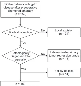
Patients who were treated with local excision (n = 34), who had indeterminate tumor regression grade of primary tumor (n = 15), or who could not be assessed for recurrence status (n = 14) were excluded. A final total of 189 patients who were diagnosed with ypT0 of resected primary tumor (PCRT group) were included in our analysis (Fig. 1). For comparison of oncologic outcomes, 77 patients diagnosed with Tis rectal cancer after radical resection without PCRT (Tis group) were
also analyzed.
This study was conducted with the approval of the Institutional Review Board for Human Research of Asan Medical Center (approval number: 2017-0791) in accordance with the Helsinki Declaration. Due to the retro spective nature of the study, informed consent was waived off.
PCRT, adjuvant treatment, and surgical resection
Preoperative radiotherapy consisted of 25 fractions at a dos- age of 45–50 Gy administered to the entire pelvis, followed by a 5.4-Gy boost in 3 fractions to the primary tumor. For concurrent chemotherapy, 2 cycles of intravenous 5-fluorouracil (375 mg/
m2/day) and leucovorin (20 mg/m2/day) were delivered in bolus over 3 days during the first and fifth weeks of radiation therapy. Alternatively, oral capecitabine (1,650 mg/m2/day) was administered twice per day during radiotherapy. Surgery was performed 6–8 weeks after completing PCRT according the principle of total mesorectal excision.
Adjuvant chemotherapy, followed by radical resection, is recommended for all medically fit patients with PCRT. The usual adjuvant treatment comprised four cycles of 5-fluorouracil and leucovorin monthly or 6 cycles of capecitabine. Oxaliplatin regimens were delivered at the discretion of the attending physician.
Postoperative surveillance
All patients received postoperative follow-up examinations, which consisted of a physical examination, serum carcino- embryonic antigen measurement, chest radiography, and ab-
Eligible patients with ypT0 disease after preoperative
chemoradiotherapy (n = 252)
Radical resection
Pathologically diagnosed total
regression
n = 189
Local excision (n = 34)
Indeterminate primary tumor regression grade
(n = 15)
Follow-up loss (n = 14) Yes
No
No
Yes
Fig. 1. CONSORT (consolidated standards for reporting of trials) diagram.
dom inal, pelvic, and chest computed tomography every 3–6 months. Most patients underwent colonoscopy at 6–12 months postoperatively and every 2–3 years thereafter. Recurrence was determined according to the radiological or histopathologic findings. Local recurrence was defined as the presence of a suspicious lesion in the areas contiguous to the bed of the primary rectal resection or the site of anastomosis, and distant metastasis was defined as the presence of any recurrence in a distant organ or dissemination to the peritoneal surface.
Recurrence-free survival (RFS) was measured from the date of surgery to the date of the first recurrence event or death.
Statistical analysis
The primary endpoint was RFS, which was calculated using the Kaplan-Meier method and compared using the log-rank test.
Multivariable Cox proportional hazard analysis was used to test the effects of potential risk factors for RFS. A P-value of <0.05 was considered statistically significant. All statistical analyses were performed using IBM SPSS Statistics ver. 21.0 (IBM Co., Armonk, NY, USA).
RESULTS
Clinicopathologic characteristics of patients
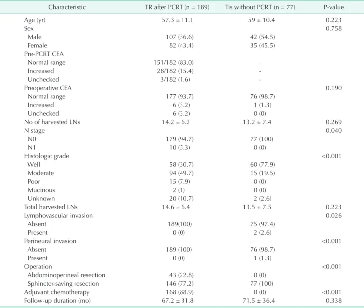
A total of 189 patients with TR of the primary tumor were included. Among them, 107 (56.6%) were men, and the mean age at diagnosis was 57.3 ± 11.1 years. The distribution of sex and age at diagnosis in the PCRT group were similar to that in the Tis group. Preoperative CEA levels were within normal range in 93.7% of the PCRT group and in 98.7% of the Tis group,
Table 1. Clinicopathologic characteristics of all patients
Characteristic TR after PCRT (n = 189) Tis without PCRT (n = 77) P-value
Age (yr) 57.3 ± 11.1 59 ± 10.4 0.223
Sex 0.758
Male 107 (56.6) 42 (54.5)
Female 82 (43.4) 35 (45.5)
Pre-PCRT CEA
Normal range 151/182 (83.0) -
Increased 28/182 (15.4) -
Unchecked 3/182 (1.6) -
Preoperative CEA 0.190
Normal range 177 (93.7) 76 (98.7)
Increased 6 (3.2) 1 (1.3)
Unchecked 6 (3.2) 0 (0)
No of harvested LNs 14.2 ± 6.2 13.2 ± 7.4 0.269
N stage 0.040
N0 179 (94.7) 77 (100)
N1 10 (5.3) 0 (0)
Histologic grade <0.001
Well 58 (30.7) 60 (77.9)
Moderate 94 (49.7) 15 (19.5)
Poor 15 (7.9) 0 (0)
Mucinous 2 (1) 0 (0)
Unknown 20 (10.7) 2 (2.6)
Total harvested LNs 14.6 ± 6.4 13.5 ± 7.5 0.223
Lymphovascular invasion 0.026
Absent 189(100) 75 (97.4)
Present 0 (0) 2 (2.6)
Perineural invasion <0.001
Absent 189 (100) 76 (98.7)
Present 0 (0) 1 (1.3)
Operation <0.001
Abdominoperineal resection 43 (22.8) 0 (0)
Sphincter-saving resection 146 (77.2) 77 (100)
Adjuvant chemotherapy 168 (88.9) 0 (0) <0.001
Follow-up duration (mo) 67.2 ± 31.8 71.5 ± 36.4 0.338
Values are presented as mean ± standard deviation or number (%).
TR, total regression; PCRT, preoperative chemoradiotherapy; LN, lymph node.
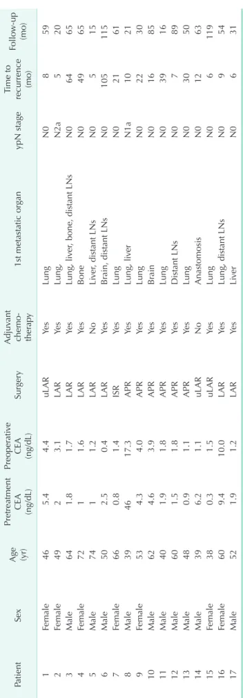
Table 2. Recurrence in patients treated with preoperative chemoradiotherapy PatientSex Age (yr)
Pretreatment
CEA (ng/dL)
Preoperative
CEA (ng/dL)
SurgeryAdjuvant chemo- therapy1st metastatic organypN stageTime to recurrence (mo)Follow-up (mo) 1Female465.44.4uLARYesLungN0859 2Female4923.1LARYesLung,N2a520 3Male641.81.7LARYesLung, liver, bone, distant LNsN06465 4Female7211.6LARYesBoneN04965 5Male7411.2LARNoLiver, distant LNsN0515 6Male502.50.4LARYesBrain, distant LNsN0105115 7Female660.81.4ISRYesLungN02161 8Male394617.3APRYesLung, liverN1a1021 9Female534.34.0APRYesLungN02230 10Male624.63.9APRYesBrainN01685 11Male401.91.8APRYesLungN03916 12Male601.51.8APRYesDistant LNsN0789 13Male480.91.1APRYesLungN03050 14Male396.21.1uLARNoAnastomosisN01263 15Female380.31.5uLARYesLungN06119 16Female609.410.0LARYesLung, distant LNsN0954 17Male521.91.2LARYesLiverN0631 uLAR, ultra-low anterior resection; LAR, low anterior resection; ISR, intersphincteric resection; APR, abdominal perineal resection; LN, lymph node.
with no significant difference between the 2 groups.
Sphincter-preserving resection was performed for 146 patients (77.2%) in the PCRT group, which was a much lower rate than that in the Tis group.
In the PCRT group, 10 patients (5.3%) had pathologically diagnosed metastatic lymph nodes (LNs). Seven patients had 1 metastatic LN and 3 patients had 2 or more metastatic LNs.
Moderately differentiated histology was the most common dif- fer entiation type in both groups. Adjuvant chemotherapy was administered to 168 patients (88.9%) of the PCRT group (Table 1).
Recurrence
The mean follow-up duration was 67.2 ± 31.8 months.
There was no recurrence in the Tis group. Tumor recurrence was observed in 17 patients (9%) of the PCRT patients. Among them, 10 were male and 7 were female. Single-site recurrence developed in 12 patients (66.7%). The most common recurrence site was lung (55.6%) followed by distant LNs (29.4%) and liver (23.5%). Sixteen patients had distant metastases, with only 1 patient showing evidence of local recurrence.
Adjuvant chemotherapy was given for 15 patients among all patients with recurrences. Among the 17 patients who developed recurrence in the PCRT group, 2 had LN metastases when the primary tumor resection was done. The mean recurrence-free interval (time interval from surgery to diagnosis of recurrence) was 56 ± 33.2 months. The recurrence developed within one year in 8 of the 17 patients (47.1%), and the latest recurrence developed after 105 months (Table 2).
RFS and associated factors in patients with TR after PCRT
The 5-year RFS in patients with TR of primary rectal cancer after PCRT (91.3%) was significantly lower than that of patients with Tis rectal cancer without PCRT (100%) (Fig. 2).Univariate analysis showed that RFS was significantly asso- ciated with post-PCRT CEA (P = 0.045) and histologic dif fer- entiation (P = 0.046). However, multivariable analysis did not show any factor independently associated with recurrence (Table 3).
12 24 36 48 72
1.0
0.8
0.6
0.4
survivalRecurrence-free 0.2
Months 60 P = 0.005
96 84 No PCRT
PCRT
0
Fig. 2. Recurrence-free survival (RFS) in patients with total re gression of primary tumor after preoperative chemo- radiotherapy (PCRT), and those with Tis rectal cancer without PCRT (No PCRT).
Table 3. Factors associated with recurrence-free survival in patients treated with preoperative chemoradiotherapy
Variable Univariate
P-value Multivariate
P-value
HR 95% CI HR 95% CI
Adjuvant chemotherapy 1.14 0.261–4.988 0.862 - -
Pre-PCRT CEA
Normal 1 - - - - -
Increased 1.057 0.303–3.688 0.931 - - -
Preoperative CEA 0.045 0.173
Normal 1 - - -
Increased 4.567 1.034–20.161 4.639 0.509–42.262
ypN stage 0.246
ypN0 1 - 1 - 0.668
ypN+ 2,402 0.547–10.549 0.606 0.061–5.990
Histologic grade 0.046 0.085
G1 1 - 1 -
G2 3.274 1.022–10.492 2.973 0.861–10.280
Sex 0.875 -
Male 1 - - -
Female 0.925 0.352–2.432 - - -
Age 0.981 0.939–1.025 0.387 - - -
HR, hazard ratio; CI, confidence interval; PCRT, preoperative chemoradiotherapy.
DISCUSSION
Our findings showed that the PCRT group had a lower RFS than did the Tis group, suggesting that TR after PCRT may not represent “no tumor” status even with tumor-confined mucosa.
We compared the oncologic outcome of the Tis tumor with that of the TR after PCRT to determine the oncologic status of the regression of primary tumor. This may provide indirect evidence to answer the clinical question of whether TR of the primary tumor can be considered as “no tumor” status.
Evaluating the oncologic status of TR after PCRT is important for determining appropriate surgical strategies after PCRT in this subgroup of patients. Rectal cancer confined within sub- mucosa has been known to have good oncologic outcomes, and 5-year RFS has been reported to exceed 95% in most studies [7,13,14]. As a result of these favorable oncologic outcomes for this group of patients, local excision for early rectal cancer has increased steadily over time [15-17]. These strategies have influenced the surgical approach for “significantly regressed”
cases of rectal cancer after neoadjuvant chemoradiotherapy which tumor cell was located within submucosa.
To date, many studies have demonstrated significant fa- vorable oncologic outcomes in patients with prominent re- gression of the primary tumor after PCRT [8,9,18,19], including significant superiority in RFS and local control of patients whose tumor showed a TR or near-TR compared with those in other regression groups [3,9,20]. Based on these findings, interest in organ-preserving surgery in patients who show a good response to PCRT has increased, in an effort to avoid morbidity and functional derangement associated with radical resection.
However, organ-preserving treatment for ypT0-1 disease after PCRT has resulted in controversial findings in many studies in terms of oncologic outcomes. Multicenter trials that include patients with cT2N0 rectal cancer treated with PCRT showed a 5-year DFS of 79.3% after local excision [4]. In this study, 49%
of patients had ypT0 or Tis disease, and oncologic outcome was not in the expected range considering the proportion of patients with regressed disease. Another research reported a wide range of oncologic outcomes after local excision for rectal cancer after PCRT. Pathologically confirmed TR was reported in 30.2%–64% of patients; local recurrence among patients with less than ypT1 disease was 2%–11.1% [4,5,6,21]; and the 10- year disease free survival was reported to be 89.5% for patients with TR after radical resection [10]. However, there is a lack of studies on long-term oncologic outcomes of patients with TR after PCRT.
Although patients with TR after PCRT have been included in many studies in which they were treated as patients with early rectal cancer, their “pretreatment tumor status” should be considered. Although surgical strategies progressed in
patients who had good response to PCRT according to their posttreatment primary TR, we still administered adjuvant chemotherapy according to their initial clinical tumor stage regardless of the final pathologic stage. Treatment irrespective of post-treatment tumor status might be caused by a lack of evidence of the long-term oncologic outcome in this group of patients. Thus, there is a need to evaluate the long-term oncologic outcomes of patients with TR after PCRT and compare with oncologic outcomes of patients with initial early rectal cancer.
Several studies have compared patient outcomes stratified by pathologic stage between patients treated with PCRT or not. It may not seem logical to compare oncologic outcomes according to the pathologic stages of patients treated with PCRT and those without PCRT, because the pathologic staging system was originally developed from results of patients not treated with neoadjuvant treatment. However, stage-stratified comparisons may help in understanding the objective oncologic status of certain subgroups of patients based on the generally accepted system.
In the present study, patients with TR after PCRT had favorable oncologic outcomes. However, the RFS of patients with TR after PCRT was “lower” than that of the initial Tis tumor. In this study, recurrence occurred in 9% of patients with TR, which is similar to findings of previous reports [4,5,6,10,21].
The most common distant metastasis site was the lung (58.8%), and recurrence occurred within 1 year after the operation in approximately half of the recurrence cases. We attempted, but were unable, to identify risk factors associated with recurrence in the TR after PCRT group. Typically, LN metastasis is suspected as a major associated factor of recurrence in patients treated with PCRT [10,22-24], even in patients who show a good response to PCRT [23,24]. Local recurrence rate occurred in only 1 patient among 17 patients with recurrences in the present study. Patients with TR after PCRT had low LN metastasis incidence and no chance of circumferential resection margin involvement which were important risk factors of local recurrence in rectal cancer. These may result in low local recurrence rate. In the present study, recurrence occurred in 20% of patients with metastatic LN and in 8.3% of patients without LN metastasis in the TR after PCRT group. However, LN metastasis was not found with multivariate analysis to be an independent risk factor of RFS. This could be the result of the small number of patients with LN metastasis among patients with TR in this study, as only 5.3% of patients with TR had LN metastasis. Future studies of are needed to evaluate the influence of LN metastasis on recurrence in a larger cohort of patients with TR after PCRT.
In many cases, the degree of LN metastasis has been known to correlate with cancer stage [25]. Patients with TR after PCRT had <10% of LN metastasis. If LN metastasis was not an
independent risk factor for recurrence in these patients, organ- preserving treatment would be accepted more easily. However, as LN metastasis has been determined as a critical risk factor for RFS, it is important to emphasize appropriate diagnosis for identifying metastatic LN before determining surgical treatment, and to use care in deciding to omit radical resection [25].
In the present study, 88.9% of patients with TR after PCRT received adjuvant chemotherapy. Considering the lack of evi- dence whether adjuvant chemotherapy is beneficial for patients with good response to PCRT, it is quite high rate. The current recommendation to use adjuvant chemotherapy after PCRT is based on the belief that the risk of recurrence is high in patients with clinical stage II or III rectal cancer, and that this is not modified by PCRT and surgery. During study period, we adapted the current concept of adjuvant chemotherapy after PCRT in our institution, and all patients who are planned to receive PCRT was informed that they have to receive adjuvant chemotherapy regardless of final pathologic results ahead of PCRT start. We thought that institutional treatment strategy and notice of adjuvant chemotherapy before PCRT were responsible for high compliance to adjuvant chemotherapy.
There are some limitations to our study. We performed a retrospective single-center database study, which may have introduced an inherent bias. Furthermore, we did not include local excision after TR, so it may not be possible to analyze the overall oncologic outcomes of the TR group after PCRT. However, comparison between patients who have undergone radical resection can provide valuable analysis of
the different outcomes within the same surgical treatment group. Additionally, we compared oncologic outcomes in the PCRT group with those in the Tis group, and it is not actual comparison with no-tumor status. Tis tumor, however, would not recur theoretically and it might be represent oncologically most favorable tumor. Although the statistical power of our analysis was limited by the abovementioned reasons, this study would show indirectly the oncologic status of TR after PCRT.
Based on the results of this study, TR after PCRT is different from Tis in terms of oncologic outcomes. Further large-scale studies should be performed to assess the long-term on- cologic outcomes of patients with TR after PCRT. It is clear that additional investigations are needed to better understand the oncologic status of patients with TR. To ensure proper treatment for patients who show a good response after PCRT, evaluation of the factors associated with recurrence and survival and a better understanding of advanced diagnostic cri- te ria before surgical treatment are necessary.
CONFLICT OF INTEREST
No potential conflict of interest relevant to this article was reported.
ACKNOWLEDGEMENTS
This study was supported by the National Research Foundation of Korea (NRF) (2016R1A2B4014039).
REFERENCES
1. Sauer R, Becker H, Hohenberger W, Rodel C, Wittekind C, Fietkau R, et al. Pre op- erative versus postoperative chemo radio- therapy for rectal cancer. N Engl J Med 2004;351:1731-40.
2. Sauer R, Liersch T, Merkel S, Fietkau R, Hohenberger W, Hess C, et al. Preoperative versus postoperative chemoradiotherapy for locally advanced rectal cancer: results of the German CAO/ARO/AIO-94 rando- mi zed phase III trial after a me dian fol - low-up of 11 years. J Clin Oncol 2012;30:
1926-33.
3. Habr-Gama A, Perez RO, Nadalin W, Sabbaga J, Ribeiro U Jr, Silva e Sousa AH Jr, et al. Operative versus nonoperative
treat ment for stage 0 distal rectal cancer fol lowing chemoradiation therapy: long- term results. Ann Surg 2004;240:711-7.
4. Garcia-Aguilar J, Renfro LA, Chow OS, Shi Q, Carrero XW, Lynn PB, et al. Organ pre ser vation for clinical T2N0 distal rec tal cancer using neoadjuvant chemo- radio ther apy and local excision (ACOSOG Z6041): results of an open-label, single- arm, multi-institutional, phase 2 trial.
Lan cet Oncol 2015;16:1537-46.
5. Perez RO, Habr-Gama A, Lynn PB, Sao Juliao GP, Bianchi R, Proscurshim I, et al.
Trans anal endoscopic microsurgery for residual rectal cancer (ypT0-2) following neo adjuvant chemoradiation therapy: an-
other word of caution. Dis Colon Rec tum 2013;56:6-13.
6. Stipa F, Picchio M, Burza A, Soricelli E, Vitelli CE. Long-term outcome of local ex- cision after preoperative chemoradi ation for ypT0 rectal cancer. Dis Colon Rec tum 2014;57:1245-52.
7. Lee SD, Park SC, Park JW, Kim DY, Choi HS, Oh JH. Laparoscopic versus open sur- gery for stage I rectal cancer: long-term on co logic outcomes. World J Surg 2013;37:
646-51.
8. Fokas E, Strobel P, Fietkau R, Ghadimi M, Liersch T, Grabenbauer GG, et al. Tumor re gression grading after preoperative che- mo radiotherapy as a prognostic factor and
in di vidual-level surrogate for disease-free sur vival in rectal cancer. J Natl Can cer Inst 2017;109.
9. Park IJ, You YN, Agarwal A, Skibber JM, Rodriguez-Bigas MA, Eng C, et al. Neo- adjuvant treatment response as an early response indicator for patients with rectal cancer. J Clin Oncol 2012;30:1770-6.
10. Fokas E, Liersch T, Fietkau R, Hohen berger W, Beissbarth T, Hess C, et al. Tumor re- gres sion grading after preoperative che- mo radiotherapy for locally advanced rec- tal carcinoma revisited: updated results of the CAO/ARO/AIO-94 trial. J Clin Oncol 2014;32:1554-62.
11. Althumairi AA, Gearhart SL. Local exci- sion for early rectal cancer: transanal endo scopic microsurgery and beyond. J Gastro intest Oncol 2015;6:296-306.
12. Chang HJ, Park CK, Kim WH, Kim YB, Kim YW, Kim HG, et al. A standardized path- ology report for colorectal cancer. Korean J Pathol 2006;40:193-203.
13. Huh JW, Kim CH, Kim HR, Kim YJ. Onco- logic outcomes of pathologic stage I lower rectal cancer with or without pre operative chemoradiotherapy: are they comparable?
Surgery 2011;150:980-4.
14. Hwang K, Park IJ, Yu CS, Lim SB, Lee JL, Yoon YS, et al. Impression of prognosis regarding pathologic stage after preoper- ative chemoradiotherapy in rectal cancer.
World J Gastroenterol 2015;21:563-70.
15. Stitzenberg KB, Sanoff HK, Penn DC, Meyers MO, Tepper JE. Practice patterns and long-term survival for early-stage rec- tal cancer. J Clin Oncol 2013;31:4276-82.
16. Allaix ME, Arezzo A, Morino M. Transanal endoscopic microsurgery for rectal cancer:
T1 and beyond? An evidence-based re- view. Surg Endosc 2016;30:4841-52.
17. Jung SM, Yu CS, Park IJ, Kim TW, Kim JH, Yoon YS, et al. Oncologic safety of local excision compared with total mesorectal excision for ypT0-T1 rectal cancer: a pro- pensity score analysis. Medicine (Balti- more) 2016;95:e3718.
18. Roh MS, Colangelo LH, O'Connell MJ, Yothers G, Deutsch M, Allegra CJ, et al.
Pre operative multimodality therapy im- proves disease-free survival in patients with carcinoma of the rectum: NSABP R-03. J Clin Oncol 2009;27:5124-30.
19. Rodel C, Martus P, Papadoupolos T, Fuzesi L, Klimpfinger M, Fietkau R, et al. Prog- nostic significance of tumor re gres sion after preoperative chemoradio ther apy for rectal cancer. J Clin Oncol 2005;23:8688- 96.
20. Maas M, Nelemans PJ, Valentini V, Das P, Rodel C, Kuo LJ, et al. Long-term outcome in patients with a pathological complete response after chemoradiation for rectal cancer: a pooled analysis of individual patient data. Lancet Oncol 2010;11:835-44.
21. Noh JM, Park W, Kim JS, Koom WS, Kim
JH, Choi DH, et al. Outcome of local ex ci sion following preoperative chemo- radio therapy for clinically T2 distal rec tal can cer: a multicenter retrospective study (KROG 12-06). Cancer Res Treat 2014;46:
243-9.
22. Yeo SG, Kim DY, Park JW, Choi HS, Oh JH, Kim SY, et al. Stage-to-stage comparison of preoperative and postoperative chemo- radiotherapy for T3 mid or distal rectal cancer. Int J Radiat Oncol Biol Phys 2012;
82:856-62.
23. Gollins S, Sun Myint A, Haylock B, Wise M, Saunders M, Neupane R, et al. Pre- op er ative chemoradiotherapy using con cur rent capecitabine and irinotecan in magnetic resonance imaging-defined lo cal ly advanced rectal cancer: impact on long-term clinical outcomes. J Clin Oncol 2011;29:1042-9.
24. Park IJ, You YN, Skibber JM, Rodriguez- Bigas MA, Feig B, Nguyen S, et al. Com- parative analysis of lymph node meta- stases in patients with ypT0-2 rectal cancers after neoadjuvant chemo radio- ther apy. Dis Colon Rectum 2013;56:135- 41.
25. Dinaux AM, Leijssen L, Bordeianou LG, Kunitake H, Amri R, Berger DL. Outcomes of persistent lymph node involvement after neoadjuvant therapy for stage III rectal cancer. Surgery 2018;163:784-8.