Received December 8, 2010, Revised April 27, 2011, Accepted for publication May 11, 2011
*This study was supported by a grant of the Korea Healthcare Technology R&D Project, Ministry of Health & Welfare, Republic of Korea (Grant No. : A103017).
Corresponding author: Jeung-Hoon Lee, M.D., Department of Der- matology, School of Medicine, Chungnam National University, 33 Munhwa-ro, Daejeon 301-040, Korea. Tel: 82-42-280-7707, Fax:
82-42-280-8459, E-mail: [email protected]
This is an Open Access article distributed under the terms of the Creative Commons Attribution Non-Commercial License (http://
creativecommons.org/licenses/by-nc/3.0) which permits unrestricted non-commercial use, distribution, and reproduction in any medium, provided the original work is properly cited.
ORIGINAL ARTICLE
Cedrol Enhances Extracellular Matrix Production in Dermal Fibroblasts in a MAPK-Dependent Manner
Mu Hyun Jin, M.S., Sun Gyoo Park, M.S., Yul-Lye Hwang, M.S.1, Min-Ho Lee, M.S.1,
Nam-Ji Jeong, M.D.2, Seok-Seon Roh, O.M.D.2, Young Lee, M.D.2, Chang Deok Kim, Ph.D.2, Jeung-Hoon Lee, M.D.2
LG Household & Health Care Ltd, 1Oriental BioMed Lab, 2Department of Dermatology and Research Institute for Medical Sciences, School of Medicine, Chungnam National University, Daejeon, Korea
Background: The extracellular matrix (ECM) produced by dermal fibroblasts supports skin structure, and degradation and/or reduced production of ECM are the main causes of wrinkle formation. Objective: The aim of this study was to identify the active ingredient that enhances ECM production in dermal fibroblasts. Methods: Polarity-based fractionation was used to isolate the active ingredient from natural ex- tracts, and the effects of cedrol (isolated from Pterocarpus indicusirginia) on ECM production in cultured human der- mal fibroblasts was investigated by reverse transcription- polymerase chain reaction, enzyme linked immunosorbent assay, and Western blot analysis. Results: Cedrol accelerated fibroblast growth in a dose-dependent manner and increased the production of type 1 collagen and elastin. Phosphory- lation of p42/44 extracellular signal-regulated kinase, p38 mitogen-activated protein kinase, and Akt was markedly increased by cedrol, indicating that enhanced ECM produc- tion is linked to activation of intracellular signaling cascades.
Conclusion: These results indicate that cedrol stimulates ECM production, with possible applications to the main- tenance of skin texture. (Ann Dermatol 24(1) 16∼21, 2012)
-Keywords-
Cedrol, Collagen, Fibroblasts
INTRODUCTION
The extracellular matrix (ECM) has important functions in the maintenance of proper skin tensile strength and elas- ticity. ECM components are found in the dermis, in which mesenchyme-derived fibroblasts produce most of the ECM components, including several types of collagen molecu- les, polymeric glycosaminoglycans, and elastic fibers1. Of these, type 1 collagen is the most abundant structural protein in skin connective tissue and has a direct influence on skin tension. During the aging process, skin gradually loses mechanical strength and elasticity, which leads to the formation of wrinkles. This phenomenon is largely due to the collapse of dermal connective tissue, which coinci- des with the loss of fibroblast proliferation and reduced ECM production2. An abnormality in type I collagen syn- thesis has been suggested to cause an imbalance in ECM metabolism and the clinical changes observed in aged skin. Therefore, it has long been believed that enhancing fibroblast activity, in the context of ECM production, may help maintain skin texture.
In the course of screening stimulators for ECM production from natural compounds we found that cedrol, derived from Pterocarpus indicusirginia (Ja Dan Hyang), activates dermal fibroblasts. In a previous study, it was demons- trated that cedrol acts on the autonomic nervous system, producing a sedative effect3. Cedrol inhalation increases parasympathetic activity and decreases sympathetic activity in healthy human subjects. Cedrol also increases barore- ceptor sensitivity in the same healthy human subjects4.
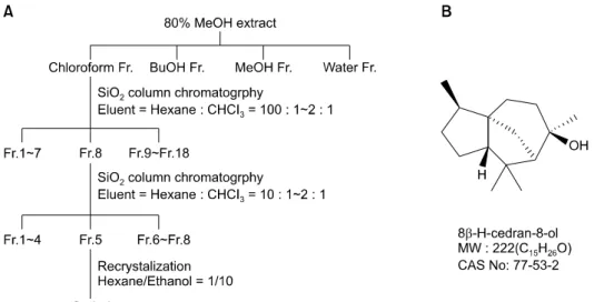
Fig. 1. (A) Isolation of cedrol from Pterocarpus indicus. (B) Chemical structure of cedrol. Cedrol was iso- lated from the lignum of P. indicu- sirginia using bioassay-directed frac- tionation. After solvent fractionation, two applications of column chroma- tography followed by recrystalliza- tion were used to isolate the active compound. The purity of cedrol was confirmed to be ∼99%, as deter- mined by high performance liquid chromatography, nuclear magnetic resonance, and electrospray ioniza- tion mass spectrometry studies.
However, it has never been reported whether cedrol affects fibroblast activity based on ECM production.
In this study, we determined the effect of cedrol on ECM production in dermal fibroblasts and investigated the puta- tive action mechanism of cedrol. We found that cedrol stimulates production of type I collagen via activation of mitogen-activated protein kinase (MAPK) signaling path- ways.
MATERIALS AND METHODS
Cell culture
Normal human skin samples were obtained from circum- cisions in accordance with the ethical committee approval process of Chungnam National University Hospital. Speci- mens were briefly sterilized in 70% ethanol, minced, and then incubated in Dulbecco's Modified Eagle's Medium supplemented with 10% FBS and antibiotics (Gibco BRL, Rockville, MD, USA). Dermal fibroblasts were produced from explants after 5∼7 days. At confluence, cells were routinely subcultured using a 1:4 split ratio. Cells were used between passages 4∼15. For treatment with cedrol, approximately 1×10⁶ cells were seeded on 100-mm culture dishes and grown to sub-confluence. Cells were serum-starved for 24 h and then treated with cedrol in serum-free medium.
Isolation of cedrol
We isolated cedrol from the lignum of Pterocarpus indi- cusirginia (Kyongdong Herbal Market, Seoul, Korea) using bioassay-directed fractionation. Air-dried P. indicusirginia (10 kg) was cut into pieces and extracted with five volumes of 80% ethanol for 5 days at room temperature. The extract was evaporated under reduced pressure conditions, then reconstituted with distilled water. The aqueous fraction
was successively extracted with chloroform and n-butanol.
The chloroform-dissolved fraction was then chromatogra- phed on silica gel with a developing solution of n-hexane and chloroform. As a result, the chloroform-dissolved frac- tion was separated into 18 sub-fractions. Of these, the eighth sub-fraction was re-chromatographed on silica gel using a gradient developing solution (n-hexane : chloroform, 10:1∼2:1), and then divided into eight additional sub-fractions. Of these, the fifth was recrystallized using a solvent of n-hexane and ethanol (Fig. 1A). The structure of the resultant crystal was determined using high performan- ce liquid chromatography, nuclear magnetic resonance (NMR), and electrospray ionization mass spectrometry, and identified as cedrol (Fig. 1B). A total of 120 mg of crystalline cedrol was obtained using this fractionation protocol.
Cell growth analysis
Fibroblast cells were seeded in a 60-mm culture dish and treated with 1 μCi of [3H]thymidine (Amersham, Bucking- hamshire, UK) for a [3H]thymidine uptake assay. Follow- ing incubation for the indicated time points, cells were washed twice with phosphate buffered saline and incubated with 0.1 N NaOH at room temperature.
Radioactivity in cell lysates was measured using a liquid scintillation counter.
Enzyme-linked immunosorbent assay (ELISA)
An ELISA kit for type 1 pro-collagen was purchased from Takara Bio Inc. (Shiga, Japan). Levels of type 1 pro-collagen secreted from fibroblasts were quantified according to the manufacturer’s recommended protocols. Measurements were repeated at least three times with independent cell batches.
Table 1. Nucleotide sequence of the primers
Name Primer Expected size (bp)
COL1A1 Forward (5’→3’) CATCAACGGGTTCACTACCG 431
Reverse (5’→3’) CCACGTAGTACACGATGGGC
COL1A2 Forward (5’→3’) AACCCCATGTGGGAATTGAT 419
Reverse (5’→3’) GCTGCTTCCACACAGCAAGT
Actin Forward (5’→3’) AAACTGGAACGGTGAAGGTG 352
Reverse (5’→3’) CTCAAGTTGGGGGACAAAAA
Fig. 2. Effect of cedrol on growth of dermal fibroblasts. Cells were treated with cedrol at the indicated concentrations for 2 d in the presence of [3H]thymidine. Radioactivity was measured using a liquid scintillation counter. Results are shown as a percentage of a control±the standard deviation (*p<0.05 vs.
control).
Western blot analysis
Cells were lysed in Proprep solution (Intron, Daejeon, Korea). After vigorous pipetting, extracts were centrifuged for 15 min at 13,000 rpm. Total protein was measured using a Bradford protein assay kit (Bio-Rad Laboratories, Hercules, CA, USA). Samples were run on sodium dodecyl sulfate-polyacrylamide gels, transferred to nitrocellulose membranes, and incubated with appropriate antibodies overnight at 4oC with gentle agitation. Blots were then incubated with peroxidase-conjugated secondary antibodies for 30 minutes at room temperature, and visualized using enhanced chemiluminescence (Intron, Scottsdale, AZ, USA). The following primary antibodies were used: collagen type 1 α1 and elastin (Santa Cruz Biotechnology, Santa Cruz, CA, USA);
phospho-p38 MAPK, total-p38 MAPK, phospho-p42/44 extracellular signal-regulated kinase (ERK), total-p42/44 ERK, phospho-Akt, total-Akt, phospho-Smad 2, total-Smad 2, phospho-Smad 3, total-Smad 3 (Cell Signaling Tech- nology, Danvers, MA, USA), and actin (Sigma, St. Louis, MO, USA).
Reverse transcription polymerase chain reaction (RT- PCR)
Two μg of total RNA was reverse-transcribed using M-MLV reverse transcriptase (ELPIS Biotech, Daejeon, Korea). Aliquots of the RT mixture were subjected to PCR with specific primer sets (Table 1).
RESULTS
We used the polarity-based sequential fractionation method to screen the putative fibroblast activators. This process involved two step chromatography based on a silica ma- trix, and one step recrystalization (Fig. 1A). Results of
1H-NMR, 13C-NMR, and electrospray Ionization mass spectrometry showed that cedrol from the lingnum of P.
indicusirginia was an active ingredient (Fig. 1B)5. The purity of cedrol was approximately 99%.
To investigate the effect of cedrol on fibroblast growth, we first performed a [3H]thymidine uptake assay. Dermal
fibroblasts were treated with various concentrations of cedrol and incubated for 2 d. As shown in Fig. 2, cedrol increased [3H]thymidine uptake of fibroblasts in a dose- dependent manner.
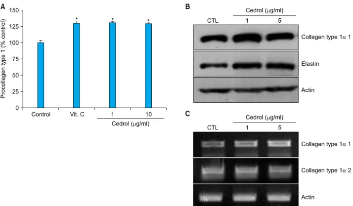
Because the proliferative property of fibroblasts is fre- quently linked to ECM production (especially collagen) in fibroblasts6, we next determined the effect of cedrol on collagen and elastin production. An ELISA showed that cedrol increased secretion of type 1 procollagen in a dose- dependent manner, comparable to results using ascorbic acid (Fig. 3A). Concomitantly, RT-PCR and Western blot analyses demonstrated that cedrol induced transcription and an increase in the intracellular protein level of colla- gen type 1 α1 (Fig. 3B, C). Cedrol also increased elastin production (similar to collagen), suggesting that cedrol activates fibroblasts.
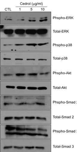
As the intracellular signaling cascades p42/44 ERK and p38 MAPK are implicated in the regulation of ECM pro- duction7-9, we investigated the effect of cedrol on MAPK activation. As shown in Fig. 4, cedrol treatment led to quick phosphorylation of p42/44 ERK and p38 MAPK.
Additionally, the phosphorylation level of Akt, an impor-
Fig. 3. Effect of cedrol on collagen and elastin production in dermal fibroblasts. (A) Cells were treated with cedrol at the indicated concentrations for 2 d. A conditioned medium was collected, and the amount of secreted procollagen type 1 was measured using an enzyme linked immunosorbent assay. Results are shown as a percentage of a control±the standard deviation (*p<0.05 vs. control).
(B) Cellular proteins were harvested and the protein levels of collagen type 1 α1 and elastin were verified using Western blot analysis.
(C) The expression levels of the COL1A1 and COL1A2 genes were determined using reverse transcription-polymerase chain reaction.
CTL: control.
tant signaling molecule for cell survival, also increased following treatment with cedrol. As the Smad pathway is critical for collagen synthesis during the wound healing process10-12, we also investigated Smad protein activation.
However, cedrol did not influence the Smad2/3phospho- rylation, suggesting that cedrol affects ECM production via the intracellular MAPK signaling cascades.
DISCUSSION
We determined the effect of cedrol on collagen produc- tion using primary cultured dermal fibroblasts. Cedrol isolated from P. indicusirginia not only enhanced fibro- blast proliferation but also increased the production of type 1 collagen and elastin. The cedrol effect was linked to activation of the ERK1/2, p38 MAPK, and Akt intra- cellular signaling pathways.
Dermis predominantly contains type I collagen synthe- sized from dermal fibroblasts as precursor molecules called procollagen. Type I collagen, the major component of ECM in skin, bone, and ligaments, is composed of glycine- and proline-rich two-α1 (I) and one-α2 chains,
which are products of the COL1A1 and COL1A2 genes13. Because collagen has a long fibrous structure and consti- tutes a major part of the extracellular space in the dermis, its primary role is to support skin structure and provide tensile strength to the skin. In this regard, fibroblasts are primarily responsible for producing collagen and other ECM. This notion is clearly supported by the observation that collagen biosynthesis by fibroblasts is markedly diminished in aged persons2. Therefore, activation of cell proliferation and enhancement of collagen production to strengthen skin texture is an attractive approach for treating connective tissue-related skin problems, such as wrinkle formation.
Elastin, physiologically important in skin elasticity, is also secreted from skin fibroblasts as the soluble precursor tropoelastin, then subsequently cross-linked into insoluble elastin14. Therefore, our results indicate that cedrol can be used to strengthen skin texture and/or treat connective tissue-related skin problems.
A number of intracellular signal transduction pathways have been reported to play a role in the regulation of many cellular functions. For example, cell growth, differen-
Fig. 4. Effect of cedrol on intracellular signaling pathways. Cells were treated with cedrol for the indicated times. Cellular proteins were prepared and phosphorylation of ERK, p38 MAPK, Akt, and Smad 2 and 3 was determined using Western blot analysis. CTL:
control, ERK: extracellular signal-regulated kinase, MAPK:
mitogen-activated protein kinase.
tiation, and apoptosis are all tightly regulated by the acti- vation status of many intracellular signaling systems15-17. Gene expression is also regulated by an intracellular signaling system. Intracellular signaling cascades including MAPK, Akt, and Smad, are involved in the regulating collagen expression13. The primary role of MAPK has been previously recognized as procollagen synthesis8,18,19. We have demonstrated that cedrol significantly increases the phosphorylation of ERK1/2, p38 MAPK and Akt. Cedrol apparently acts as an intracellular signal modulator based on the association between collagen synthesis and MAPK activation. As cedrol has a two ring-structure and shows oily characteristics, cedrol probably penetrates the plasma membrane and directly affects intracellular signaling.
However, the precise intracellular action mechanism of cedrol remains to be determined.
In summary, we have demonstrated that cedrol stimulates
collagen and elastin production in dermal fibroblasts.
Cedrol should be investigated for treatment of connective tissue-related skin problems as a co-modality together with first-line treatments.
REFERENCES
1. Pieraggi MT, Bouissou H, Angelier C, Uhart D, Magnol JP, Kokolo J. The fibroblast. Ann Pathol 1985;5:65-76.
2. Uitto J. Connective tissue biochemistry of the aging dermis.
Age-related alterations in collagen and elastin. Dermatol Clin 1986;4:433-446.
3. Yada Y, Sadachi H, Nagashima Y, Suzuki T. Overseas survey of the effect of cedrol on the autonomic nervous system in three countries. J Physiol Anthropol 2007;26:349-354.
4. Umeno K, Hori E, Tsubota M, Shojaku H, Miwa T, Nagashi- ma Y, et al. Effects of direct cedrol inhalation into the lower airway on autonomic nervous activity in totally laryngecto- mized subjects. Br J Clin Pharmacol 2008;65:188-196.
5. Joseph-Nathan P, Santillan RL, Gutiérrez A. Gutierrez, Car- bon-13 NMR. Study of Cedrol, 6-Isocedrol, and α-Cedrene.
J Nat Prod 1984;47:924-933.
6. Roh SS, Lee MH, Hwang YL, Song HH, Jin MH, Park SG, et al. Stimulation of the extracellular matrix production in dermal fibroblasts by velvet antler extract. Ann Dermatol 2010;22:173-179.
7. Ignotz RA, Massagué J. Transforming growth factor-beta stimulates the expression of fibronectin and collagen and their incorporation into the extracellular matrix. J Biol Chem 1986;261:4337-4345.
8. Lim IJ, Phan TT, Tan EK, Nguyen TT, Tran E, Longaker MT, et al. Synchronous activation of ERK and phosphatidylino- sitol 3-kinase pathways is required for collagen and extracel- lular matrix production in keloids. J Biol Chem 2003;278:
40851-40858.
9. Lee DJ, Rosenfeldt H, Grinnell F. Activation of ERK and p38 MAP kinases in human fibroblasts during collagen matrix contraction. Exp Cell Res 2000;257:190-197.
10. Jinnin M. Mechanisms of skin fibrosis in systemic sclerosis. J Dermatol 2010;37:11-25.
11. Bhogal RK, Stoica CM, McGaha TL, Bona CA. Molecular aspects of regulation of collagen gene expression in fibrosis.
J Clin Immunol 2005;25:592-603.
12. Chen SJ, Yuan W, Mori Y, Levenson A, Trojanowska M, Varga J. Stimulation of type I collagen transcription in hu- man skin fibroblasts by TGF-beta: involvement of Smad 3. J Invest Dermatol 1999;112:49-57.
13. Ghosh AK. Factors involved in the regulation of type I collagen gene expression: implication in fibrosis. Exp Biol Med (Maywood) 2002;227:301-314.
14. Sephel GC, Davidson JM. Elastin production in human skin fibroblast cultures and its decline with age. J Invest Dermatol 1986;86:279-285.
15. Neumann E, Gay RE, Gay S, Müller-Ladner U. Functional genomics of fibroblasts. Curr Opin Rheumatol 2004;16:
238-245.
16. Wiedłocha A, Sørensen V. Signaling, internalization, and intracellular activity of fibroblast growth factor. Curr Top Microbiol Immunol 2004;286:45-79.
17. Chiquet M, Renedo AS, Huber F, Flück M. How do fibro- blasts translate mechanical signals into changes in extracel- lular matrix production? Matrix Biol 2003;22:73-80.
18. Nagai Y, Miyata K, Sun GP, Rahman M, Kimura S, Miyatake A, et al. Aldosterone stimulates collagen gene expression
and synthesis via activation of ERK1/2 in rat renal fibroblasts.
Hypertension 2005;46:1039-1045.
19. Sato M, Shegogue D, Gore EA, Smith EA, McDermott PJ, Trojanowska M. Role of p38 MAPK in transforming growth factor beta stimulation of collagen production by scleroderma and healthy dermal fibroblasts. J Invest Dermatol 2002;118:
704-711.
![Fig. 2. Effect of cedrol on growth of dermal fibroblasts. Cells were treated with cedrol at the indicated concentrations for 2 d in the presence of [ 3 H]thymidine](https://thumb-ap.123doks.com/thumbv2/123dokinfo/5170402.106190/3.892.78.817.1014.1144/effect-cedrol-fibroblasts-treated-indicated-concentrations-presence-thymidine.webp)