CMV infection is known to be common in organ transplant recipients, patients with malignant neoplasms, and patients with ac- quired immunodeficiency syndrome(AIDS)1.
The manifestations of CMV infection fre- quently include ocular, gastrointestinal, and pulmonary symptoms but cutaneous lesions have been reported rarely2. In Korea, only two cases were previously reported3,4. We re- port a case of 68-year-old female with CMV inclusion disease presenting as perianal ulcers which developed about 3 weeks after a cadaveric renal transplantation.
CASE REPORT
A 68-year-old Korean woman was con- sulted to the department of dermatology to evaluate ulcerated lesions on the perianal area. She had suffered from diabetes mellitus
and hypertension for more than 20 years and had been diagnosed as having a chronic renal failure 2 years before. She had been treated with hemodialysis. She underwent a cadaveric allogenic renal transplantation due to her end stage renal disease and thereafter she was taking medicines including my- cophenolate mofetil, cyclosporin, and pred- nisolone. About 3 weeks later, multiple ulcers developed on her perianal area (Fig. 1).
Results of the following laboratory studies were within normal limits or negative; a blood cell count, urinalysis, liver function tests, chemical battery, and CMV cultures in the blood and urine. The blood CMV IgG was positive, 175 in titer but not IgM. On chest X ray, there were no abnormal findings.
Histopathologic examination showed Owl’s eye-appearing endothelial cells, which had large nuclei with basophilic intranuclear in- clusions surrounded by perinuclear halo in the upper dermis (Fig. 2) and PCR study re- vealed positive CMV DNA from the tissue (Fig. 3). Diagnosis of CMV inclusion disease was done. She was treated with intravenous ganciclovir 250 mg two times daily and the lesions healed slowly.
Cutaneous Cytomegalovirus Infection Presenting as Perianal Ulcers
Won Sin Lee, M.D., Sung Eun Chang, M.D., Jee Ho Choi, M.D., Kyung Jeh Sung, M.D., Kee Chan Moon, M.D., Jai Kyoung Koh, M.D.
Department of Dermatology, Asan Medical Center, College of Medicine, University of Ulsan, Seoul, Korea
A 68-year-old Korean woman was consulted to the department of dermatology to evaluate ulcer- ated lesions on the perianal area, which developed about 3 weeks after a cadaveric renal transplan- tation. Histopathologic examination showed large atypical cytomegalic cells in the upper dermis. Poly- merase chain reaction(PCR) study revealed positive cytomegalovirus(CMV) deoxyribonucleic acid(DNA) from the skin tissue.
We herein present a case of cutaneous CMV infection presenting as perianal ulcers.
(Ann Dermatol 14(1) 56-58, 2002).
Key Words : Cytomegalovirus, Renal transplantation, Perianal ulcer
Received May 28, 2001.
Accepted for publication September 18, 2001.
Reprint request to : Kee Chan Moon, M.D., Depart- ment of Dermatology, Asan Medical Center, College of Medicine, University of Ulsan,
388-1 Poongap-dong, Songpa-gu Seoul, 138-736 Korea Tel. (02)2224-3460, Fax. (02)486-7381
56
Cutaneous Cytomegalovirus Infection Presenting as Perianal Ulcers 57
DISCUSSION
CMV is a DNA virus and is a member of the herpes family of viruses, which also in- cludes herpes simplex virus, varicella-zoster virus, and Epstein-Barr virus1. Like the other herpes viruses, CMV normally exists in certain tissues in a latent state after the pri- mary infection, and the virus may be excret- ed for months or years and may manifest periodic episodes of reactivation1,5,6. The virus can be isolated from urine, breast milk, semen, tears, feces, saliva, blood, cervi- cal secretions, and lymphocytes in healthy
persons1,3,6.
It has been estimated that the majority of the population has been infected with CMV by puberty, with the incidence of exposure continuing to rise thereafter, probably related to venereal spread1. After primary CMV infection, which is most often subclinical and asymptomatic, the virus persists in a lifelong latent stage with the omnipresent potential for reactivation. Symptomatic clini- cal infection in immunocompetent hosts is rare. Clinically apparent disease may manifest itself in neonates and in the immunosup- pressed or immunocompromised hosts6,7.
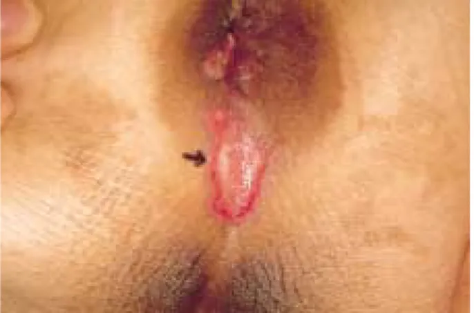
With increasing numbers of organ trans- plant recipients and individuals infected with the human immunodeficiency virus (HIV), the incidence of symptomatic CMV-associated morbidity and mortality has also increased3,6. More than 80% of the primary infections and more than 20% of the reactivation infection in renal transplant patients are symptomatic8. Symptomatic CMV infection may present as mononucleosis, pneumonitis, hepatitis, encephalitis, choriore- tinitis, or gastroenteritis but cutaneous lesions have been reported rarely2. The most specif- ic cutaneous manifestation of CMV is ulcer- ation, especially in the perianal area. Ulcera- tions on the buttocks, perineum, and thigh with visceral involvement have also been well described. Other types of cutaneous le- sions are indurated hyperpigmented nodules or Fig. 1. Well-defined irregular perianal ulcers with
overlying white crust and erythematous border.
Fig. 2. Owl’s eye-appearing atypical cytomegalic cell(arrow) (H&E, ×400).
Fig. 3. Polymerase chain reaction.
Line 1: 1kb marker.
Line 2: positive CMV DNA from the positive control.
Line 3: negative control.
Line 4 & 5: positive CMV DNA from the tissue.
plaques, a widespread exanthem that may become papular and purpuric, and vesiculob- ullous lesions2,9,10.
Light and electronmicroscopy, immunofluo- rescence and immunoperoxidase techniques, and cytopathic effects in human fibroblast tissue cultures allow reliable microscopic de- tection of CMV11. In situ DNA hybridization and PCR technique are also useful methods6.
The characteristic finding in infected tissue is the presence of cytomegalic cells with large intranuclear inclusions surrounded by a clear halo. Occasionally cytoplasmic inclu- sions are also observed1,3,6,10. Cytomegalic cells may appear in urine1. The diagnosis of CMV infection by histologic findings alone is not always possible since the typical CMV- associated inclusions may be subtle and rela- tively sparse even in highly infected tis- sues12.
Patients with cutaneous CMV have a very poor prognosis, with a mortality rate that approximates 85 percent within 6 months, although most of these cases were reported before the general use of improved antiviral agents6,12. CMV is the major cause of morbidity and mortality in renal transplant recipients. Especially, CMV in- fection is very critical during first 3 postop- erative months, presumably a consequence of the intense immunosuppression required during this period to prevent allograft rejec- tion8.
High-dose acyclovir can be used effectively as prophylaxis against CMV infection, al- though it is ineffective against active viral disease. Ganciclovir, a necleoside analogue of acyclovir, is known to be 50 times more effective than acyclovir in vitro against CMV and inhibits viral DNA polymerase6.
Because skin lesions may be clues to the presence of disseminated CMV infections and early diagnosis and treatment may lead to a successful outcome, recognition of these
infections assumes increased significance1,6.
REFERENCES
1. Lesher JL: Cytomegalovirus infections and the skin.
J Am Acad Dermatol 18:1333-1338, 1988.
2. Bournerias I, Boisnic S, Patey O, et al.: Unusual cu- taneous cytomegalovirus involvement in patients with acquired immunodeficiency syndrome. Arch Dermatol 125:1243-1246, 1989.
3. Kim SD, Kim HB, Youn SW, et al: A case of cy- tomegalovirus induced perineal ulcer in an AIDS patient. Korean J Dermatol 37:257-261, 1999.
4. Chang SE, Jung EC, Choi JH, et al: A case of cy- tomegalovirus induced purpura in a bone marrow transplant recipient. Korean J Dermatol 38:966-968, 2000.
5. Stagno S, Whitley RJ: Herpesvirus infections of pregnancy. N Engl J Med 313:1270-1273, 1985.
6. Bowers KE: Cytomegalovirus infection. In Fitz- patrick TB, Eisen AZ, Wolff K, et al. (eds): Derma- tology in general medicine. 5th ed. McGraw-Hill, New York, 1999, pp2450-2457.
7. Vilmer C, Perol Y: Cutaneous manifestations of cy- tomegalovirus infections. Ann Dermatol Venereol 111:119-125, 1984.
8. Lee JY: Cytomegalovirus infection involving the skin in immunocompromised hosts. A clinicopatho- logic study. Am J Clin Pathol 92:96-100, 1989.
9. Smith KJ, Skeleton III HG, James WD, Peter Angritt: Concurrent epidermal involvement of cy- tomegalovirus and herpes simplex virus in two HIV-infected patients. J Am Acad Dermatol 25:
500-506, 1991.
10. Parisier RJ: Histologically specific skin lesions in disseminated cytomegalovirus infection. J Am Acad Dermatol 9:937-946, 1983.
11. Horn TD, Hood AF: Clinically occult cytome- galovirus present in skin biopsy specimens I im- munosuppressed hosts. J Am Acad Dermatol 21:
781-784, 1989.
12. Toome BK, Bowers KE, Scott GA: Diagnosis of cy- tomegalovirus infection: A review and report of a case. J Am Acad Dermatol 24:860-867, 1991.
Cutaneous Cytomegalovirus Infection Presenting as Perianal Ulcers 58