ISSN 2234-3806 • eISSN 2234-3814
https://doi.org/10.3343/alm.2017.37.5.459 www.annlabmed.org 459
Ann Lab Med 2017;37:459-461
https://doi.org/10.3343/alm.2017.37.5.459
Letter to the Editor
Clinical Microbiology
Multiple Brain Abscesses Caused by Nocardia asiatica in a Patient With Systemic Lupus Erythematosus: The First Case Report and Literature Review
Ji Hun Jeong, M.D.1, Song Mi Moon, M.D. 2, Pil Whan Park, M.D.1, Jeong Yeal Ahn, M.D.1, Kyung Hee Kim, M.D.1, Ja Young Seo, M.D.1, Hwan Tae Lee, M.D.1, Kwoun Woo Jae, M.D. 1, and Yiel Hea Seo, M.D.1
Departments of Laboratory Medicine1 and Infectious Disease2, Gachon University Gil Medical Center, Incheon, Korea
Dear Editor,
Nocardia species are uncommon pathogens that affect immu- nosuppressed patients; although cerebral nocardiosis is a rare condition, it is associated with significant morbidity and mortality [1]. Because Nocardia species exhibit different antibiotic suscep- tibilities, accurate species identification is important for progno- ses. To the best of our knowledge, this is the first case of Nocar- dia asiatica brain abscesses reported in a systemic lupus ery- thematosus (SLE) patient.
A 51-yr old man visited our emergency department on May 2016 complaining of left leg weakness, dysarthria, dizziness, nau- sea, vomiting, and uncontrolled fever lasting three days. His past medical history consisted of SLE (diagnosed in August 2002) treated intermittently with steroid and platelet transfusion be- cause of severe thrombocytopenia. In addition, in April 2015, he was diagnosed as having diabetes; however, no medical treat- ment had been undertaken. His last admission to hospital, due to severe thrombocytopenia (6×10⁹/L), was two months prior to this presentation. He was subsequently treated with danazol (400 mg twice daily), hydroxychloroquine (200 mg twice daily), meth- otrexate (15 mg/week), and prednisolone (15 mg/day).
At presentation, the patient’s temperature was 39.1°C, and blood tests indicated a white blood cell count of 11.03×10⁹/L
with a differential count of 76.2% neutrophils. Serum C-reactive protein (71.9 mg/L) and erythrocyte sedimentation rate (28 mm/
hr) were elevated. Brain magnetic resonance imaging (MRI) re- vealed multiple contrast-enhanced lesions in both cerebral and cerebellar hemispheres (Fig. 1A). A subsequent brain abscess aspiration removed 5 mL of a yellowish aspirate; Gram staining of the aspirate revealed gram-positive filamentous branched ba- cilli, and specimen culturing on blood agar plates for 48 hr at 37°C under aerobic conditions yielded white, rough, and dry col- onies, which also presented gram-positive filamentous branched bacilli and were modified acid fast bacilli stain-positive (Fig. 1B- E). 16S rRNA gene sequencing was performed for isolate iden- tification according to the CLSI guidelines with primer pair for- ward 4F and reverse 801R [2]. The isolate 16S rRNA sequence (671 bp; GenBank accession number KY417120) showed 100%
homology with N. asiatica (KC333452.1) and N. abscessus (GU- 471235.1). Alternative gene targets, such as the secA1 gene, are necessary for accurate species discrimination in the Nocar- dia asteroides group, because several N. asiatica, N. abscessus, N. asteroides, and N. arthritidis strains share ≥99.6% identity [2]. Thus, gene amplification and additional sequencing of secA1 were performed with primer pair forward F47 and reverse ConR.
The results (497; KY417121) showed 99.4% (494/497) and
Received: November 17, 2016 Revision received: January 18, 2017 Accepted: April 28, 2017
Corresponding author: Yiel Hea Seo
Department of Laboratory Medicine, Gachon University Gil Medical Center, 21, Namdong-daero 774 Beon-gil, Namdong-gu, Incheon 21565, Korea Tel: +82-32-460-3863, Fax: +82-32-460-3415
E-mail: [email protected]
© Korean Society for Laboratory Medicine.
This is an Open Access article distributed under the terms of the Creative Commons Attribution Non-Commercial License (http://creativecommons.org/licenses/by-nc/4.0) which permits unrestricted non-commercial use, distribution, and reproduction in any medium, provided the original work is properly cited.
1 / 1 CROSSMARK_logo_3_Test
2017-03-16 https://crossmark-cdn.crossref.org/widget/v2.0/logos/CROSSMARK_Color_square.svg
Jeong JH, et al.
Nocardia asiatica-derived brain abscesses
460 www.annlabmed.org https://doi.org/10.3343/alm.2017.37.5.459 Fig. 1. Brain infection from Nocardia asiatica. (A) Brain magnetic resonance images; multiple peripheral enhancing lesions with diffusion restriction in bilateral cerebral and cerebellar hemispheres, suggestive of abscess, (B) microscopic morphology of filamentous, branching, gram-positive bacilli in abscess aspirates, (C) colony morphology on a blood agar plate, (D) Gram stain of a cultured colony and (E) modi- fied acid fast bacilli stain of a cultured colony.
A
B C
D E
Jeong JH, et al.
Nocardia asiatica-derived brain abscesses
https://doi.org/10.3343/alm.2017.37.5.459 www.annlabmed.org 461
94.2% (468/497) similarity with N. asiatica (JQ773453.1) and N.
abscessus (GU179083.1), respectively. The organism was finally identified as N. asiatica. Following treatment with trimetho prim/
sulfamethoxazole (TMP-SMX, 480 mg/day) and ceftriaxone (4 g/
day) for one month, clinical conditions and brain MRI findings improved. Due to severe thrombocytopenia and the elevation of aspartate aminotransferase and alanine transaminase level, he was not taken the sufficient antibiotic treatment. Eventually he visited emergency room due to brain multifocal hemorrhage with septic emboli and aggravation of pulmonary aspergillosis. At ten months of follow up, the patient died during treatment.
Nocardia infection in SLE has been reported to have a high mortality rate (35%), which more than doubles (75%) when the CNS is involved [3]. Nocardial brain abscesses can be misdiag- nosed as malignant brain tumors [4] and can mimic the pre- sentations of underlying disorder flare-ups in SLE patients [5].
Therefore, the possibility of Nocardia infection should be con- sidered during the differential diagnosis of a cerebral lesion to ensure early diagnosis and treatment.
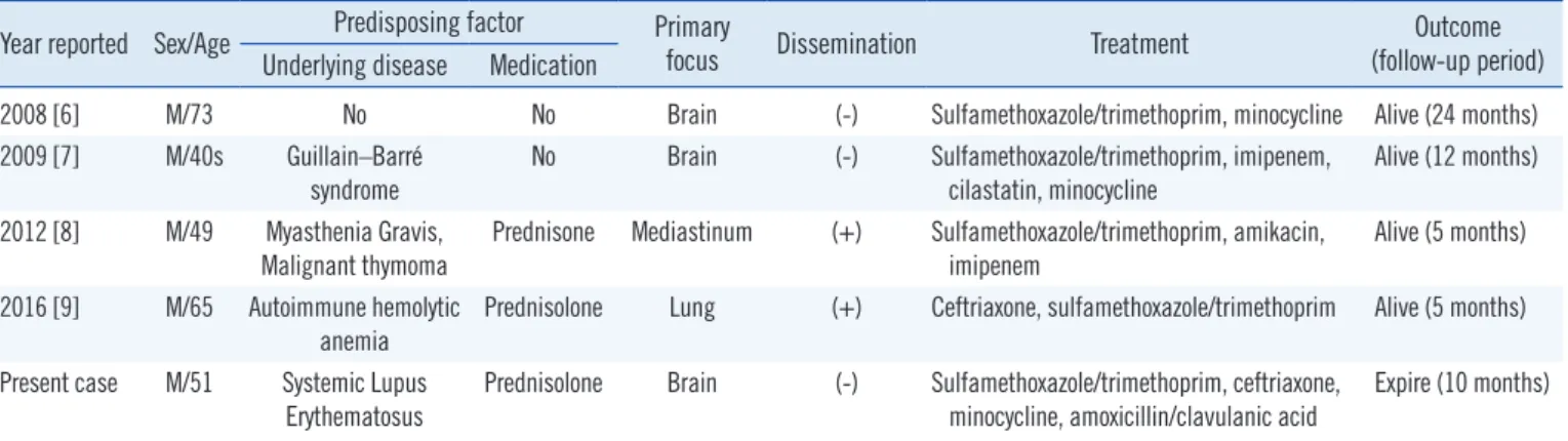
Several cases of N. asiatica infections have been reported in the literature; however, only four have involved brain abscess (Table 1) [6-9]. TMP-SMX is active against most Nocardia spe- cies; however, susceptibility is Nocardia species-dependent; N.
otitidiscaviarum is commonly resistant to TMP-SMX, while N.
nova and N. farcinica are occasionally resistant [1]. Therefore, identification to the species level is required to determine appro- priate treatment. Furthermore, species formerly included in the N. asteroides complex are now considered distinct species. Im- portantly, our case demonstrates that secA1 sequence analysis provides better resolution to the species level in N. asteroides than 16S rRNA sequence analysis [2].
Here, we present the first case of multiple brain abscesses caused by N. asiatica in an SLE patient. The possibility of No- cardia infection should be considered in SLE patients, and early
and accurate identification of Nocardia species is essential for successful treatment. And the administration of prolonged oral antimicrobial treatment after primary infection is also necessary for good prognosis.
Authors’ Disclosures of Potential Conflicts of Interest
No potential conflicts of interest relevant to this article were re- ported.
REFERENCES
1. Wilson JW. Nocardiosis: updates and clinical overview. Mayo Clin Proc 2012;87:403-7.
2. Clinical and Laboratory Standards Institute. Interpretive criteria for iden- tification of bacteria and fungi by DNA target sequencing. Approved guideline. Wayne, PA: CLSI document MM18-A. Clinical and Laboratory Standards Institute, 2008.
3. Mc-Nab P, Fuentealba C, Ballesteros F, Pacheco D, Alvarez M, Dabanch J, et al. Nocardia asteroides infection in a patient with systemic lupus erythematosus. Rev Med Chil 2000;128:526-8.
4. Yamada SM, Nakai E, Toyonaga S, Nakabayashi H, Park KC, Shimizu K.
A rapidly enlarging nocardial brain abscess mimicking malignant glio- ma. J Nippon Med Sch 2005;72:308-11.
5. Cheng HM, Huang DF, Leu HB. Disseminated nocardiosis with initial manifestation mimicking disease flare-up of systemic lupus erythemato- sus in an SLE patient. Am J Med 2005;118:1297-8.
6. Wakui D, Ito H, Ikeda R, Yoshida Y, Furuya Y, Tanaka K, et al. A compli- cated case of Nocardia brain abscess for differential diagnosis. No Shin- kei Geka 2008;36:1011-6.
7. Ryu A, Kawahara R, Mori K, Tamura M, Nakano K, Yoshioka A, et al. A case of brain abscess caused by Nocardia asiatica. J Jpn Soc Clin Mi- crobiol 2009;19:163-70.
8. El-Herte RI, Kanj SS, Araj GF, Chami H, Gharzuddine W. First report of Nocardia asiatica presenting as an anterior mediastinal mass in a pa- tient with myasthenia gravis: a case report and review of the literature.
Case Rep Infect Dis 2012;2012:325767.
9. Uneda A, Suzuki K, Okubo S, Hirashita K, Yunoki M, Yoshino K. Brain abscess caused by Nocardia asiatica. Surg Neurol Int 2016;7:74.
Table 1. Summary of brain abscess cases due to Nocardia asiatica Year reported Sex/Age Predisposing factor Primary
focus Disse mination Treatment Outcome
(follow-up period) Underlying disease Medication
2008 [6] M/73 No No Brain (-) Sulfamethoxazole/trimethoprim, minocycline Alive (24 months)
2009 [7] M/40s Guillain–Barré
syndrome No Brain (-) Sulfamethoxazole/trimethoprim, imipenem,
cilastatin, minocycline Alive (12 months) 2012 [8] M/49 Myasthenia Gravis,
Malignant thymoma Prednisone Mediastinum (+) Sulfamethoxazole/trimethoprim, amikacin,
imipenem Alive (5 months)
2016 [9] M/65 Autoimmune hemolytic anemia
Prednisolone Lung (+) Ceftriaxone, sulfamethoxazole/trimethoprim Alive (5 months) Present case M/51 Systemic Lupus
Erythematosus
Prednisolone Brain (-) Sulfamethoxazole/trimethoprim, ceftriaxone, minocycline, amoxicillin/clavulanic acid
Expire (10 months)