I Altunay, et al
442 Ann Dermatol
Received October 28, 2013, Revised September 14, 2014, Accepted for publication December 29, 2014
Corresponding author: Asli Aksu Çerman, Department of Dermatology, Şişli Etfal Training and Research Hospital, Halaskargazi Cad., Etfal S., 34371, Şişli, Istanbul, Turkey. Tel: 90-542-6196919, Fax: 90-212- 2240772, E-mail: [email protected]
This is an Open Access article distributed under the terms of the Creative Commons Attribution Non-Commercial License (http://
creativecommons.org/licenses/by-nc/4.0) which permits unrestricted non-commercial use, distribution, and reproduction in any medium, provided the original work is properly cited.
Ann Dermatol Vol. 27, No. 4, 2015 http://dx.doi.org/10.5021/ad.2015.27.4.442
CASE REPORT
Marjolin’s Ulcer Presenting with In-Transit Metastases:
A Case Report and Literature Review
Ilknur Altunay, Asli Aksu Çerman, Damlanur Sakiz1, Bilge Ates
Departments of Dermatology and 1Pathology, Şişli Etfal Training and Research Hospital, Istanbul, Turkey
Marjolin’s ulcer is an aggressive cutaneous malignancy common in previously traumatized or chronically inflamed skin. It has high regional metastasis and fatality rates. Our patient presented with subcutaneous nodules and ulcer- ations on the right limb. He had a history of osteomyelitis of the fifth toe. Histopathological examination of the nod- ule and ulceration demonstrated squamous cell carcinoma.
The nodules and ulcerations were in-transit metastases of Marjolin’s ulcer. Here, we present a case of squamous cell carcinoma arising at a site of a chronic osteomyelitis with resultant in-transit metastases. (Ann Dermatol 27(4) 442∼
445, 2015) -Keywords-
Metastasis, Leg ulcer, Squamous cell carcinoma
INTRODUCTION
Marjolin’s ulcer refers to a malignant transformation of chronic wounds; it is a rare but aggressive squamous cell carcinoma (SCC) that is most commonly associated with chronic burn wounds and chronic fistulae. It has also been reported with chronic osteomyelitis, venous and decubitus ulcers, hidradenitis suppurativa, vaccination scars, piloni- dal sinus, skin graft donor sites, chronic pressure ulcers,
and discoid lupus erythematosus1,2. Marjolin’s ulcers are usually more aggressive than other primary SCCs and have higher regional metastasis and fatality rates.
In-transit metastasis is defined as intralymphatic metastasis more than 2 cm away from the primary tumor but before the first echelon of regional lymph nodes3. The concept of in-transit metastasis has been well described in melanoma, but in-transit metastasis in cutaneous SCC was only recently reported in organ transplant recipients4. It is associated with aggressive disease and a poor prognosis.
Here, we report a case of in-transit cutaneous metastases from a Marjolin’s ulcer on the right leg.
CASE REPORT
A 53-year-old man presented with a 1-month history of multiple subcutaneous nodules and ulcerations over the right lower extremity and unilateral lymphedema. Regarding his medical history, he had a 6-year history of right fifth toe injury that was surgically repaired but had never healed.
The toe had been amputated because of osteomyelitis 1 year previously. However, the ulceration had not been re- solved despite long-term antibiotic therapy. In recent months, he developed right lower-extremity edema, nodules on the leg, and ulcerations on the dorsum of the right foot, with gradually increasing severity.
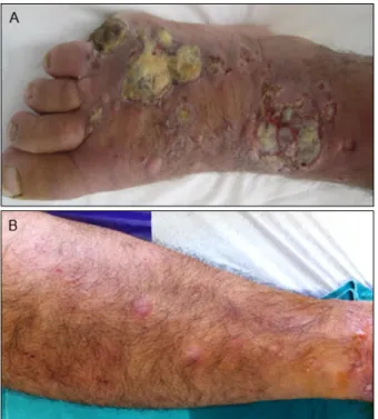
On dermatologic examination, he had an amputated fifth toe and 3 irregularly shaped deep ulcerations on the dor- sum of the right foot, approximately 5 cm in diameter (Fig.
1A). He had multiple erythematous subcutaneous nodules all over his right lower extremity (Fig. 1B). During hospi- talization, he developed 10∼12 new ulcerations 0.5∼1 cm in diameter on the right lower extremity (Fig. 2). As the right lower limb was edematous from the foot to the groin, lymph node palpation could not be performed. On magnetic resonance imaging, few sub-centimeter lympha- denopathies were observed.
Marjolin’s Ulcer with In-Transit Metastases
Vol. 27, No. 4, 2015 443 Fig. 1. (A) Irregularly shaped deep ulcerations on the dorsum
of the right foot. (B) Multiple erythematous subcutaneous nodules on the right lower extremity.
Fig. 2. Development of new ulcerations on the dorsum of the (A) foot and (B) thigh due to in-transit metastases.
Fig. 3. Dermal infiltration of atypical squamous cells (H&E, ×100).
Incisional biopsy was performed on a nodule and the mar- gins of an ulceration for the differential diagnosis of sporo- trichosis, actinomycosis, and atypical mycobacteria infec- tions. Simultaneous tissue cultures revealed methicillin- sensitive Staphylococcus aureus and Acinetobacter species.
Therefore, intravenous ampicillin/sulbactam 1.5 g 4 times/
day and oral ciprofloxacin 500 mg twice daily were admi- nistered. Meanwhile, ulcerations on the dorsum of the right foot gradually enlarged and became necrotic despite wound care with wet and antiseptic wound dressings as well as antibiotics. Myiasis developed on the 15th day of hospi- talization. Screwworms were removed in 4 days by using irrigation with diluted alcohol solution.
Histopathological examination of the specimens showed poorly differentiated SCC with deep invasion of the der- mis and subcutaneous adipose tissue. There was no neu- rovascular invasion (Fig. 3). Laboratory evaluations includ- ing full blood counts, sedimentation, and biochemistry were normal. Cranial, thoracic, and abdominal computed to- mography did not show any organ involvement. After con- sultation with the plastic surgery, orthopedic surgery, and medical oncology departments, systemic chemotherapy was suggested as first-line treatment, but the patient refused.
Therefore, hemipelvectomy surgery was discussed with the patient as second-line treatment, but he refused this option as well. After he was discharged from the hospital, he did not attend follow-up appointments. He died 2 months later, but no information about the actual cause of death was available.
DISCUSSION
Marjolin’s ulcer is a SCC that develops in posttraumatic scars and chronic wounds. However, a diagnosis of SCC with Marjolin’s ulcer is uncommon. The incidence of chronic osteo-
I Altunay, et al
444 Ann Dermatol
myelitis developing into SCC ranges from 0.2%∼1.7%5. Most of these tumors are located on the extremities, par- ticularly the lower limbs. Malignant transformation occurs after a mean of 43 years, ranging from 10 to 70 years.
Tenopyr and Silverman6 state that at least 3 years is re- quired for the progression of an ulcer to malignant trans- formation1. Although the malignant transformation of chron- ic wounds requires further clarification, various factors are implicated, including toxins released from damaged tis- sue, immunological factors, repeated irritation, poor lym- phatic regeneration, co-carcinogens, DNA mutations, and local toxins7.
SCCs originating from these lesions are more aggressive than other primary SCCs. Marjolin’s ulcer has a higher tendency for local recurrence and distant metastasis via the lymphatic system5,7. The most significant prognostic factors predicting recurrence are histological staging and tumor grade. Anatomic location also appears to play an important role in the metastatic potential of the tumor7,8. Tumors of the lower extremities, as in the present case, al- so have a higher risk of recurrence than tumors at other locations. The metastasis rate of lower-extremity lesions is reported to be 30%, whereas Novick et al.9 report an over- all metastasis rate of 53.8%. Once metastasis has occurred, the mortality rate is also high: up to 32.6%8.
In-transit metastasis is frequently reported in malignant mel- anoma but is rarely reported in other primary cutaneous malignancies. The American Joint Committee on Cancer (AJCC) staging system for cutaneous melanoma defines in- transit metastasis as, “intralymphatic metastasis occurring more than 2 cm from the primary tumor.” In-transit meta- stasis is a poor prognostic feature in melanoma; like nodal involvement, it signifies stage III disease10. Reports of in- transit metastases from primary cutaneous SCC are limited to single case reports and one multicenter study of 21 pa- tients, 15 of whom were iatrogenically immunosuppressed organ-transplant recipients. Risk factors for the develop- ment of in-transit metastases are immunosuppressed sta- tus, large primary lesions, lesions on the head and neck, histo- logically poorly differentiated lesions, and recurrent tu- mors4,10.
The prognostic significance of in-transit metastases in SCC is less clear than that in melanoma. Intralymphatic meta- stasis is not mentioned in the current AJCC system for SCC11. Dinehart and Peterson12 recently pointed out some deficiencies of the current AJCC staging system for cuta- neous SCC; they state that satellite and in-transit meta- stases, which are locally invasive tumors, are different from lymph node metastases and that the prognosis of a patient with a locally invasive tumor will never be similar to one with a nodal metastasis. Therefore, they proposed a
new classification system11,12. Additional studies in this area may elucidate the prognostic significance of in-transit metastasis in cutaneous SCC.
Epidermotropic and sporotrichoid cutaneous metastases from cutaneous SCC have been described in the literature.
Clinically, in-transit metastases from cutaneous SCC are mostly subcutaneous or dermal papules with occasional exophytic features4,13. The 2 cases reported by Weidner and Foucar14 were SCC of the lower lip and hand that had in-transit metastases to the submental region of the skin and an upper extremity, respectively. Meanwhile, Copcu et al.13 report 2 cases of in-transit cutaneous metastasis with an acantholytic pattern from a skin tumor on the face.
Wain et al.10 report a case of extensive cutaneous in-trans- it metastasis in an immunocompetent man with cutaneous SCC of the toe. In the present case, the patient had multi- ple subcutaneous nodules arising from the lymphedema in the right lower extremity. Therefore, our initial suspicion was sporotrichosis, actinomycosis, or atypical mycobacteria in- fection. However, new ulcers occurred rapidly, and histo- pathologic examination revealed SCC. Meanwhile, the pa- tient had ulcers and subcutaneous nodules compatible with the definition of in-transit metastases. The lymphedema in our case may have been contributing to the development of in-transit metastases; a possible explanation for this is the increased extravasation of tumor cells within the lymphe- dematous limb and delayed lymphatic return.
Treatment options for multiple in-transit metastases include intralesional or systemic chemotherapy, radiotherapy, ex- cision, amputation, and hyperthermic isolated limb perfu- sion10,15. In particular, hyperthermic isolated limb perfu- sion is an isolated regional therapy that can control un- resectable advanced local or in-transit disease, avoiding amputation and preserving limb functionality15. Single cas- es responding to cetuximab and topical miltefosine have also been reported16,17.
Histopathologic examination is important, particularly in osteomyelitis patients with chronic ulcers, and SCC should be suspected in these cases. Clinicians should be aware that a Marjolin’s ulcer is more aggressive and more prone to metastasis than other skin cancers of the same cell type.
Delayed presentation or misdiagnosis can lead to systemic and potentially fatal metastasis. Therefore, a policy of low threshold to biopsy for chronic non-healing ulcers should be adopted; repeated biopsies may be necessary.
In summary, we report a case of extensive cutaneous in- transit metastasis in a patient with a Marjolin’s ulcer.
Marjolin’s Ulcer with In-Transit Metastases
Vol. 27, No. 4, 2015 445
REFERENCES
1. Tavares E, Martinho G, Dores JA, Vera-Cruz F, Ferreira L.
Marjolin’s ulcer associated with ulceration and chronic osteomyelitis. An Bras Dermato 2011;86:366-369.
2. Ashraf M, Biswas J. Chronic ringworm infestation and Marjolin’s ulcer, an association unknown in the literature.
Rare Tumors 2010;2:e31.
3. Hayes AJ, Clark MA, Harries M, Thomas JM. Management of in-transit metastases from cutaneous malignant melanoma.
Br J Surg 2004;91:673-682.
4. Carucci JA, Martinez JC, Zeitouni NC, Christenson L, Coldiron B, Zweibel S, et al. In-transit metastasis from primary cutaneous squamous cell carcinoma in organ transplant re- cipients and nonimmunosuppressed patients: clinical cha- racteristics, management, and outcome in a series of 21 patients. Dermatol Surg 2004;30:651-655.
5. Ogawa B, Chen M, Margolis J, Schiller FJ, Schnall SB.
Marjolin’s ulcer arising at the elbow: a case report and literature review. Hand (N Y) 2006;1:89-93.
6. Tenopyr J, Silverman I. The relation of chronic varicose ulcer to epithelioma. Ann Surg 1932;95:754-758.
7. Copcu E, Aktas A, Sişman N, Oztan Y. Thirty-one cases of Marjolin’s ulcer. Clin Exp Dermatol 2003;28:138-141.
8. Sabin SR, Goldstein G, Rosenthal HG, Haynes KK. Aggressive squamous cell carcinoma originating as a Marjolin’s ulcer.
Dermatol Surg 2004;30:229-230.
9. Novick M, Gard DA, Hardy SB, Spira M. Burn scar carcinoma:
a review and analysis of 46 cases. J Trauma 1977;17:809-817.
10. Wain EM, Webber NK, Stefanato CM, Banerjee P, Morris
SL. Multiple in-transit cutaneous metastases from a primary cutaneous squamous cell carcinoma. Clin Exp Dermatol 2009;34:522-524.
11. Weinberg AS, Ogle CA, Shim EK. Metastatic cutaneous squamous cell carcinoma: an update. Dermatol Surg 2007;
33:885-899.
12. Dinehart SM, Peterson S. Evaluation of the American Joint Committee on cancer staging system for cutaneous squamous cell carcinoma and proposal of a new staging system. Dermatol Surg 2005;31:1379-1384.
13. Copcu E, Dikicioglu E, Sivrioglu N, Meteoglu I. Subcutaneous nodules on the face: acantholytic in-transit cutaneous meta- stasis. Dermatol Surg 2004;30:1415-1419.
14. Weidner N, Foucar E. Epidermotropic metastatic squamous cell carcinoma. Report of two cases showing histologic con- tinuity between epidermis and metastasis. Arch Dermatol 1985;121:1041-1043.
15. Olieman AF, Liénard D, Eggermont AM, Kroon BB, Lejeune FJ, Hoekstra HJ, et al. Hyperthermic isolated limb perfusion with tumor necrosis factor alpha, interferon gamma, and melphalan for locally advanced nonmelanoma skin tumors of the extremities: a multicenter study. Arch Surg 1999;134:
303-307.
16. Bauman JE, Eaton KD, Martins RG. Treatment of recurrent squamous cell carcinoma of the skin with cetuximab. Arch Dermatol 2007;143:889-892.
17. Mahieu-Renard L, Richard MA, Dales JP, Buscaylet S, Lagrassa S, Grob JJ. Treatment of cutaneous metastases of a squamous cell carcinoma of the leg with topical miltefosine.
Ann Dermatol Venereol 2005;132:346-348.