JH Kim, et al
624 Ann Dermatol
Received July 16, 2012, Revised October 25, 2012, Accepted for publication October 26, 2012
Corresponding author: Bark-Lynn Lew, Department of Dermatology, Kyung Hee University Hospital at Gangdong, 892 Dongnam-ro, Gangdong-gu, Seoul 134-727, Korea. Tel: 82-2-440-7329, Fax: 82-2- 440-7336, E-mail: [email protected]
This is an Open Access article distributed under the terms of the Creative Commons Attribution Non-Commercial License (http://
creativecommons.org/licenses/by-nc/3.0) which permits unrestricted non-commercial use, distribution, and reproduction in any medium, provided the original work is properly cited.
Ann Dermatol Vol. 26, No. 5, 2014 http://dx.doi.org/10.5021/ad.2014.26.5.624
CASE REPORT
Fig. 1. Subcutaneous nodule covered with erythematous, hairless patch on the frontal scalp.
Alopecia Neoplastica due to Gastric Adenocarcinoma Metastasis to the Scalp, Presenting as Alopecia: A Case Report and Literature Review
Jung-Hee Kim, Min-Joong Kim, Woo-Young Sim, Bark-Lynn Lew
Department of Dermatology, Kyung Hee University College of Medicine, Seoul, Korea
Alopecia neoplastica is defined as hair loss secondary to a visceral malignancy that has metastasized to the scalp. The scalp is a relatively common site of cutaneous metastasis, usually presenting as a single or multiple firm scalp nodules. Alopecia neoplastica is a well-recognized but rare presentation, and its pathogenesis is incompletely understood. Atrophy of the hair follicles due to tumor invasion of the scalp plays a role in the development of alopecia. Herein, we describe a 33-year-old woman with gastric adenocarcinoma who developed alopecia neoplastica while receiving cancer chemotherapy. Scalp biopsy revealed metastatic adenocarcinoma cells interspersed be- tween collagen bundles and around hair follicles. Immunohisto- chemical analysis indicated that the tumor cells originated from the primary gastric adenocarcinoma. Therefore, she was diagnosed with alopecia neoplastica due to gastric adenocarci- noma. The findings from this report may be helpful for under- standing the mechanism of alopecia neoplastica. (Ann Dermatol 26(5) 624∼627, 2014)
-Keywords-
Alopecia neoplastica, Gastric adenocarcinoma, Scalp metastasis
INTRODUCTION
The scalp is a relatively common site of cutaneous metas- tasis, usually presenting as a single or multiple firm scalp nodules1,2. Alopecia neoplastica is a well-recognized but rare presentation that, manifests as a single or multiple areas of cicatricial alopecia. Herein we report a rare case of alopecia neoplastica due to metastatic gastric adenocarci- noma and review the relavant literature.
CASE REPORT
A 33-year-old woman was referred for a subcutaneous nodule on the surface of an erythematous-, hairless patch on the frontal scalp observed 3 months previously, to rule out metastasis from her known gastric adenocarcinoma diagnosed in January 2008. She had undergone total gastrectomy for the gastric carcinoma diagnosed in May 2007; she subsequently underwent 6 cycles of chemotherapy and total abdominal
Gastric Adenocarcinoma Metastasis to the Scalp
Vol. 26, No. 5, 2014 625 Fig. 2. (A) Histologic examination revealed decreased pilosebaceous units and scattered, infiltrated tumor cells around hair follicles, upper and mid-dermis (H&E, ×40). (B) Metastatic adenocarcinoma cells were interspersed between collagen bundles and around hair follicles (H&E, ×200). (C) Tumor cells were positively stained against tumor marker MSH-2 (MSH-2, ×200).
Fig. 3. (A) Total gastrectomy specimen shows many signet ring cells (H&E, ×200). Signet ring cells are magnified in inset (H&E,
×400). (B) There are poorly differentiated tumor cells either (H&E, ×200). (C) Part of poorly differentiated tumor cells were positively stained against tumor marker MSH-2 (MSH-2, ×200). (D) Whole body fusion positron emission tomography scan performed after diagnosed with stomach cancer shows abnormal FDG uptake on stomach and rectosigmoid. Following colonoscopy and colon biopsy revealed no other malignancy.
hysterectomy with bilateral salphingo-oophorectomy after being diagnosed with metastatic adenocarcinoma (Krukenberg cancer) in November 2007.
Examination revealed no abnormalities besides a scalp lesion exhibiting a hard, movable, non-tender subcutaneous nodule covered with a slightly erythematous alopecic patch (Fig. 1). The patient did not report any previous derma- tological diseases at the site of alopecia. Routine laboratory test results including full blood count, liver function, renal function, electrolytes, chest radiography and electrocar- diogram were all normal. Histopathological examination of the scalp lesion showed decreased hair follicle cells, as well as metastatic adenocarcinoma cells interspersed between collagen bundles and around hair follicles (Fig. 2A, B).
Similar to the original gastric cancer, tumor cells stained positively for tumor marker MSH-2, the DNA mismatch repair protein (Fig. 2C). The total gastrectomy specimen showed signet ring cells (Fig. 3A) and poorly differentiated adenocarcinoma cells (Fig. 3B) which stained positive for MSH-2 (Fig. 3C). MSH-2 is a marker of a major mismatch repair gene, MSH-2. Polymorphisms in the MSH-2 gene were recently suggested to modulate an individual’s suscep- tibility to gastric cancer3. Although there were no signet ring
cells, the scalp specimen showed scattered, poorly differen- tiated, MSH-2−positive carcinoma cells. Whole body positron emission tomography (PET) scanning showed no other abnormal uptake than in the stomach (Fig. 3D).
Following colonoscopy with biopsy also revealed no malignancy. PET scanning performed after total abdominal hysterectomy with bilateral salphingo-oophorectomy in November 2007 revealed no remaining malignancy. There- fore, we concluded the scalp metastasis originated from the gastric cancer. Cutaneous metastasis usually exhibits fea- tures consistent with the underlying malignancy. However, the metastasis may exhibit less differentiation and be more anaplastic. Therefore, we can infer that atrophy of the hair follicles and gastric cancer invaded the collagenous stroma, influencing the development of alopecia. On the basis of both clinical and histopathological findings, the patient was diagnosed with alopecia neoplastica due to gastric adeno- carcinoma. Despite performing the cancer chemotherapy, no hair regrowth was observed.
DISCUSSION
The overall incidence of cutaneous metastasis from visceral
JH Kim, et al
626 Ann Dermatol
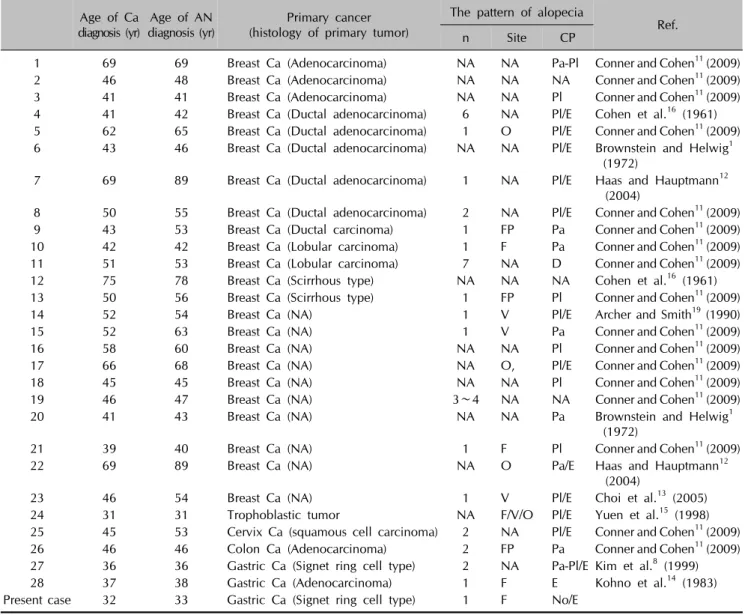
Table 1. Review of the previously reported cases of alopecia neoplastica Age of Ca
diagnosis (yr) Age of AN
diagnosis (yr) Primary cancer (histology of primary tumor)
The pattern of alopecia n Site CP Ref.
1 69 69 Breast Ca (Adenocarcinoma) NA NA Pa-Pl Conner and Cohen11 (2009)
2 46 48 Breast Ca (Adenocarcinoma) NA NA NA Conner and Cohen11 (2009)
3 41 41 Breast Ca (Adenocarcinoma) NA NA Pl Conner and Cohen11 (2009)
4 41 42 Breast Ca (Ductal adenocarcinoma) 6 NA Pl/E Cohen et al.16 (1961) 5 62 65 Breast Ca (Ductal adenocarcinoma) 1 O Pl/E Conner and Cohen11 (2009) 6 43 46 Breast Ca (Ductal adenocarcinoma) NA NA Pl/E Brownstein and Helwig1
(1972)
7 69 89 Breast Ca (Ductal adenocarcinoma) 1 NA Pl/E Haas and Hauptmann12
(2004)
8 50 55 Breast Ca (Ductal adenocarcinoma) 2 NA Pl/E Conner and Cohen11 (2009)
9 43 53 Breast Ca (Ductal carcinoma) 1 FP Pa Conner and Cohen11 (2009)
10 42 42 Breast Ca (Lobular carcinoma) 1 F Pa Conner and Cohen11 (2009)
11 51 53 Breast Ca (Lobular carcinoma) 7 NA D Conner and Cohen11 (2009)
12 75 78 Breast Ca (Scirrhous type) NA NA NA Cohen et al.16 (1961)
13 50 56 Breast Ca (Scirrhous type) 1 FP Pl Conner and Cohen11 (2009)
14 52 54 Breast Ca (NA) 1 V Pl/E Archer and Smith19 (1990)
15 52 63 Breast Ca (NA) 1 V Pa Conner and Cohen11 (2009)
16 58 60 Breast Ca (NA) NA NA Pl Conner and Cohen11 (2009)
17 66 68 Breast Ca (NA) NA O, Pl/E Conner and Cohen11 (2009)
18 45 45 Breast Ca (NA) NA NA Pl Conner and Cohen11 (2009)
19 46 47 Breast Ca (NA) 3∼4 NA NA Conner and Cohen11 (2009)
20 41 43 Breast Ca (NA) NA NA Pa Brownstein and Helwig1
(1972)
21 39 40 Breast Ca (NA) 1 F Pl Conner and Cohen11 (2009)
22 69 89 Breast Ca (NA) NA O Pa/E Haas and Hauptmann12
(2004)
23 46 54 Breast Ca (NA) 1 V Pl/E Choi et al.13 (2005)
24 31 31 Trophoblastic tumor NA F/V/O Pl/E Yuen et al.15 (1998)
25 45 53 Cervix Ca (squamous cell carcinoma) 2 NA Pl/E Conner and Cohen11 (2009)
26 46 46 Colon Ca (Adenocarcinoma) 2 FP Pa Conner and Cohen11 (2009)
27 36 36 Gastric Ca (Signet ring cell type) 2 NA Pa-Pl/E Kim et al.8 (1999)
28 37 38 Gastric Ca (Adenocarcinoma) 1 F E Kohno et al.14 (1983)
Present case 32 33 Gastric Ca (Signet ring cell type) 1 F No/E
Ca: cancer, AN: alopecia neoplastica, CP: clinical presentation, NA: information not available, Pa-Pl: both patch and plaque, Pl: plaque, E: erythema, O: occipital, FP: frontoparietal, Pa: patch, F: frontal, D: diffuse, V: vertex, No: nodule.
carcinomas ranges from 0.7%∼9%4. The scalp is a relati- vely common site of cutaneous metastasis. Brownstein and Helwig1 report that the scalp is the site of 4% of all skin metastases, usually presenting as single or multiple no- dules5-8. The most frequent manifestation of scalp metastasis is the occurrence of single or multiple non-tender nodules that usually appear suddenly and grow rapidly9,10. Alopecia neoplastica is a well-recognized but rarer manifestation that presents as a single or multiple areas of cicatricial alopecia.
To our knowledge, 29 patients with alopecia neoplastica including the one described herein have been reported in the literature (Table 1)1,8,11-16. A review of these cases revealed that alopecia neoplastica is usually a presentation of breast cancer metastasis, and other primary sites are ex-
tremely rare. Yuen et al.15 reported a case in which the primary tumor was a placental trophoblastic tumor. Fur- thermore, metastases from colon and cervical cancer have also been reported. Kohno et al.14 and Kim et al.8, each of them reported 2 cases of alopecia neoplastica due to metastasis from gastric carcinoma. Meanwhile, only 3 cases of alopecia neoplastica due to gastric carcinoma, including this case, have been reported, among them 2 cases were alopecia neoplastica due to signet ring cell-type gastric carcinoma. Because of the small number of cases, it remains unclear if the metastatic potential of signet ring cell-type gastric carcinoma is greater to the skin or scalp.
However, a recent study about the characteristics of gastric adenocarcinoma shows that signet ring-cell gastric cancers
Gastric Adenocarcinoma Metastasis to the Scalp
Vol. 26, No. 5, 2014 627 are more differentiated and less aggressive than non-signet
ring-cell gastric adenocarcinoma17. Nevertheless, the me- chanism of alopecia neoplastica is uncertain. Histopa- thological examination of the scalp in alopecia neoplastica demonstrates metastatic carcinoma cells in a dense collage- nous stroma, which is observed in the dermis and subcuta- neous tissue with a loss of pilosebaceous units18. However, most malignant tumors evoke a stromal response, which is usually in the form of fibroplasia. Moreover, it is unclear if fibrosis or the release of cytokines from tumor cells leads to the disappearance of hair follicles. It is difficult to explain the complete disappearance of pilosebaceous units, but a loss of hair follicles may be the result of fibrosis; Cohen et al.16 consider this to be the major mechanism. In particular, lobular breast carcinoma, which accounts for 10% of invasive breast carcinomas, usually elicits reactive fibrosis, which leads to cicatricial alopecia. Factors independent of fibrosis, such as tumor invasion of the hair sheaths, may play a role in the development of alopecia, because hair may be regrown in alopecic areas after effective cancer treatment19,20. In the present case, metastatic adenocarci- noma cells were interspersed between collagen bundles and around hair follicles. Immunohistochemical staining was positive, which explains tumor cell infiltration around hair follicles. These findings corroborate the hypothesis that dermal infiltration of cancer cells causes the disappearance of hair follicles. Furthermore, infiltration of metastatic cancer cells into the dermis and subcutaneous tissue may put pressure on and destroy hair follicles, eventually causing alopecia.
In summary, we report a case of alopecia neoplastica due to gastric adenocarcinoma, which is a well-recognized but extremely rare manifestation of cancer metastasis to the skin. This report may be helpful for understanding the mechanism of alopecia neoplastica. Regardless, further study is needed to elucidate the specific pathologic mechanism of this disease.
REFERENCES
1. Brownstein MH, Helwig EB. Patterns of cutaneous metastasis.
Arch Dermatol 1972;105:862-868.
2. Bergfeld WF. Hair disorders. In: Moschella SL, Hurley HJ, editors. Dermatology. 3rd ed. Philadelphia: W.B.Saunders Company, 1992:1551-1552.
3. Wang D, Zhou J, Wang T, Li X, Li S, Chen S, et al. Polymor- phisms in MSH2 gene and risk of gastric cancer, and inter-
actions with lifestyle factors in a Chinese population. Cancer Epidemiol 2012;36:e171-e176.
4. Lookingbill DP, Spangler N, Sexton FM. Skin involvement as the presenting sign of internal carcinoma. A retrospective study of 7316 cancer patients. J Am Acad Dermatol 1990;
22:19-26.
5. Gül U, Kiliç A, Akbaş A, Aslan E, Demiriz M. Alopecia neoplastica due to metastatic colon adenocarcinoma. Acta Derm Venereol 2007;87:93-94.
6. Sgambati SA, Barrows GH. Cutaneous metastasis of colon carcinoma: a case report. Conn Med 1993;57:665-667.
7. Proffer LH, Czarnik KL, Sartori CR. Colon carcinoma cutis: a case report. Cutis 1999;63:301-302.
8. Kim HJ, Min HG, Lee ES. Alopecia neoplastica in a patient with gastric carcinoma. Br J Dermatol 1999;141:1122-1124.
9. Chae YS , Suh KS, Kim ST. Alopecia neoplastica due to brea- st cancer. Ann Dermatol 1990;2:89-92.
10. Oh JU, Park JI, Kim HO, Park SR. A case of alopecia neopla- stica metastasis from breast carcinoma. Korean J Dermatol 1999;37:225-228.
11. Conner KB, Cohen PR. Cutaneous metastasis of breast carcinoma presenting as alopecia neoplastica. South Med J 2009;102:385-389.
12. Haas N, Hauptmann S. Alopecia neoplastica due to metastatic breast carcinoma vs. extramammary Paget's disease: mimicry in epidermotropic carcinoma. J Eur Acad Dermatol Venereol 2004;18:708-710.
13. Choi HB, Rho JH, Joh OJ, Park SR, Song KY. A case of alopecia neoplastica from breast cancer. Korean J Dermatol 2005;43:1416-1418.
14. Kohno A, Saruta T, Kimura H. A case of alopecia neoplastica due to cutaneous metastasis from stomach. Rhinsho Derm 1983;25:334-335.
15. Yuen YF, Lewis EJ, Larson JT, Wilke MS, Rest EB, Zachary CB. Scalp metastases mimicking alopecia areata. First case report of placental site trophoblastic tumor presenting as cutaneous metastasis. Dermatol Surg 1998;24:587-591.
16. Cohen I, Levy E, Schreiber H. Alopecia neoplastica due to breast carcinoma. Arch Dermatol 1961;84:490-492.
17. Murakami H, Nakanishi H, Tanaka H, Ito S, Misawa K, Ito Y, et al. Establishment and characterization of novel gastric signet-ring cell and non signet-ring cell poorly differentiated adenocarcinoma cell lines with low and high malignant potential. Gastric Cancer 2013;16:74-83.
18. Johnson WC. Metastatic carcinoma of the skin. In: Elder D, Elenitas R, Jaworsky C, Johnson B Jr, editors. Lever's histo- pathology of the skin. 10th ed. Philadelphia: Lippincott, 2009:1155-1156.
19. Archer CB, Smith NP. Alopecia neoplastica responsive to tamoxifen. J R Soc Med 1990;83:647-648.
20. Scheinfeld N. Review of scalp alopecia due to a clinically unapparent or minimally apparent neoplasm (SACUMAN).
Acta Derm Venereol 2006;86:387-392.