Brief Report
Vol. 29, No. 4, 2017 511
Received May 17, 2016, Revised August 18, 2016, Accepted for publication August 19, 2016
Corresponding author: Young Lee, Department of Dermatology, Chungnam National University School of Medicine, 282 Munhwa-ro, Jung-gu, Daejeon 35015, Korea. Tel: 82-42-280-7706, Fax: 82-42-280-7706, E-mail: [email protected]
This is an Open Access article distributed under the terms of the Creative Commons Attribution Non-Commercial License (http://creativecommons.org/
licenses/by-nc/4.0) which permits unrestricted non-commercial use, distribution, and reproduction in any medium, provided the original work is properly cited.
Copyright © The Korean Dermatological Association and The Korean Society for Investigative Dermatology
ification by GA. Further studies are required to elucidate the biological and physiological significance of Glycer-AGEs in skin, but our study suggests that they may influence normal human keratinocytes and fibroblasts by different mechanisms and accumulate to a different degree than GO-induced AGEs.
CONFLICTS OF INTEREST
The authors have nothing to disclose.
REFERENCES
1. Gkogkolou P, Böhm M. Advanced glycation end products:
Key players in skin aging? Dermatoendocrinol 2012;4:259-270.
2. Zhu P, Ren M, Yang C, Hu YX, Ran JM, Yan L. Involvement of RAGE, MAPK and NF-κB pathways in AGEs-induced MMP-9 activation in HaCaT keratinocytes. Exp Dermatol 2012;21:123-129.
3. Miura J, Yamagishi Si, Uchigata Y, Takeuchi M, Yamamoto H, Makita Z, et al. Serum levels of non-carboxymethyllysine advanced glycation endproducts are correlated to severity of
microvascular complications in patients with Type 1 diabetes.
J Diabetes Complicat 2003;17:16-21.
4. Takino J, Yamagishi S, Takeuchi M. Cancer malignancy is enhanced by glyceraldehyde-derived advanced glycation end-products. J Oncol 2010;2010:739852.
5. Abe R, Shimizu T, Sugawara H, Watanabe H, Nakamura H, Choei H, et al. Regulation of human melanoma growth and metastasis by AGE-AGE receptor interactions. J Invest Dermatol 2004;122:461-467.
6. Takeuchi M, Makita Z, Bucala R, Suzuki T, Koike T, Kameda Y. Immunological evidence that non-carboxymethyllysine advanced glycation end-products are produced from short chain sugars and dicarbonyl compounds in vivo. Mol Med 2000;6:114-125.
7. Kawabata K, Yoshikawa H, Saruwatari K, Akazawa Y, Inoue T, Kuze T, et al. The presence of N(ε)-(Carboxymethyl) lysine in the human epidermis. Biochim Biophys Acta 2011;1814:
1246-1252.
8. Park HY, Kim JH, Jung M, Chung CH, Hasham R, Park CS, et al. A long-standing hyperglycaemic condition impairs skin barrier by accelerating skin ageing process. Exp Dermatol 2011;20:969-974.
https://doi.org/10.5021/ad.2017.29.4.511
Scar Sarcoidosis Developed after Blepharoplasty in Acute Lymphoblastic Leukemia Patient
Sue-Jeong Kim, Ji-Young Kim, Myung Im, Young-Joon Seo, Jeung-Hoon Lee, Young Lee
Department of Dermatology, Chungnam National University School of Medicine, Daejeon, Korea
Dear Editor:
A 46-year-old woman presented with linear erythematous papules that developed from a 20-year-old scar on her left upper eyelid 2 weeks earlier. She had undergone an up-
per eyelid blepharoplasty 20 years earlier. She was treated with a topical steroid for 1 week, without improvement.
She felt some discomfort while opening her eyes. On physical examination, erythematous, firm, non-tender pap-
Brief Report
512 Ann Dermatol
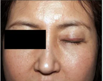
Fig. 1. Erythematous linear papules along left upper eyelid.
Fig. 2. (A, B) Histologic specimen (H&E) shows dense, numerous non- caseating granulomatous infiltrations in entire dermis with epithelioid cells (A: ×10, B: ×100) and (C) asteroid body (×400).
ules were seen along the blepharoplasty scar on her left upper eyelid (Fig. 1). She was diagnosed with precursor B cell lymphoblastic leukemia in 2012 and achieved com- plete remission after allogeneic peripheral blood stem cell transplantation. For a diagnosis and exclusion of leukemia cutis, we performed a skin biopsy. Histological examina- tion showed a dense, non-caseating granulomatous in- filtration throughout the dermis, with numerous epi- thelioid cells. There were no atypical lymphoid cells or immature granulocytes (Fig. 2). These histological findings were consistent with sarcoidosis. A chest X-ray and labo- ratory studies showed no evidence of systemic sarcoidosis.
After 4 months, the lesion improved spontaneously with- out any treatment.
Sarcoidosis is a multiorgan granulomatous disease with unknown etiology1. Scar sarcoidosis is a rare form of cuta- neous sarcoidosis in which pre-existing scars are infiltrated
by non-caseating granulomas. Scar sarcoidosis can devel- op after various events; although there are a few reports on scar sarcoidosis developing after blepharoplasty2. Moreover, there has been no report on scar sarcoidosis occurring on an old blepharoplasty scar in a leukemia pa- tient after a surgical procedure. The patient had blephar- oplasty on both upper eyelids, but the sarcoidosis appeared only on the left side. We don’t know the exact mechanism for this, but different pre-operation state of upper eyelids, surgeon’s skill or different post-operation care could be the reason for the asymmetric appearance of scar sarcoidosis.
Similar to our case, Mantese et al.3 reported the patients who was diagnosed with scar sarcoidosis that developed only part of the scars. Although the patient had blephar- oplasty, hysterectomy and caesarean, she had sarcoidosis on left upper eyelid and on the left lateral side of in- fra-umbical scar.
There have been several reports on sarcoidosis related to hematological malignancies. Among these, only four re- ports described sarcoidosis that developed after acute leu- kemia, after a period ranging from 11 months to 17 years4. There are several hypotheses to explain the relationship between acute leukemia and sarcoidosis. First, the gran- ulomatous inflammation of sarcoidosis may develop in re- action to the tumor-associated antigens found in acute leu- kemia2. Second, the transmission of sarcoidosis or sarcoi- dosis-inducing pathogens via bone marrow transplantation may be associated with the development of sarcoidosis5. Scar sarcoidosis can resolve slowly and spontaneously6, although many treatments have been used for cutaneous sarcoidosis. For patients with only cutaneous sarcoidosis, with no systemic involvement, systemic treatment is not necessary and topical and intralesional steroids can be helpful. When there is systemic involvement, systemic treatment with hydroxychloroquine, prednisolone, or me-
Brief Report
Vol. 29, No. 4, 2017 513
Received April 11, 2016, Revised July 1, 2016, Accepted for publication August 22, 2016
Corresponding author: Won-Soo Lee, Department of Dermatology, Institute of Hair and Cosmetic Medicine, Yonsei University Wonju College of Medicine, 20 Ilsan-ro, Wonju 26426, Korea. Tel: 82-33-741-1345, Fax: 82-33-748-2650, E-mail: [email protected]
This is an Open Access article distributed under the terms of the Creative Commons Attribution Non-Commercial License (http://creativecommons.org/
licenses/by-nc/4.0) which permits unrestricted non-commercial use, distribution, and reproduction in any medium, provided the original work is properly cited.
Copyright © The Korean Dermatological Association and The Korean Society for Investigative Dermatology
thotrexate can be helpful.
This case is a rare occurrence of sarcoidosis that devel- oped on an old upper blepharoplasty scar in a leukemia patient. We suggest a skin biopsy to exclude scar sarcoi- dosis when firm papules appear on a scar in leukemia patients.
ACKNOWLEDGMENT
This research was supported by the Basic Science Research Program through the National Research Foundation of Korea (NRF) funded by the Ministry of Education, Science, and Technology (2015R1A2A2A01004664).
CONFLICTS OF INTEREST
The authors have nothing to disclose.
REFERENCES
1. Kang MJ, Kim HS, Kim HO, Park YM. Cutaneous sarcoidosis presenting as multiple erythematous macules and patches.
Ann Dermatol 2009;21:168-170.
2. Shumway NK, Snitzer L, Alamdari H, Martin K. Resident rounds: part III--cutaneous sarcoidosis in a blepharoplasty scar. J Drugs Dermatol 2015;14:1169-1171.
3. Mantese SA, Berbert AL, Cesário TS, Silva HB. Sarcoidosis on skin scars: a case report. An Bras Dermatol 2010;85:
903-905.
4. Baskaran V, Goodwin A, Athithan L, Addeo A, Rinaldi C. A case of abdominal sarcoidosis in a patient with acute myeloid leukemia. Case Rep Hematol 2013;2013:379898.
5. Padilla ML, Schilero GJ, Teirstein AS. Donor-acquired sarcoidosis. Sarcoidosis Vasc Diffuse Lung Dis 2002;19:
18-24.
6. Mañá J, Marcoval J. Skin manifestations of sarcoidosis.
Presse Med 2012;41:e355-e374.
https://doi.org/10.5021/ad.2017.29.4.513
The Association between Exercise and Androgenetic Alopecia: A Survey-Based Study
Jaewoong Choi, Myungsoo Jun, Solam Lee, Sung-Soo Oh
1, Won-Soo Lee
Department of Dermatology, Institute of Hair and Cosmetic Medicine, Yonsei University Wonju College of Medicine, 1Department of Occupational and Environmental Medicine, Yonsei University Wonju College of Medicine, Wonju, Korea
Dear Editor:
Androgenetic alopecia (AGA) is the most common type of hair loss1. Both genetic and non-genetic factors are known to play a role in development of AGA2. Many studies have focused on the non-genetic factors of AGA, but few have investigated the association between AGA and exercise.
We perform a survey-based study to shed a light on the as- sociation among AGA and exercise. This study was ap- proved by the institutional review board of Yonsei University Wonju College of Medicine (YWMR-15-0-071).
The subjects visited occupational medical clinic of Wonju Severance Christian Hospital for regular medical check up