Surface treatment of feldspathic porcelain:
scanning electron microscopy analysis
Azam Valian, Elham Moravej-Salehi*
Department of Restorative Dentistry, School of Dentistry, Shahid Beheshti University of Medical Sciences, Tehran, Iran
PURPOSE. Topographic analysis of treated ceramics provides qualitative information regarding the surface texture affecting the micromechanical retention and locking of resin-ceramics. This study aims to compare the surface microstructure following different surface treatments of feldspathic porcelain. MATERIALS AND
METHODS. This in-vitro study was conducted on 72 porcelain discs randomly divided into 12 groups (n=6). In 9 groups, feldspathic surfaces were subjected to sandblasting at 2, 3 or 4 bar pressure for 5, 10 or 15 seconds with 50 μm alumina particles at a 5 mm distance. In group 10, 9.5% hydrofluoric acid (HF) gel was applied for 120 seconds. In group 11, specimens were sandblasted at 3 bar pressure for 10 seconds and then conditioned with HF. In group 12, specimens were first treated with HF and then sandblasted at 3 bar pressure for 10 seconds. All specimens were then evaluated under scanning electron microscopy (SEM) at different magnifications. RESULTS.
SEM images of HF treated specimens revealed deep porosities of variable sizes; whereas, the sandblasted surfaces were more homogenous and had sharper peaks. Increasing the pressure and duration of sandblasting increased the surface roughness. SEM images of the two combined techniques showed that in group 11 (sandblasted first), HF caused deeper porosities; whereas in group 12 (treated with HF first) sandblasting caused irregularities with less homogeneity. CONCLUSION. All surface treatments increased the surface area and caused porous surfaces. In groups subjected to HF, the porosities were deeper than those in sandblasted only groups. [J Adv Prosthodont 2014;6:387-94]
KEY WORDS: Air abrasion; Dental porcelain; Hydrofluoric acid; Scanning electron microscopy; Surface treatment
INTRODUCTION
A reliable bond between the tooth structure and restorative materials has always been a concern in restorative dentistry.1 Dentists are always searching for methods to increase the longevity and function of restorations.2 A durable bond
between the resin cement, tooth structure and restorative material is required to form a uniform coherent structure and ensure the long-term survival and clinical success of restorations.3
Both micromechanical and chemical bonding of ceramic surfaces reinforce the fracture resistance of the restored teeth and restorations, provide high retention, prevent microleakage and improve marginal adaptation.4 Resin- ceramics bond strength is influenced by the ceramic surface treatment technique depending on its chemical composi- tion.5-7
Feldspathic porcelains are a type of silica-based porce- lains containing quartz and feldspar.6 Feldspar bonds to some metal oxides to form a glass phase with an amor- phous structure.6,8 This type of porcelain has a high glass content and can best simulate optical properties of enamel and dentin and thus, is commonly used for manufacturing indirect restorations namely laminates, inlays, onlays and overlays.6,8 They are also used for veneering of ceramic copings and metal frameworks.9 In these porcelains, quartz
Corresponding author:
Elham Moravej-Salehi
Department of Restorative Dentistry, School of Dentistry, Shahid Beheshti University of Medical Sciences, Daneshjou Blvd, Evin, Tehran 19834, Iran Tel. 989121263015: e-mail, e.msalehi@yahoo.com
Received 12 February, 2014 / Last Revision 7 July, 2014 / Accepted 8 July, 2014
© 2014 The Korean Academy of Prosthodontics
This is an Open Access article distributed under the terms of the Creative Commons Attribution Non-Commercial License (http://creativecommons.
org/licenses/by-nc/3.0) which permits unrestricted non-commercial use, distribution, and reproduction in any medium, provided the original work is properly cited.
This investigation was supported by Deputy of research, School of Dentistry, Shahid Beheshti University of Medical Sciences, Tehran, Iran. This article was based on a research project (number 310/2598).
constitutes the crystalline phase.6 In addition to high esthet- ics, these ceramics have high resistance to wear and com- pressive loads and are biocompatible.6,9 However, their mechanical strength is lower than other ceramic types.6
These ceramic restorations require internal surface preparation in order to bond to tooth structure with the use of adhesive cements8 and may also undergo chipping or fracture due to various reasons.10,11 Therefore, they require surface treatment before repair with composite resin to pre- vent replacement of restoration, further damage to the tooth and high cost for patients2 and a simpler, more con- servative and quicker treatment is performed as such.12 On the other hand, with increasing number of adults requiring orthodontic treatment, there is a need for a reliable method for bonding brackets to ceramic surfaces.13,14
In all conditions mentioned above, a strong resin bond should be achieved by pre-treatment and formation of micromechanical interlocking to ceramic surfaces. This pro- cess requires micro-roughness and cleaning of the surface for adequate retention.15,16
Acid etching with HF, sandblasting with alumina parti- cles or sometimes a combination of both are among the most common surface treatments for silica-based ceramics.
The bond strength following these surface treatments has been assessed in several studies.10,17,18
HF affects the feldspathic porcelain through reaction with the glass phase2 and its optimal concentration and application time have been specified.19 The mechanism of action of sandblasting is abrasion of the surface with alu- minum oxide particles under air pressure through a chair- side device that increases surface area.20 Function of this process depends on various parameters namely size of par- ticles, applied pressure and duration of application.21,22 According to Dravell’s study,23 large abrasive particles can create greater surface abrasion because surface abrasion is increased by the square of particle diameter. Studies regard- ing the effect of other parameters of sandblasting on sur- face roughness of feldspathic porcelain are scarce.24 Moreover, using a combination of HF etching and sand- blasting, may be the order of applied methods due to them different mechanisms of action, may yield variable results in terms of porcelain surface roughness and micromechani- cal retention and thus are in need of further investigation.
By analyzing the surface topography of treated ceram- ics, we can obtain qualitative data regarding the surface tex- ture that can affect the mechanism of micromechanical retention.25 Scanning electron microscopy (SEM) provides high-resolution information regarding the surface texture for the assessment of surface topography.26
This study aims to compare the surface texture of feld- spathic porcelain following different treatment techniques.
The null hypothesis was that the surface roughness follow- ing the use of different sandblasting pressures and times and HF acid etching is not significantly different. Another hypothesis was that surface roughness in the HF acid etch- ing and sandblasting combined techniques is not influenced by the order of applied methods.
MATERIALS AND METHODS
For the assessment of surface topography, 72 porcelain discs measuring 6 mm × 2 mm (CeramCo 3, Dentsply, Burlington, NJ, USA) with metal base (Ni-Cr alloy) were prepared, fired and polished according to the manufactur- er’s instructions(Fig. 1A). Specimens were evaluated under a magnifier at 10× magnification to ensure absence of cracks or chipping. Specimen surfaces were polished with 400 and 600 grit SiC discs under running water, rinsed and dried with oil-free air. Specimens were randomly divided into 12 groups of 6 and coded. The following surface treatments were performed (Table 1):
Group (G) 1. The surface of specimens was sandblasted using microsandblaster (Microetcher II, Danville Materials Inc., San Raman, CA, USA) (Fig. 1B) with 50 μm alumina particles (Ronvig, Denmark) at 2 bar pressure for 5 seconds from a 5 mm distance at 90° angulation of the nozzle in a circular motion. (For the surface preparation, a special jig was fabricated for this purpose for all specimens in 12 groups.) Specimens were then rinsed with oil-free water and dried with oil-free air.
G 2. Specimens were sandblasted similar to G 1 but sandblasting was performed at 2 bar pressure for 10 seconds.
G 3. Specimens were sandblasted similar to G 1 but sandblasting was performed at 2 bar pressure for 15 seconds.
G 4. Specimens were sandblasted similar to G 1 but sandblasting was performed at 3 bar pressure for 5 seconds.
G 5. Specimens were sandblasted similar to G 1 but sandblasting was performed at 3 bar pressure for 10 seconds.
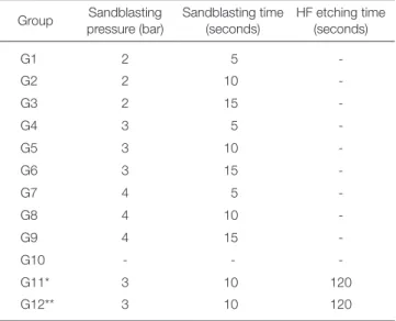
Table 1. Surface treatment parameters in different study groups
Group Sandblasting pressure (bar)
Sandblasting time (seconds)
HF etching time (seconds)
G1 2 5 -
G2 2 10 -
G3 2 15 -
G4 3 5 -
G5 3 10 -
G6 3 15 -
G7 4 5 -
G8 4 10 -
G9 4 15 -
G10 - - -
G11* 3 10 120
G12** 3 10 120
*In this group, specimens were first sandblasted and then etched with HF acid gel.
**In this group, specimens were first etched with HF acid gel and then sandblasted.
G 6. Specimens were sandblasted similar to G 1 but sandblasting was performed at 3 bar pressure for 15 seconds.
G 7. Specimens were sandblasted similar to G 1 but sandblasting was performed at 4 bar pressure for 5 seconds.
G 8. Specimens were sandblasted similar to G 1 but sandblasting was performed at 4 bar pressure for 10 seconds.
G 9. Specimens were sandblasted similar to G 1 but sandblasting was performed at 4 bar pressure for 15 seconds.
G 10 (control). Buffered 9.5% HF gel (Porcelain etchant gel, Bisco, Schamburg, IL, USA) (Fig. 1C) was applied to the specimen surfaces for 2 minutes and then rinsed with oil free water and dried with oil free air spray.
G 11. Specimens were first sandblasted as in G 5 and then HF acid was applied as in G 10.
G 12. Specimens were first treated with HF acid gel as in G 10 and then sandblasted as in G 5.
All specimens were then gold-coated by sputter-coater machine (K950X, EMTECH, England) (Fig. 1D) with 15 nm thickness and SEM images were obtained of 5 random areas in each specimen under electron microscopy (VEGA, TESCAN, Czech Republic) at 500×, 2,000×, 4,000× and 10,000× magnifications.
RESULTS
SEM analysis showed that the sandblasted surfaces were porous with steep peaks. Increasing the pressure and dura- tion of sandblasting increased surface roughness. In some SEM images of sandblasted surfaces, tiny alumina chips were evident. By increasing the sandblasting pressure from 2 to 4 bar surface porosities increased as well and a slight increase was observed in the depth of the porosities (Fig. 2, Fig. 3, Fig. 4).
SEM analysis of the HF treated feldspathic porcelain surfaces revealed a porous surface of variable-sized porosi- ties (Fig. 5). SEM showed that 9.5% HF for 2 minutes caused deeper porosities than sandblasting even at 4 bar pressure for 15 seconds. Sandblasted surfaces yielded a rougher microstructure with deep and uniform size porosi- ties and steeper peaks than the surfaces treated with HF acid. Increasing the pressure and duration of sandblasting enhanced uniformity of the size of the porosities.
SEM analysis of specimens treated with the combined technique of etching with HF and subsequent sandblasting showed increased surface roughness and porosities (Fig. 6,
Fig. 1. Materials and equipment used. (A) feldspathic porcelain disc measuring 6 x 2 mm, (B) microsandblaster, (C) buffered 9.5% HF gel, (D) sputter-coater machine.
A B
C D
A: G1 B: G2 C: G3
Fig. 2. SEM analysis of the feldspathic porcelain surface topography following sandblasting with 50 μm alumina particles from a 5 mm distance at 2 bar pressure for 5 s (A), 10 s (B) and 15 s (C). (2,000X magnification).
Fig. 3. SEM analysis of the feldspathic porcelain surface topography following sandblasting with 50 μm alumina particles from a 5 mm distance at 3 bar pressure for 5 s (A), 10 s (B) and 15 s (C). (2,000X magnification).
A: G4 B: G5 C: G6
Fig. 4. SEM analysis of the feldspathic porcelain surface topography following sandblasting with 50 μm alumina particles from a 5 mm distance at 4 bar pressure for 5 s (A), 10 s (B) and 15 s (C). (2,000X magnification).
A: G7 B: G8 C: G9
Fig. 7). SEM analysis of specimens treated with the com- bined technique of sandblasting and subsequent treatment with HF acid showed more prominent deep porosities due to HF acid. In the former technique (HF application and subsequent sandblasting), sandblasting removed a greater surface area and provided less uniform porosities compared to the other sandblasted groups.
DISCUSSION
Microscopic analysis can greatly help study the characteris- tics of materials.25 Qualitative assessment of micro-mor-
phologic surface changes following different ceramic sur- face treatments can enhance our understanding of the sur- face changes affecting the bond strength.27 Surface treat- ments roughen the porcelain and enhance the formation of optimal micromechanical bond between the porcelain and resin.28 Thus, ceramic surface preparation (by etching or sandblasting)14 is a critical part for clinical success of indi- rect bonded ceramic restorations, direct ceramic repair pro- cedures25 and bonding of orthodontic brackets to ceramic surfaces.13 This study aimed to assess the quality of micro- structure obtained by different surface treatments of feld- spathic porcelain.
Fig. 5. SEM analysis of the feldspathic porcelain surface topography following acid etching with 9.5% HF acid for 120 s. (2,000X magnification).
G10
Fig. 6. SEM analysis of the feldspathic porcelain surface topography following sandblasting with 50 μm alumina particles at 3 bar pressure for 10 s and then HF acid etching for 120 s; (A): 500X magnification; (B): 2,000X magnification.
A: G11 B: G11
A B
Fig. 7. SEM analysis of the feldspathic porcelain surface topography following 9.5% HF acid etching for 120 s and then sandblasting with 50 μm alumina particles at 3 bar pressure for 10 s from a 5 mm distance; (A): 500X magnification; (B): 2,000X magnification.
A: G12 B: G12
SEM images following the application of HF acid to feldspathic porcelain surface (control group) showed small and large micro-porosities as a three-dimensional network of canals and voids, increasing the surface microroughness.
These porosities had a tunnel-like micro-retentive pattern resembling a honey-comb pattern. This finding is in accord with the results of studies by Borges et al.,29 Bottino et al.27 and Kukiattrakoon and Thammasitboon.2 This pattern results from chemical interaction of HF with glass matrix forming insoluble hexafluorosilicate and partially removing the glass matrix. Subsequently, the crystalline structure is exposed and a surface containing tunnel-like undercuts is created.30 Due to the increased surface energy, the wettabili- ty of silane improves as well.27 Sandblasting is another tech- nique for successful surface treatment of feldspathic porce- lain.31 SEM images of sandblasted surfaces in our study revealed increased surface irregularity and micro-roughness and porosities with sharper peaks but shallower than those of surfaces treated with HF acid alone. Different SEM pat- terns in sandblasted and HF treated groups are due to the different types of surface treatment28 because, as stated ear- lier, HF chemically reacts with the silica phase and pene- trates into the depth30,32 while in sandblasting, the surface is mechanically bombarded by alumina particles under air pressure,20,33 resulting in removal of weak ceramic phases25 and subsequent surface irregularity and increased surface area.25,32 A clean active bonding area is formed as such in the porcelain increasing resin and ceramic wettability and improving the micromechanical interlocking and bond strength.34
SEM images revealed that by increasing the pressure and duration of sandblasting, irregularities and surface microhardness increased as well. Moreover, increased pres- sure slightly increased the depth of impact. Increased micro-roughness is enhanced by the mechanism of action of the microsandblaster device. In a study by Aminsalehi et al, increased time of sandblasting revealed slight increase in feldspathic porcelain surface porosities.24 SEM analysis in our study demonstrated that surface roughness caused by sandblasting especially at higher pressures and longer time periods was more than that following HF acid application.
Other studies have demonstrated greater surface roughness due to sandblasting rather than HF acid etching in glass matrix ceramics using profilometry, SEM and AFM.15,34,35 However, some researchers like Saraç et al.28 stated that air abrasion caused less surface roughness than application of 9.6% HF gel for 2 minutes. Thus, different sandblasting parameters should be taken into account in such studies.
Saraç et al.28 used 25 μm alumina particles at 2.5 bar pres- sure and a 10 mm distance for 4 seconds. Such sandblasting settings might be responsible for the less surface roughness achieved in their study in comparison to ours.
SEM images in our study showed different micro-mor- phology and higher surface roughness due to sandblasting compared to HF application. However, some studies found no significant difference in bond strength following sand- blasting and HF acid treatment.13,15,24 Ersu et al.15 stated that
although sandblasting provides a rougher surface, this roughness does not improve bond strength. Results in this regard are controversial and some others have reported higher bond strength following sandblasting compared to HF application.28
Considering all the above, a few points should be taken into account: First, aside from surface roughness, there are several other factors that affect the resin-ceramics bond strength.2 More importantly, although sandblasting increas- es surface roughness, such surface irregularities are not in the form of retentive undercuts (like in HF-treated surfac- es) and bond strength may even decrease in the long- term2,36 because hydrolytic changes are intensified by aging.
This effect is more significant in sandblasted specimens compared to the HF-treated ones because porosities caused by HF application are deeper and thus, resin is better pro- tected from the degradation process.37 However, application of silane as the promoter of adhesion, in the clinical pro- cess of reinforcing the chemical resin-ceramic bond can enhance the clinical success of mechanical surface treat- ment methods38; because silane helps achieve a durable res- in-ceramic bond.38 The bond strength following HF appli- cation or sandblasting has been reported to be clinically acceptable.24,39,40 On the other hand, it should be noted that there is a threshold for surface porosity limiting its effect on bond strength.2 In some studies like the one by Amin Salehi et al.,24 increasing the sandblasting time of feldspathic porcelain increased surface roughness but could not increase the bond strength. Kern et al.,41 in their study in 2009 reported that by increasing the zirconia sandblasting pressure, surface roughness increased but no increase in bond strength was noted. Thus, in case of presence of ade- quate bond strength, there is no need to increase surface roughness because porcelain roughness may decrease por- celain restoration strength28 and even its flexural strength because superficial cracks can lead to failure at lower stress levels.42 On the other hand, bond strength depends on its indication. For example, for bonding orthodontic brackets, 6 to 8 MPa bond strength is clinically sufficient43 because porcelain roughness following debonding of orthodontic brackets may increase plaque accumulation, gingival inflam- mation and soft tissue adverse reactions.28 Surface rough- ness can affect the gloss and color of porcelain and lead to staining. Porcelain esthetics can be negatively affected as such.2 Thus, there is no need for high pressure or long duration of sandblasting either alone or in combination with HF despite increasing the surface roughness because based on previous studies, excessive sandblasting can induce chipping or significant loss of porcelain material.7,34
On the other hand, although HF is effective for porce- lain surface treatment and creation of micromechanical retention,2,44 some previous studies show that treatment with HF provides statistically higher bond strength than other types of surface treatments.2 However, it has an acid- ic and corrosive nature4 and can decrease the flexural strength of ceramics.45 It has the potential to severely trau- matize soft tissue and tooth structure.38 Intraoral applica-
tion of HF acid is not easy and its contact with the tooth is sometimes inevitable.46 It has been shown that exposure of the dentin surface to HF can cause collapse of collagen fibers and formation of a hybrid layer with 3 μm thickness.
Deposition of relatively insoluble CaF2 on dentin prevents the effect of phosphoric acid on the tooth surface and neg- atively affects the process of bond formation.46 HF can also cause soft tissue burns and rash.2 Its inhalation is tox- ic47 and in the long-term it can even adversely affect the nerves.46 Thus, care must be taken when using HF acid38 and some studies even suggest that this method not be used for intra-oral repair of restorations.18,34,47 In our study, SEM images showed that the combined method of HF etching and sandblasting with alumina particles increased the surface roughness and porosities related in both proce- dures. In some studies, application of HF after sandblasting caused no significant difference in surface roughness com- pared to sandblasting alone.28,35 Thus, it seems that sand- blasting may be superior to HF only because of the harm- ful nature and stimulatory effects of HF.28 On the other hand, in our study, following primary application of HF and then sandblasting for surface preparation of feldspath- ic porcelain, sandblasting was found to be more aggressive than other methods (based on SEM images) and removed more volume from the porcelain surface. Because HF reacts with silica phase, removes some of porcelain surface and weakens porcelain structure.30 Since sandblasting impact is on loose phases of ceramic,25 application of sand- blasting following HF etching results in more degradation of surface.
All methods used in this study increased the surface roughness but HF was also able to cause micro-retentive areas in addition to increasing the surface roughness and surface area compared to those of sandblasting. Sandblasting at higher pressure and longer periods caused greater surface roughness. Use of sandblasting following HF etching yield- ed more aggressive results. Although surface roughness, to some extent, promotes the micromechanical retention, but excessive roughness should be avoided due to adverse effects.
Limitation of this study is the lack of quantitative evalu- ation of surface texture. Future studies are needed to evalu- ate other surface treatments such as various lasers and their parameters.
CONCLUSION
Within the limitations of this study, the following results were obtained:
SEM analysis showed that the porosities caused by HF acid etching were deeper and of various sizes while sand- blasting porosities were uniform in size and more homoge- nous. The sandblasted surface was rougher with sharper peaks.
Increasing the sandblasting time increased surface roughness. Increasing the pressure increased surface rough- ness and depth of pores.
Combination of HF etching and sandblasting increased
surface roughness and porosities. More deeper-pores were evidently observed in surfaces that were subjected to sand- blasting. In case of primary HF etching, sandblasting removed a greater volume of porcelain surface and the porosities were less uniform in size and shape.
REFERENCES
1. Leinfelder KF. Dentin adhesives for the twenty-first century.
Dent Clin North Am 2001;45:1-6.
2. Kukiattrakoon B, Thammasitboon K. Optimal acidulated phosphate fluoride gel etching time for surface treatment of feldspathic porcelain: on shear bond strength to resin com- posite. Eur J Dent 2012;6:63-9.
3. Gökçe B. Effects of Er: YAG laser irradiation on dental hard tissues and all-ceramic materials: SEM evaluation. Available from: http://www.intechopen.com/books/scanning-electron- microscopy/effects-of-laser-irradiation-on-dental-hard-tis- sues-and-dental-materials-sem-evaluation-at 12 September, 2014.
4. Zarone F, Sorrentino R, Vaccaro F, Traini T, Russo S, Ferrari M. Acid etching surface treatment of feldspathic, alumina and zirconia ceramics: a micromorphological SEM analysis.
Int Dent South Afr 2006;8:20-6.
5. Torres SM, Borges GA, Spohr AM, Cury AA, Yadav S, Platt JA. The effect of surface treatments on the micro-shear bond strength of a resin luting agent and four all-ceramic systems. Oper Dent 2009;34:399-407.
6. de Carvalho RF, Martins ME, de Queiroz JR, Leite FP, Ozcan M. Influence of silane heat treatment on bond strength of resin cement to a feldspathic ceramic. Dent Mater J 2011;30:392-7.
7. Blatz MB, Sadan A, Kern M. Resin-ceramic bonding: a re- view of the literature. J Prosthet Dent 2003;89:268-74.
8. Kelly JR. Dental ceramics: what is this stuff anyway? J Am Dent Assoc 2008;139:4S-7S.
9. Vargas MA, Bergeron C, Diaz-Arnold A. Cementing all-ce- ramic restorations: recommendations for success. J Am Dent Assoc 2011;142:20S-4S.
10. de Melo RM, Valandro LF, Bottino MA. Microtensile bond strength of a repair composite to leucite-reinforced feld- spathic ceramic. Braz Dent J 2007;18:314-9.
11. El-Hosary MMK, Shokry TE, Zaki DY, El-Shakour ASA.
Bond strength of different intraoral repair systems for metal- ceramic restorations. J Am Sci 2011; 7:383-8.
12. Reston EG, Filho SC, Arossi G, Cogo RB, Rocha Cdos S, Closs LQ. Repairing ceramic restorations: final solution or al- ternative procedure? Oper Dent 2008;33:461-6.
13. Falkensammer F, Jonke E, Bertl M, Freudenthaler J, Bantleon HP. Rebonding performance of different ceramic brackets conditioned with a new silane coupling agent. Eur J Orthod 2013;35:103-9.
14. Yadav S, Upadhyay M, Borges GA, Roberts WE. Influence of ceramic (feldspathic) surface treatments on the micro- shear bond strength of composite resin. Angle Orthod 2010;80:577-82.
15. Ersu B, Yuzugullu B, Ruya Yazici A, Canay S. Surface rough-
ness and bond strengths of glass-infiltrated alumina-ceramics prepared using various surface treatments. J Dent 2009;37:
848-56.
16. Osorio E, Toledano M, da Silveira BL, Osorio R. Effect of different surface treatments on In-Ceram Alumina rough- ness. An AFM study. J Dent 2010;38:118-22.
17. Kupiec KA, Wuertz KM, Barkmeier WW, Wilwerding TM.
Evaluation of porcelain surface treatments and agents for composite-to-porcelain repair. J Prosthet Dent 1996;76:119-24.
18. Pollington S, Fabianelli A, van Noort R. Microtensile bond strength of a resin cement to a novel fluorcanasite glass-ce- ramic following different surface treatments. Dent Mater 2010;26:864-72.
19. Barghi N, Fischer DE, Vatani L. Effects of porcelain leucite content, types of etchants, and etching time on porcelain- composite bond. J Esthet Restor Dent 2006;18:47-52.
20. Kara HB, Dilber E, Koc O, Ozturk AN, Bulbul M. Effect of different surface treatments on roughness of IPS Empress 2 ceramic. Lasers Med Sci 2012;27:267-72.
21. Retief DH. Standardizing laboratory adhesion tests. Am J Dent 1991;4:231-6.
22. Della Bona A, Donassollo TA, Demarco FF, Barrett AA, Mecholsky JJ Jr. Characterization and surface treatment ef- fects on topography of a glass-infiltrated alumina/zirconia- reinforced ceramic. Dent Mater 2007;23:769-75.
23. Darvell BW. Materials science for dentistry. 9th ed. Elsevier, UK; 2009. p. 450-70.
24. Amin Salehi E, Heshmat H, Moravej Salehi E, Kharazifard M. In vitro evaluation of the effect of different sandblasting times on the bond strength of feldspathic porcelain to com- posite resin. J Islam Dent Assoc IRAN 2013;25:22-30.
25. Della Bona A, Anusavice KJ. Microstructure, composition, and etching topography of dental ceramics. Int J Prosthodont 2002;15:159-67.
26. Subaşı MG, İnan Ö. Evaluation of the topographical surface changes and roughness of zirconia after different surface treatments. Lasers Med Sci 2012;27:735-42.
27. Bottino MC, Ozcan M, Coelho PG, Valandro LF, Bressiani JC, Bressiani AH. Micro-morphological changes prior to ad- hesive bonding: high-alumina and glassy-matrix ceramics.
Braz Oral Res 2008;22:158-63.
28. Saraç YS, Elekdag-Turk S, Saraç D, Turk T. Surface condi- tioning methods and polishing techniques effect on surface roughness of a feldspar ceramic. Angle Orthod 2007;77:723-8.
29. Borges GA, Sophr AM, de Goes MF, Sobrinho LC, Chan DC. Effect of etching and airborne particle abrasion on the microstructure of different dental ceramics. J Prosthet Dent 2003;89:479-88.
30. Dilber E, Yavuz T, Kara HB, Ozturk AN. Comparison of the effects of surface treatments on roughness of two ceramic systems. Photomed Laser Surg 2012;30:308-14.
31. Cal-Neto JP, Castro S, Moura PM, Ribeiro D, Miguel JA.
Influence of enamel sandblasting prior to etching on shear bond strength of indirectly bonded lingual appliances. Angle Orthod 2011;81:149-52.
32. Ozcan M. Evaluation of alternative intra-oral repair tech- niques for fractured ceramic-fused-to-metal restorations. J
Oral Rehabil 2003;30:194-203.
33. Kulunk S, Kulunk T, Ural C, Kurt M, Baba S. Effect of air abrasion particles on the bond strength of adhesive resin ce- ment to zirconia core. Acta Odontol Scand 2011;69:88-94.
34. Yavuz T, Dilber E, Kara HB, Tuncdemir AR, Ozturk AN.
Effects of different surface treatments on shear bond strength in two different ceramic systems. Lasers Med Sci 2013;28:1233-9.
35. Schmage P, Nergiz I, Herrmann W, Ozcan M. Influence of various surface-conditioning methods on the bond strength of metal brackets to ceramic surfaces. Am J Orthod Dentofacial Orthop 2003;123:540-6.
36. Wood DJ, Bubb NL, Millar BJ, Dunne SM. Preliminary in- vestigation of a novel retentive system for hydrofluoric acid etch-resistant dental ceramics. J Prosthet Dent 1997;78:275-80.
37. Torres SM, Borges GA, Spohr AM, Cury AA, Yadav S, Platt JA. The effect of surface treatments on the micro-shear bond strength of a resin luting agent and four all-ceramic systems. Oper Dent 2009;34:399-407.
38. Saraç YŞ, Külünk T, Elekdağ-Türk S, Saraç D, Türk T.
Effects of surface-conditioning methods on shear bond strength of brackets bonded to different all-ceramic materi- als. Eur J Orthod 2011;33:667-72.
39. Lacy AM, LaLuz J, Watanabe LG, Dellinges M. Effect of porcelain surface treatment on the bond to composite. J Prosthet Dent 1988;60:288-91.
40. Falkensammer F, Freudenthaler J, Pseiner B, Bantleon HP.
Influence of surface conditioning on ceramic microstructure and bracket adhesion. Eur J Orthod 2012;34:498-504.
41. Kern M, Barloi A, Yang B. Surface conditioning influences zirconia ceramic bonding. J Dent Res 2009;88:817-22.
42. Herion DT, Ferracane JL, Covell DA Jr. Porcelain surface al- terations and refinishing after use of two orthodontic bond- ing methods. Angle Orthod 2010;80:167-74.
43. Türk T, Saraç D, Saraç YS, Elekdağ-Türk S. Effects of sur- face conditioning on bond strength of metal brackets to all- ceramic surfaces. Eur J Orthod 2006;28:450-6.
44. Kara HB, Ozturk AN, Aykent F, Koc O, Ozturk B. The effect of different surface treatments on roughness and bond strength in low fusing ceramics. Lasers Med Sci 2011;26:599-604.
45. Zogheib LV, Bona AD, Kimpara ET, McCabe JF. Effect of hydrofluoric acid etching duration on the roughness and flex- ural strength of a lithium disilicate-based glass ceramic. Braz Dent J 2011;22:45-50.
46. Loomans BA, Mine A, Roeters FJ, Opdam NJ, De Munck J, Huysmans MC, Van Meerbeek B. Hydrofluoric acid on den- tin should be avoided. Dent Mater 2010;26:643-9.
47. Fabianelli A, Pollington S, Papacchini F, Goracci C, Cantoro A, Ferrari M, van Noort R. The effect of different surface treatments on bond strength between leucite reinforced feld- spathic ceramic and composite resin. J Dent 2010;38:39-43.