ABSTRACT
Purpose: This study investigated whether the metabolic avidity of primary tumors and/
or metastatic lymph nodes (LNs) measured by 18F-fluorodeoxyglucose (18F-FDG) positron emission tomography/computed tomography (PET/CT) was related to survival after surgery in patients with advanced gastric cancer (AGC).
Materials and Methods: One hundred sixty-eight patients with AGC who underwent preoperative 18F-FDG PET/CT and curative resection were included. The 18F-FDG avidity of the primary gastric tumor and LNs was determined quantitatively and qualitatively. The diagnostic performance of 18F-FDG PET/CT was calculated, and the prognostic significance of
18F-FDG avidity for recurrence-free survival (RFS) and overall survival (OS) was assessed.
Results: In all, 51 (30.4%) patients experienced recurrence, and 32 (19.0%) died during follow-up (median follow-up duration, 35 months; range, 3–81 months); 119 (70.8%) and 33 (19.6%) patients showed 18F-FDG-avid primary tumors and LNs, respectively. 18F-FDG PET/
CT showed high sensitivity (73.8%) for the detection of advanced pathologic T (pT ≥3) stage and high specificity (92.2%) for the detection of advanced pN (≥2) stage. 18F-FDG avidity of LNs was significantly associated with RFS (P=0.012), whereas that of primary tumors did not show significance (P=0.532). Univariate and multivariate analyses revealed that 18F-FDG avidity of LNs was an independent prognostic factor for RFS (hazard ratio=2.068; P=0.029).
Conclusions: 18F-FDG avidity of LNs is an independent prognostic factor for predicting RFS.
Preoperative 18F-FDG PET/CT can be used to determine the risk and prognosis of patients with AGC after curative resection.
Keywords: Gastric cancer; 18F-fluorodeoxyglucose; Positron Emission Tomography Computed Tomography; Recurrence
INTRODUCTION
Advanced gastric cancer (AGC) is one of the most common malignancies resulting in cancer- related deaths in Asian populations [1-3]. Radical surgery is the standard treatment option for patients with resectable disease; however, the prognosis remains poor with a 5-year survival of approximately 30% [3]. Recurrence after curative surgery often occurs in 12%−54% of cases and is the main cause of cancer-related death [4]. Hence, stratifying the patient's risk
Original Article
Received: May 1, 2018 Revised: Jul 15, 2018 Accepted: Jul 23, 2018 Correspondence to Sungeun Kim
Department of Nuclear Medicine, Korea University Anam Hospital, Korea University College of Medicine, 73 Inchon-ro, Seongbuk-gu, Seoul 02841, Korea.
E-mail: [email protected] Sungsoo Park
Department of Surgery, Korea University Anam Hospital, Korea University College of Medicine, 73 Inchon-ro, Seongbuk-gu, Seoul 02841, Korea.
E-mail: [email protected]
*Sungsoo Park and Sungeun Kim equally contributed to this work.
Copyright © 2018. Korean Gastric Cancer Association
This is an Open Access article distributed under the terms of the Creative Commons Attribution Non-Commercial License (https://
creativecommons.org/licenses/by-nc/4.0) which permits unrestricted noncommercial use, distribution, and reproduction in any medium, provided the original work is properly cited.
ORCID iDs Hyun Woo Kwon
https://orcid.org/0000-0002-0597-837X Liang An
https://orcid.org/0000-0002-5466-7834 Hye Ryeong Kwon
https://orcid.org/0000-0002-4060-8294 Sungsoo Park
https://orcid.org/0000-0002-1779-8683
Hyun Woo Kwon 1, Liang An 2,Hye Ryeong Kwon 1, Sungsoo Park 2,*, Sungeun Kim 1,*
1Department of Nuclear Medicine, Korea University Anam Hospital, Seoul, Korea
2Department of Surgery, Korea University Anam Hospital, Seoul, Korea
Preoperative Nodal 18 F-FDG Avidity
Rather than Primary Tumor Avidity
Determines the Prognosis of Patients
with Advanced Gastric Cancer
and prediction of prognosis are key issues in the postoperative management of AGC patients.
The tumor, node, metastasis (TNM) classification system by the American Joint Committee on Cancer (AJCC) has been used to predict patients' prognoses. However, the conventional clinicopathological approach does not adequately represent the complexity of gastric cancer biology [5,6].
Current guidelines for staging AGC include gastrofiberscopy; contrast-enhanced computed tomography (CECT) of the thorax, abdomen, and pelvis; and laparoscopy.
18F-fluorodeoxyglucose (18F-FDG) positron emission tomography/computed tomography (PET/CT) is preferred to evaluate metastatic disease [7,8]. 18F-FDG PET/CT has been shown to be useful in AGC patients in many aspects. 18F-FDG PET/CT can determine the operability of locally advanced gastric cancer [9] and facilitate the detection of recurrence during follow-up after surgery [10,11]. Several studies have shown the usefulness of 18F-FDG PET/CT for the prognosis of AGC patients undergoing chemotherapy [12,13]. However, the independent role of preoperative 18F-FDG PET/CT in AGC still needs to be investigated.
Lymph node (LN) metastasis is a substantial factor in the staging and prognosis of patients with AGC [14]. Although 18F-FDG PET/CT has low diagnostic sensitivity for LN metastasis, evaluation of LN metabolism can be useful to predict prognosis [15]. The advantage of
18F-FDG PET/CT is the simultaneous assessment and comparison of metabolism in primary tumors and metastatic LNs, which provide prognostic information [16,17]. A comprehensive evaluation of the metabolic aggressiveness of metastatic LNs in the preoperative setting can be used as a potential biomarker in AGC.
In this study, we evaluated the metabolic characteristics of primary tumors and/or metastatic LNs and compared them with survival data after surgery in patients with AGC to determine the clinical impact of preoperative 18F-FDG PET/CT.
MATERIALS AND METHODS
Patients
We analyzed the medical records of consecutive AGC patients in our hospital between July 2010 and December 2014. Among them, 168 patients with AGC who underwent curative surgical resection were enrolled in this study. The inclusion criteria were: 1) initial diagnosis of gastric cancer, 2) preoperative staging with 18F-FDG PET/CT in our hospital, 3) pathologic staging of T2–T4, and 4) a histological type of common epithelial tumor according to the Japanese Gastric Cancer Association system [18]. The exclusion criteria were: 1) early gastric cancers, 2) double primary malignancies, 3) distant organ metastasis, 4) loss to follow- up within 3 months after surgery, and 5) treatment with palliative/non-curative surgery.
Patients' clinical and histopathologic characteristics analyzed in this study were acquired prospectively at the time of surgery. The Institutional Review Board of our hospital approved this retrospective clinical study (AN17196-001), and the requirement for informed consent was waived. All procedures performed in this study involving human participants were in accordance with the 1964 Helsinki declaration and its later amendments or comparable ethical standards.
All patients underwent total or subtotal gastrectomy according to the anatomic location of the primary tumor, accompanied by lymphadenectomy according to the General Rules Sungeun Kim
https://orcid.org/0000-0002-3392-0263 Funding
This work was supported by a Korea University Grant.
Author Contributions
Conceptualization: P.S., K.S.; Data curation:
K.H.W., A.L., K.H.R., P.S.; Investigation:
K.H.W., K.H.R.; Project administration: P.S., K.S.; Writing - original draft: K.H.W.; Writing - review & editing: P.S., K.S.
Conflict of Interest
No potential conflict of interest relevant to this article was reported.
for Gastric Cancer [18]. Curative resection was defined by the complete absence of visible tumor tissue with negative margins. In cases of subtotal gastrectomy, tumor-free margins of >3 cm and 5 cm were obtained in patients with well-marginated and infiltrative primary tumors, respectively. Histopathologic classification of the primary tumors included subtype (signet ring cell vs. non-signet ring cell, which included adenocarcinoma with papillary, tubular, poorly differentiated, and mucinous subtypes), Lauren classification (intestinal vs.
non-intestinal, which included diffuse and mixed types), and Bormann classification. After surgery, patients underwent adjuvant chemotherapy based on their histopathologic results and performance status. Subsequently, patients were regularly followed using serum tumor markers, gastrofiberscopy, and/or CECT.
18F-FDG PET/CT
18F-FDG PET/CT was performed using a dedicated scanner (Gemini TF 16, Philips Medical Systems, Andover, MA, USA). Patients fasted for 6 hours, and blood glucose level was
<200 mg/dL at the time of 18F-FDG injection. PET/CT image acquisition was performed 60 minutes after the administration of 18F-FDG (approximately 5.18 MBq/kg). A low-dose CT scan was acquired for attenuation correction (50 mA, 120 kVp, slice thickness 4 mm, matrix size 512×512). Emission PET images were acquired for 1 minute in each bed position. PET images were reconstructed using a 3-dimensional iterative algorithm (row action maximum likelihood algorithm) with TOF function (3 iterations, 33 subsets, matrix size 144×144).
Image analysis
All 18F-FDG PET/CT images were reviewed by 2 experienced nuclear medicine physicians in consensus. 18F-FDG avidity for both primary tumors and LNs were determined using quantitative and qualitative methods. Quantitative parameters included the maximum standardized uptake value (SUVmax), metabolic tumor volume (MTV), total lesion glycolysis (TLG), and the LN-to- tumor ratio (LTR). The SUVmax of the lesion was measured in volume of interest (VOI). VOI of the primary tumor was determined from the hottest point in the lesion in transaxial PET/CT images. In the case of a non-discernible primary lesion on PET/CT, VOI was drawn on the stomach wall according to the tumor location based on CECT and gastrofiberscopy. SUV was calculated as (radioactivity [kBq] per unit volume [mL]/injected 18F-FDG activity [kBq]
per body mass [g]). The MTV was defined as the volume (cm3) within the VOI. The TLG was calculated as the product of the MTV and mean SUV within the VOI. The ratio of the SUVmax of the LTR was calculated. The LTR in non-discernible LNs was calculated by coding the SUVmax
of the LNs as 1. All image analyses were performed using a dedicated workstation (Extended Brilliance Workspace 4.0, Philips Healthcare, Amsterdam, The Netherlands).
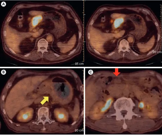
Criteria for determining 18F-FDG avidity for both primary tumors and LNs were defined as 1) focally increased 18F-FDG uptake by visual analysis and 2) a reasonable isocontouring result of MTV with a cutoff value of 50% of the SUVmax (Fig. 1).
Clinicopathologic and survival data
Clinical data obtained from medical records included age at surgery, sex, preoperative serum carcinoembryonic antigen (CEA), histopathologic results, and pathologic T (pT) and N (pN) staging. Pathologic staging was based on the 7th edition of the AJCC System. Recurrence- free survival (RFS) was defined as the interval from the date of surgery to the date of cross- sectional imaging when recurrence (by pathologic confirmation or clinical decision) was first noted. Overall survival (OS) was defined as the interval from the date of surgery to the date of the last hospital visit.
Statistical analyses
Numeric data were represented as the mean±standard deviation. Differences among variables between groups were compared using Student's t-test, the Kruskal-Wallis test, and the χ2 test. Diagnostic performance of 18F-FDG avidity for detecting primary tumors and metastatic LNs was evaluated by calculating the sensitivity, specificity, positive predictive value (PPV), and negative predictive value (NPV). Survival time was estimated using the log-rank test.
Univariate Cox regression analysis was performed for evaluating the statistical significance of clinicopathologic parameters. Continuous variables were analyzed directly or dichotomized using the optimal cutoff value by receiver operating characteristics curve analysis.
Multivariate Cox regression analysis was performed to evaluate the effect of LN-related parameters on other significant parameters. Statistical analyses were performed using SPSS statistics software (version 18.0, SPSS Inc., Chicago, IL, USA), and a P value of less than 0.05 was considered statistically significant.
RESULTS
Patient characteristics
Of the 168 enrolled patients, 51 (30.4%) experienced recurrence during the follow-up period (median follow-up duration, 35 months; range, 3–81 months), and 32 (19.0%) were documented deaths. Age, sex, and operation type were not significantly different between patients with and without recurrence (Table 1). The history of adjuvant chemotherapy, pT stage, pN stage,
CB C
A
25 cm
20 cm
20 cm
Fig. 1. Representative cases of 18F-FDG-avid primary tumor (A, left) and LNs (A, right), non-avid primary tumor (B), and non-avid LN (C). The metabolic tumor volume of the primary tumor and LNs in the patient (A) was 18.8 cm3 and 0.2 cm3, respectively (yellow arrow: primary tumor, red arrow: LN).
18F-FDG = 18F-fluorodeoxyglucose; LN = lymph node.
TNM staging, tumor size, and the presence of lymphovascular invasion (LVI) were significantly different according to recurrence (P<0.05). For histopathologic results, the frequency of signet ring cell-type was not significantly different according to recurrence (P=0.288). The frequency of intestinal-type was higher in the non-recurring group (P=0.003). However, the Bormann classification results did not vary significantly in terms of recurrence (P=0.062).
18F-FDG avidity of tumor lesions: diagnostic evaluation
We detected 18F-FDG-avid primary tumors in 119 (70.8%) patients. However, the presence of 18F-FDG-avid primary tumors was not significantly different between patients with (n=37, 72.5%) and without (n=82, 70.1%) recurrence (P=0.747). The SUVmax, MTV, and TLG values of 18F-FDG-avid primary tumors showed no significant difference according to recurrence (P=0.561, 0.551, and 0.800, respectively). The SUVmax of all primary tumors (including non-avid tumors) also showed no difference between patients with and without recurrence (5.73±4.26 vs. 5.75±5.08; P=0.979). We found that 33 (19.6%) patients showed more than one 18F-FDG-avid LN on 18F-FDG PET/CT. 18F-FDG-avid LNs tended to be more frequent in recurring cases (n=14, 27.5%) than in non-recurring cases (n=19, 16.2%; P=0.093). Similar Table 1. Patient characteristics
Characteristics Total (n=168) Recurrence (n=51) No recurrence (n=117) P
Age (yr) 63±12 61±13 64±11 0.102
Male (%) 117 (69.6) 31 (60.7) 86 (73.5) 0.161
Extent of gastric resection 0.283
Total 85 29 56
Subtotal 83 22 61
Adjuvant Tx 0.001
Yes 92 38 54
No 76 13 63
pT stage <0.001
T2 42 1 41
T3 79 21 58
T4 47 29 18
pN stage <0.001
N0 46 4 42
N1 31 4 27
N2 40 12 28
N3 51 31 20
TNM stage <0.001
I 20 0 20
II 58 5 53
III 90 46 44
Tumor size (cm) 6.0±2.9 7.6±3.5 5.3±2.3 <0.001
LVI <0.001
Yes 121 47 74
No 47 4 43
Pathology 0.288
Signet ring cell 14 6 8
Non-signet ring cell 154 45 109
Lauren* 0.003
Intestinal 86 16 70
Non-intestinal 79 34 45
Bormann type† 0.062
1 6 1 5
2 20 4 16
3 117 34 83
4 20 11 9
Tx = therapy; pT = pathologic tumor; pN = pathologic node; TNM = tumor, node, metastasis; LVI = lymphovascular invasion.
*Three, †5 unclassified.
to primary tumors, the SUVmax, MTV, and TLG values of 18F-FDG-avid LNs were not different between patients with and without recurrence (P=0.854, 0.864, and 0.661, respectively).
The calculated LTR was also not significantly different between patients with and without recurrence (0.38±0.26 vs. 0.36±0.29; P=0.749).
The diagnostic evaluation of 18F-FDG PET/CT for high T stage (pT 3–4) primary tumors showed a sensitivity of 73.8%, a specificity of 38.1%, a PPV of 78.2%, and an NPV of 32.7%
(Table 2). For the detection of high N stage (pN 2–3), the sensitivity, specificity, PPV, and NPV of 18F-FDG PET/CT were 29.7%, 92.2%, 81.8%, and 52.6%, respectively. For the detection of metastatic LNs (pN 1–3), the sensitivity, specificity, PPV, and NPV of 18F-FDG PET/CT were 26.2%, 97.8%, 97.0%, and 33.3%, respectively (Table 3).
Prognostic factors for RFS and OS
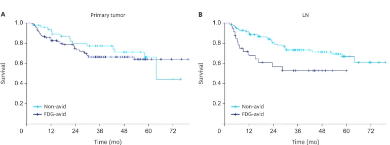
The median RFS and OS were 34.9 and 36.5 months, respectively. The significance of 18F-FDG avidity for recurrence was evaluated (Fig. 2). The 18F-FDG avidity of primary tumors was not significantly correlated with RFS or OS (P=0.532 and 0.658, respectively). However, the
18F-FDG avidity of LNs showed significant correlation with a shorter RFS (P=0.012). The mean RFS of patients with 18F-FDG-avid LNs was 36.5±4.5 months, whereas that of patients with non-avid LNs was 60.4±2.7 months. The relationship between 18F-FDG avidity of LNs and OS did not show statistical significance (P=0.181).
Table 2. 18F-FDG avidity of primary tumor according to pT stage
18F-FDG avidity pT2 pT3–4 Total
18F-FDG-avid 26 93 119
Non-avid 16 33 49
Total 42 126 168
18F-FDG = 18F-fluorodeoxyglucose; pT = pathologic tumor.
Table 3. 18F-FDG avidity of metastatic LNs according to pN stage
18F-FDG avidity pN0 pN1 pN2–3 Total
18F-FDG-avid 1 5 27 33
Non-avid 45 26 64 135
Total 46 31 91 168
18F-FDG = 18F-fluorodeoxyglucose; LN = lymph node; pT = pathologic node.
Primary tumor
Time (mo) 0.6
0 24 72
Survival
1.0
0.8
0.4
12 36 48 60
A
0.2
0.6 1.0
0.8
0.4
0.2
LN
Time (mo)
0 24 72
Survival
12 36 48 60
B
Non-avid
FDG-avid Non-avid
FDG-avid
Fig. 2. Cumulative RFS curve according to the 18F-FDG avidity of (A) primary tumors and (B) LNs in enrolled patients (red: non-avid group, blue: 18F-FDG-avid group).
RFS = recurrence-free survival; LN = lymph node.
Univariate analysis was performed to evaluate the prognostic significance of the
clinicopathologic parameters (Table 4). Age and sex were not correlated with patients' survival.
The size of the primary tumor and the number of metastatic LNs proven histopathologically were significantly correlated with both RFS and OS (P≤0.001). High pT (≥3) and pN (≥2) stages were also significantly correlated with patients' survival. The histopathologic subtype of signet ring cell cancer was a prognostic factor for OS (P=0.044), but not for RFS (P=0.377).
Non-intestinal subtype and LVI were significant prognostic factors for both RFS and OS.
Preoperative CEA level was not related to RFS (P=0.536) and OS (P=0.351). 18F-FDG avidity of primary tumors was not correlated with RFS and OS, but 18F-FDG avidity of LNs was a significant prognostic factor for RFS (P=0.015). The correlation between 18F-FDG avidity of LNs and OS was not significant (P=0.187). Other 18F-FDG-PET/CT-driven parameters, including the SUVmax of primary tumors and the LTR, were not significantly correlated with patients' survival.
Construction of the multivariate survival models was based on significant parameters driven from univariate analysis, and metastatic LN-related parameters were combined to compare the effect in each model: 1) Model with 18F-FDG avidity of LNs, 2) Model with number of metastatic LNs, and 3) Model with pN stage (≥2) (Tables 5 and 6). For RFS, the size of the primary tumor was an independent prognostic factor in all models (Table 5). Non-intestinal subtype and LVI were independent factors in models 1 and 2, but significance was not observed in model 3. 18F-FDG avidity of LNs was an independent prognostic factor for predicting RFS. For OS, the size of the primary tumor was an independent prognostic factor in all models (Table 6).
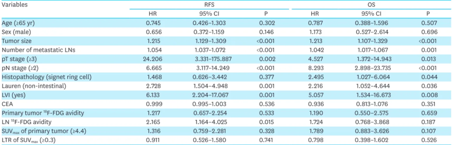
Table 4. Univariate Cox regression analysis for prognostic factors related to survival
Variables RFS OS
HR 95% CI P HR 95% CI P
Age (≥65 yr) 0.745 0.426–1.303 0.302 0.787 0.388–1.596 0.507
Sex (male) 0.656 0.372–1.159 0.146 1.173 0.527–2.614 0.696
Tumor size 1.215 1.129–1.309 <0.001 1.213 1.107–1.329 <0.001
Number of metastatic LNs 1.054 1.037–1.072 <0.001 1.042 1.017–1.067 0.001
pT stage (≥3) 24.206 3.331–175.887 0.002 4.527 1.372–14.943 0.013
pN stage (≥2) 6.665 3.117–14.249 <0.001 8.293 2.898–23.735 <0.001
Histopathology (signet ring cell) 1.468 0.626–3.442 0.377 2.495 1.027–6.064 0.044
Lauren (non-intestinal) 2.728 1.504–4.948 0.001 2.216 1.052–4.644 0.036
LVI (yes) 6.133 2.204–17.067 0.001 5.057 1.534–16.673 0.008
CEA 0.999 0.995–1.003 0.536 0.936 0.813–1.076 0.351
Primary tumor 18F-FDG avidity 1.217 0.657–2.254 0.533 1.190 0.550–2.575 0.659
LN 18F-FDG avidity 2.165 1.164–4.025 0.015 1.724 0.768–3.868 0.187
SUVmax of primary tumor (≥4.4) 1.316 0.759–2.281 0.328 1.789 0.883–3.626 0.107
LTR of SUVmax (≥0.3) 0.911 0.526–1.580 0.741 0.798 0.398–1.602 0.526
RFS = recurrence-free survival; OS = overall survival; HR = hazard ratio; CI = confidence interval; LN = lymph node; pT = pathologic tumor; pN = pathologic node;
LVI = lymphovascular invasion; CEA = carcinoembryonic antigen; 18F-FDG = 18F-fluorodeoxyglucose; SUVmax = maximum standardized uptake value; LTR = lymph node-to-tumor ratio.
Table 5. Multivariate Cox regression models for RFS
Variables Model with LN avidity Model with LN number Model with pN stage
HR (95% CI) P HR (95% CI) P HR (95% CI) P
Size 1.158 (1.071–1.252) <0.001 1.114 (1.028–1.208) 0.009 1.125 (1.043–1.215) 0.002
Lauren (non-intestinal) 2.378 (1.252–4.517) 0.005 1.946 (1.030–3.677) 0.040 1.861 (0.990–3.498) 0.054
LVI 3.704 (1.278–10.741) 0.017 3.657 (1.273–10.505) 0.016 2.099 (0.630–6.998) 0.228
LN 18F-FDG avidity 1.964 (1.012–3.811) 0.046 N/A N/A
Number of metastatic LNs N/A 1.027 (1.006–1.048) 0.011 N/A
pN stage N/A N/A 3.128 (1.251–7.818) 0.015
RFS = recurrence-free survival; LN = lymph node; pN = pathologic node; HR = hazard ratio; CI = confidence interval; LVI = lymphovascular invasion; 18F-FDG =
18F-fluorodeoxyglucose; N/A = not assessed.
Other histopathologic factors did not show statistical significance. Among the LN-related parameters, only pN stage was an independent factor for predicting OS.
Subgroup analysis was performed for patients with histopathologically proven adenocarcinoma (n=154), and histologic grade of the primary tumor was added in the survival analysis. As in the total patient population, tumor size, number of metastatic LNs, pathologic staging, and LVI showed significance for predicting RFS and OS (Supplementary Table 1). Poorly differentiated subtype was a significant prognostic factor for RFS (P=0.005) but not for OS (P=0.175). Lauren nonintestinal subtype was also a significant factor for RFS (P=0.001) but not for OS (P=0.137).
The 18F-FDG avidity of LNs was a significant factor for RFS in the univariate analysis (P=0.031) but not in the multivariate analysis (P=0.120, Supplementary Table 2).
DISCUSSION
This study evaluated the diagnostic and prognostic value of preoperative 18F-FDG PET/CT in patients with resectable AGC. Current evidence suggests that primary tumor resection and standardized D2 lymphadenectomy under best post-operative care lower recurrence, death rate, and morbidity/mortality [19]. Standardized preoperative workup is recommended to evaluate the disease status based on patient examination, laboratory profile, endoscopy procedure, and CECT [7]. However, the 5-year survival rate after curative resection remains disappointing.
Randomized clinical trials have reported varying results for adjuvant chemotherapy according to regional differences [20,21]. These findings suggest the heterogeneous biology of gastric cancer and the need for effective prognostic biomarkers for AGC patients [22]. Evaluation of tumor metabolism by 18F-FDG PET/CT may play a potential role as a surrogate marker of tumor aggressiveness, which is correlated with patients' prognoses.
The diagnosis of LN metastasis is one of the most important prognostic factors in AGC patients after curative surgery [23]. LN metastasis predicts accurate and stage-specific survival in the subclassification of patients with a similar pT stage [24]. The number of metastatic LNs is directly correlated with survival time [25]. Hence, a comprehensive analysis of LN metastasis is needed to evaluate AGC patients, especially those contemplating curative surgery. Endoscopic ultrasonography showed a high predictive value (approximately 92.6%) for node-negative, early gastric cancer but played a limited role in predicting extra-perigastric LN metastasis [26,27]. CECT has been widely used for the evaluation of the degree of LN metastasis; however, the size criteria for determining LN metastasis often have limited sensitivity and yield false-negative results [28]. The diagnostic accuracy of preoperative
18F-FDG PET/CT for LN metastasis in patients with AGC has long been discussed. The Table 6. Multivariate Cox regression models for OS
Variables Model with LN avidity Model with LN number Model with pN stage
HR (95% CI) P HR (95% CI) P HR (95% CI) P
Size 1.170 (1.054–1.300) 0.003 1.149 (1.030–1.282) 0.013 1.130 (1.017–1.255) 0.023
Lauren (non-intestinal) 0.337 (0.643–3.639) 0.337 1.304 (0.562–3.027) 0.537 1.163 (0.506–2.675) 0.722
LVI 3.063 (0.871–10.780) 0.081 3.217 (0.928–11.149) 0.065 1.210 (0.261–5.611) 0.807
Histopathology (signet ring cell) 2.202 (0.826–5.871) 0.115 2.175 (0.814–5.813) 0.121 1.979 (0.749–5.228 0.168
LN 18F-FDG avidity 1.702 (0.694–4.174) 0.245 N/A N/A
Number of metastatic LNs N/A 1.014 (0.984–1.045) 0.365 N/A
pN stage N/A N/A 4.862 (1.220–19.373) 0.025
OS = overall survival; LN = lymph node; pN = pathologic node; HR = hazard ratio; CI = confidence interval; LVI = lymphovascular invasion; 18F-FDG =
18F-fluorodeoxyglucose; N/A = not assessed.
diagnostic sensitivity for metastasis of N1 LNs was below 50%, which was lower than for CECT. However, 18F-FDG PET/CT outperformed CECT for the specificity of N staging.
Especially in advanced N stage, the specificity by 18F-FDG PET/CT was 96% and 99% in N2 and N3 lymph nodes, respectively [29-31]. In view of resectability, accurate staging of advanced disease is essential for planning the surgical approach and extent of en bloc resection. This study also showed comparable accuracy with high specificity (92.2%) for the diagnosis of advanced N stage, although 18F-FDG avidity of LNs was determined by combining qualitative and quantitative criteria. We suggest that the diagnostic value of 18F-FDG PET/CT might be related directly with prognostic information to determine patients' survival.
Until now, a few studies have reported the correlation between 18F-FDG uptake by LNs and the prognoses of patients with AGC. A recent study revealed that a high SUVmax of LNs is an independent factor for patients' RFS and OS [15]. Metabolic information for LNs was correlated with patients' OS, whereas the size of lymphadenopathies did not show a significant relationship. Additionally, 18F-FDG PET/CT revealed metastatic disease that was not detected on CT [32]. In this study, the 18F-FDG avidity of LNs in patients with AGC was significantly correlated with disease recurrence as well as other histopathologic parameters for determining metastatic LNs. Patients with non-avid LNs had a 2-year RFS rate of 82.2%
(111/135), whereas patients with 18F-FDG-avid LNs showed a 2-year RFS rate of 60.6% (20/33).
The mean difference in RFS according to the 18F-FDG avidity of LNs was approximately 24 months. The results suggest the need for revisiting the role of preoperative 18F-FDG PET/CT for accurate prediction of recurrence in patients with AGC.
Studies for evaluating the utility of 18F-FDG PET/CT in AGC focused on 18F-FDG uptake by primary tumors. High 18F-FDG uptake by primary tumors was associated with 2-year RFS after curative resection [33,34]. In this study, the 18F-FDG avidity of primary tumors was not significantly correlated with recurrence. 18F-FDG uptake by primary tumors is partly affected by background gastric wall activity and histopathologic subtype [35]. Furthermore, several parameters for measuring 18F-FDG uptake by primary tumors have been proposed, including SUVmax, MTV, and tumor-to-liver ratio [11,36]. Further studies for optimizing 18F-FDG PET/
CT-driven parameters are needed to investigate the correlation between glucose metabolism in gastric cancer and patients' prognoses. In the present study, the 18F-FDG avidity of LNs was determined dichotomously instead of using continuous SUVmax values. The SUVmax of small perigastric LNs is often hard to standardize because of the partial-volume effect and spill- over effect [37]. 18F-FDG avidity of LNs used in this study had high specificity for determining nodal metastases and represents an independent prognostic factor for RFS.
This study has several limitations. First, the 18F-FDG avidity of LNs was correlated with patients' RFS but not with OS, probably due to the relatively small sample and event size.
Second, the prognosis-predicting model in this study was not entirely based on pre-operative clinical parameters. Further studies are needed to evaluate the significance of preoperative
18F-FDG PET/CT in terms of clinical staging. Finally, this retrospective single-center study might be inevitably associated with a patient selection bias.
In conclusion, this study demonstrates that 18F-FDG avidity of LNs evaluated by PET/CT scan is an independent factor contributing to RFS following curative resection in patients with AGC. We suggest that preoperative 18F-FDG PET/CT should be used for predicting prognosis aimed at appropriate risk monitoring of AGC patients during follow-up after surgery.
SUPPLEMENTARY MATERIALS
Supplementary Table 1
Univariate Cox regression analysis for prognostic factors related with survival (subgroup analysis) Click here to view
Supplementary Table 2
Multivariate Cox regression models for RFS (subgroup analysis) Click here to view
REFERENCES
1. Jung KW, Won YJ, Oh CM, Kong HJ, Lee DH, Lee KH, et al. Cancer statistics in Korea: incidence, mortality, survival, and prevalence in 2014. Cancer Res Treat 2017;49:292-305.
PUBMED | CROSSREF
2. Katanoda K, Hori M, Matsuda T, Shibata A, Nishino Y, Hattori M, et al. An updated report on the trends in cancer incidence and mortality in Japan, 1958–2013. Jpn J Clin Oncol 2015;45:390-401.
PUBMED | CROSSREF
3. Siegel RL, Miller KD, Jemal A. Cancer statistics, 2016. CA Cancer J Clin 2016;66:7-30.
PUBMED | CROSSREF
4. Maehara Y, Hasuda S, Koga T, Tokunaga E, Kakeji Y, Sugimachi K. Postoperative outcome and sites of recurrence in patients following curative resection of gastric cancer. Br J Surg 2000;87:353-357.
PUBMED | CROSSREF
5. Deng N, Goh LK, Wang H, Das K, Tao J, Tan IB, et al. A comprehensive survey of genomic alterations in gastric cancer reveals systematic patterns of molecular exclusivity and co-occurrence among distinct therapeutic targets. Gut 2012;61:673-684.
PUBMED | CROSSREF
6. Dulak AM, Schumacher SE, van Lieshout J, Imamura Y, Fox C, Shim B, et al. Gastrointestinal adenocarcinomas of the esophagus, stomach, and colon exhibit distinct patterns of genome instability and oncogenesis. Cancer Res 2012;72:4383-4393.
PUBMED | CROSSREF
7. Ajani JA, D'Amico TA, Almhanna K, Bentrem DJ, Chao J, Das P, et al. Gastric cancer, version 3.2016, NCCN clinical practice guidelines in oncology. J Natl Compr Canc Netw 2016;14:1286-1312.
PUBMED | CROSSREF
8. Smyth EC, Verheij M, Allum W, Cunningham D, Cervantes A, Arnold D, et al. Gastric cancer: ESMO clinical practice guidelines for diagnosis, treatment and follow-up. Ann Oncol 2016;27:v38-v49.
PUBMED | CROSSREF
9. Smyth E, Schöder H, Strong VE, Capanu M, Kelsen DP, Coit DG, et al. A prospective evaluation of the utility of 2-deoxy-2-[18F]fluoro-D-glucose positron emission tomography and computed tomography in staging locally advanced gastric cancer. Cancer 2012;118:5481-5488.
PUBMED | CROSSREF
10. Baiocchi GL, Marrelli D, Verlato G, Morgagni P, Giacopuzzi S, Coniglio A, et al. Follow-up after gastrectomy for cancer: an appraisal of the Italian research group for gastric cancer. Ann Surg Oncol 2014;21:2005-2011.
PUBMED | CROSSREF
11. Lee JW, Jo K, Cho A, Noh SH, Lee JD, Yun M. Relationship between 18F-FDG uptake on PET and recurrence patterns after curative surgical resection in patients with advanced gastric cancer. J Nucl Med 2015;56:1494-1500.
PUBMED | CROSSREF
12. Lordick F, Ott K, Krause BJ, Weber WA, Becker K, Stein HJ, et al. PET to assess early metabolic response and to guide treatment of adenocarcinoma of the oesophagogastric junction: the MUNICON phase II trial. Lancet Oncol 2007;8:797-805.
PUBMED | CROSSREF
13. Ott K, Weber WA, Lordick F, Becker K, Busch R, Herrmann K, et al. Metabolic imaging predicts response, survival, and recurrence in adenocarcinomas of the esophagogastric junction. J Clin Oncol 2006;24:4692-4698.
PUBMED | CROSSREF
14. Washington K. 7th edition of the AJCC cancer staging manual: stomach. Ann Surg Oncol 2010;17:3077-3079.
PUBMED | CROSSREF
15. Song BI, Kim HW, Won KS, Ryu SW, Sohn SS, Kang YN. Preoperative standardized uptake value of metastatic lymph nodes measured by 18F-FDG PET/CT improves the prediction of prognosis in gastric cancer. Medicine (Baltimore) 2015;94:e1037.
PUBMED | CROSSREF
16. Cerfolio RJ, Bryant AS. Ratio of the maximum standardized uptake value on FDG-PET of the mediastinal (N2) lymph nodes to the primary tumor may be a universal predictor of nodal malignancy in patients with nonsmall-cell lung cancer. Ann Thorac Surg 2007;83:1826-1830.
PUBMED | CROSSREF
17. Park J, Byun BH, Noh WC, Lee SS, Kim HA, Kim EK, et al. Lymph node to primary tumor SUV ratio by
18F-FDG PET/CT and the prediction of axillary lymph node metastases in breast cancer. Clin Nucl Med 2014;39:e249-e253.
PUBMED | CROSSREF
18. Japanese Gastric Cancer Association. Japanese classification of gastric carcinoma: 3rd English edition.
Gastric Cancer 2011;14:101-112.
PUBMED | CROSSREF
19. Songun I, Putter H, Kranenbarg EM, Sasako M, van de Velde CJ. Surgical treatment of gastric cancer: 15- year follow-up results of the randomised nationwide Dutch D1D2 trial. Lancet Oncol 2010;11:439-449.
PUBMED | CROSSREF
20. Ajani JA, Rodriguez W, Bodoky G, Moiseyenko V, Lichinitser M, Gorbunova V, et al. Multicenter phase III comparison of cisplatin/S-1 with cisplatin/infusional fluorouracil in advanced gastric or gastroesophageal adenocarcinoma study: the FLAGS trial. J Clin Oncol 2010;28:1547-1553.
PUBMED | CROSSREF
21. Sakuramoto S, Sasako M, Yamaguchi T, Kinoshita T, Fujii M, Nashimoto A, et al. Adjuvant chemotherapy for gastric cancer with S-1, an oral fluoropyrimidine. N Engl J Med 2007;357:1810-1820.
PUBMED | CROSSREF
22. Abbas M, Habib M, Naveed M, Karthik K, Dhama K, Shi M, et al. The relevance of gastric cancer biomarkers in prognosis and pre- and post- chemotherapy in clinical practice. Biomed Pharmacother 2017;95:1082-1090.
PUBMED | CROSSREF
23. Deng JY, Liang H. Clinical significance of lymph node metastasis in gastric cancer. World J Gastroenterol 2014;20:3967-3975.
PUBMED | CROSSREF
24. Sarela AI, Turnbull AD, Coit DG, Klimstra D, Brennan MF, Karpeh MS. Accurate lymph node staging is of greater prognostic importance than subclassification of the T2 category for gastric adenocarcinoma. Ann Surg Oncol 2003;10:783-791.
PUBMED | CROSSREF
25. Ichikura T, Tomimatsu S, Okusa Y, Uefuji K, Tamakuma S. Comparison of the prognostic significance between the number of metastatic lymph nodes and nodal stage based on their location in patients with gastric cancer. J Clin Oncol 1993;11:1894-1900.
PUBMED | CROSSREF
26. Ahn HS, Lee HJ, Yoo MW, Kim SG, Im JP, Kim SH, et al. Diagnostic accuracy of T and N stages with endoscopy, stomach protocol CT, and endoscopic ultrasonography in early gastric cancer. J Surg Oncol 2009;99:20-27.
PUBMED | CROSSREF
27. Hwang SW, Lee DH. Is endoscopic ultrasonography still the modality of choice in preoperative staging of gastric cancer? World J Gastroenterol 2014;20:13775-13782.
PUBMED | CROSSREF
28. Kim SH, Kim JJ, Lee JS, Kim SH, Kim BS, Maeng YH, et al. Preoperative N staging of gastric cancer by stomach protocol computed tomography. J Gastric Cancer 2013;13:149-156.
PUBMED | CROSSREF
29. Kim SK, Kang KW, Lee JS, Kim HK, Chang HJ, Choi JY, et al. Assessment of lymph node metastases using
18F-FDG PET in patients with advanced gastric cancer. Eur J Nucl Med Mol Imaging 2006;33:148-155.
PUBMED | CROSSREF
30. Kim EY, Lee WJ, Choi D, Lee SJ, Choi JY, Kim BT, et al. The value of PET/CT for preoperative staging of advanced gastric cancer: comparison with contrast-enhanced CT. Eur J Radiol 2011;79:183-188.
PUBMED | CROSSREF
31. Yun M, Lim JS, Noh SH, Hyung WJ, Cheong JH, Bong JK, et al. Lymph node staging of gastric cancer using 18F-FDG PET: a comparison study with CT. J Nucl Med 2005;46:1582-1588.
PUBMED
32. Coupe NA, Karikios D, Chong S, Yap J, Ng W, Merrett N, et al. Metabolic information on staging FDG-PET-CT as a prognostic tool in the evaluation of 97 patients with gastric cancer. Ann Nucl Med 2014;28:128-135.
PUBMED | CROSSREF
33. Lee JW, Lee SM, Lee MS, Shin HC. Role of 18F-FDG PET/CT in the prediction of gastric cancer recurrence after curative surgical resection. Eur J Nucl Med Mol Imaging 2012;39:1425-1434.
PUBMED | CROSSREF
34. Mochiki E, Kuwano H, Katoh H, Asao T, Oriuchi N, Endo K. Evaluation of 18F-2-deoxy-2-fluoro-D-glucose positron emission tomography for gastric cancer. World J Surg 2004;28:247-253.
PUBMED | CROSSREF
35. Stahl A, Ott K, Weber WA, Becker K, Link T, Siewert JR, et al. FDG PET imaging of locally advanced gastric carcinomas: correlation with endoscopic and histopathological findings. Eur J Nucl Med Mol Imaging 2003;30:288-295.
PUBMED | CROSSREF
36. Kim J, Lim ST, Na CJ, Han YH, Kim CY, Jeong HJ, et al. Pretreatment F-18 FDG PET/CT parameters to evaluate progression-free survival in gastric cancer. Nucl Med Mol Imaging 2014;48:33-40.
PUBMED | CROSSREF
37. Soret M, Bacharach SL, Buvat I. Partial-volume effect in PET tumor imaging. J Nucl Med 2007;48:932-945.
PUBMED | CROSSREF