I. Introduction
In clinical settings, bone is frequently needed to adequately repair defects due to periodontal dis- ease, trauma, or congenital abnormalities1,2). Most bone grafts performed today utilize either autograft, allograft or alloplast with moderate clinical success3-
5). Autografts have the advantage of optimal biologic incorporation, histocompatibility and little chance of disease transmission; lack of availability limits its use, and the harvesting of graft tissue may have attendant patient morbidity. In contrast, allografts enjoy much wider availability and no patient mor- bidity is associated with graft procurement.
However, problems with unreliable graft incorpora- tion, immune response, and possible disease trans- mission represent clear drawbacks to their use.
While several synthetic graft materials have also been introduced, most alloplastic materials function primarily as a biocompatible defect filler6).
One possible approach toward addressing the respective problems inherent in autograft and allo- graft material is employing tissue engineering which
is actively researched today. That is, after creation of synthetic graft materials(scaffolding material) that posses the biological advantages of autograft tissue and supply advantages of allograft tissue, osteoblasts like cells are seeded and cultured. Finally, the cell- scaffold complex is transplanted to the defect. The term "tissue engineering" was originally coined to denote the construction in the laboratory of a device containing viable cells and biologic mediators in a synthetic or biologic matrix that could be implanted in patients to facilitate regeneration of particular tis- sues. Tissue engineering is a science in which mate- rial properties of synthetic compounds are manipu- lated to enable delivery of an aggregate of dissociat- ed cells into host in a manner that will result in the formation of new tissue7). Thus, the major goal of tissue engineering is in vitro construction of trans- plantable vital tissue.
The engineering of bone tissue requires appropri- ate carriers that allow a 3- dimensional distribution of cells. Ishaug et al. suggested several prerequisites for a scaffold material for bone formation8). First, the scaffolding material for bone formation must allow
Biological Activities of Calcium Polyphosphate
Yang-Jo Seol1·Jae-Il Lee2·Yong-Moo Lee1·Yoon-Tak Lim3 Seok-Young Kim3·Young Ku1·In-Chul Rhyu1·Byung-Do Hahm1
Soo-Boo Han1·Sang-Mook Choi1·Chong-Pyoung Chung1
1Department of Periodontology, College of Dentistry, Seoul National University
2Department of Oral Pathology, College of Dentistry, Seoul National University
3Department of Material Technology, College of Engineering, Yeungnam University
대한치주과학회지 : Vol. 30, No. 2, 2000
This study was supported by a grant of the Korea Health 21 R&D Project, Ministry of Health & Welfare, Republic of Korea(HMP-99- E-10-0003).
for the attachment of osteoblasts because they are anchorage dependent cells that require a supportive matrix in order to survive. Second, the scaffold must provide an appropriate environment for prolifera- tion and function of osteoblasts. Third, the scaffold should allow for ingrowth of vascular tissue to ensure the survival of transplanted cells. Fourth, the materials should be biodegradable and its degraded molecules should be easily metabolized and excret- ed. Finally, it should be processable into irregular 3- dimensional shapes.
Several studies have reported the tissue engi- neered bone regeneration using various scaffolds in vitro. Casser-Bette et al. had induced bone like tis- sue formation in vitro 56-day culture of 3-dimen- sional matrix of collagen type I onto which cells of the clonal osteogenic cell line MC3T3-E1 was seed- ed9). Laurencin et al. studied osteoblast proliferation and bone formation using rat calvaria cells on the surface of porous poly(lactide/glycolide)/hydroxy apatite 3-dimensional polymer matrix in vitro10). Ishaug et al. investigated bone formation in vitro by culturing stromal osteoblasts in 3-dimensional, biodegradable poly(DL-lactide-co-glycolic acid) foams8). In addition to in vitro tissue construction, many investigators have tried to induce intentional bone formation by transplantation of cell-scaffold construct. Caplan group reported many investiga- tions in relation to bone and cartilage formation using porous ceramic with marrow and periosteal cells11-16). Vacanti group used embossed nonwoven mesh of polyglycolic acid as a scaffold for cartilage and bone formation7,17-19). Puelacher et al., who are members of Vacanti group, showed that implanta- tion of periosteum derived cells seeded onto syn- thetic polymer scaffolds resulted in bone formation in surgically induced athymic rat femur defect18). Their study is of particular interest because their report supports potential application of the tech-
nique of tissue engineered growth of bone to a non- healing defect in weight bearing bone. Recently, Breitbart et al. demonstrated that resorbable polyg- lycolic acid scaffold seeded with periosteum derived cells induced bone regeneration in critical size cal- varial defect in a rabbit model and confirmed that the cultured periosteal cells contributed to this bone formation by detection of prelabelled cells in the newly formed bone20).
Porous alloplastic implants have been studied extensively for their use in oral and maxillofacial applications2). The use of these materials allows for recovery of the cosmetics and continuity of the sur- rounding bony structures without the concerns asso- ciated with the use of autogenic implants. These include but are not limited to increased potential for graft resorption, donor site morbidity, and immuno- genic reaction to bank bone. Other advantages of porous alloplastic implants in periodontal and cran- iofacial applications include an increase in resistance to fatigue fracturing and greater resistance to separa- tion21). Ceramic, porous block hydroxyapatite(HA), which is one such alloplastic implant, has been shown to be an effective implant material in short- and long-term applications22). With advances in ceramics technology, the application of calcium phosphate materials have received considerable attention as bone substitutes for several decades.
Calcium phosphate bone substitutes are believed to be biocompatible and osteoconductive when implanted in bone defects23-26). Numerous animal studies provide histologic evidence of the long-term biocompatibility of porous HA and of its favorable interaction with soft tissue and bone1,27). In addition, these studies indicate the lack of an inflammatory response to HA implants28,29).
A substrate such as hydroxyapatite that provides a three-dimensional guideline for bone shape facili- tates bony ingrowth and subsequent positional sta-
bility as discussed in Wolford et al30,31). The porous structure of HA provides a template for fibrovascular ingrowth which is followed by osteoblast differentia- tion that results in the deposition of new lamellar bone. Porous materials are highly favorable over nonporous materials owing to the accessibility of the interior of the material to tissue ingrowth. If the pores appropriately sized, they can provide a frame- work for bone growth into the matrix of the materi- al.
The purpose of this study was to investigate the applicability of calcium polyphosphate(CPP) as a bone graft material and tissue engineering scaffold for bone formation in 3-dimensional osteoblasts cul- ture and to test mutagenicity of calcium polyphos- phate(CPP).
II. Materials and Methods
1. Manufacturing galcium polyphosphateInterconnected porous calcium polyphosphate (CPP) blocks were prepared by condensation of anhydrous Ca(H2PO4)2(Duksan Chemical Co., Inc.) to form non-crystalline Ca(PO3)2. From the latter, an homogenous melt was created by thermal treat- ment, quenched in distilled water, and the block was then milled to produce CPP powder. And macroporous 3-dimensional scaffolds were made using a polyurethane(PU) sponge method32,33)with addition of 5% Na2O. The PU was burnt out and the resultant inorganic scaffold was sintered at 900℃ for 1h to create CPP. Two kinds of CPP blocks were prepared according to the pore size, 45ppi and 60ppi. Pore size of CPP(45ppi) is approximately 450-550㎛ and that of CPP(60ppi) is approximately 200-300㎛. The manufactured CPP matrices were made into shapes of 5×5×1 mm3for cell seeding
and culture of cell-CPP matrix construct.
2. In vitro culture of cell-matrix constructs
1) Isolation of rat bone marrow cell
Stromal osteoblastic cells were obtained from the bone marrow of young adult Sprague Dawley rats(approximate weight:100g) according to the method described by Ishague et al8). Briefly, follow- ing euthanasia by ethyl ether inhalation, femora were aseptically excised, cleaned of soft tissue, and washed in HBSS. Then, the metaphyseal ends were cut off and the marrow flushed from the midshaft with 5ml of α-MEM using a syringe equipped with a 22-gauge needle and collected in a sterile petri dish.
Cell clumps were broken up by repeatedly pipetting the cell suspension. The cells then were centrifuged at 400 x g for 10 min at 4℃. The resulting cell pel- lets were resuspended in 12ml of primary media and plated in flasks. After 3 days, hematopoietic cells and other unattached cells were removed from the flasks by repeated washing with α-MEM. When confluent monolayers were reached the cells were enzymatically lifted from the flask using a 0.25%
trypsin in 4mM EDTA(Gibco, Grand Island, NY, USA). The cells were concentrated by centrifugation at 400 x g for 10 min at 4℃. The cell pellets were resuspended into 35mm tissue culture dishes at a density of 4 x 10⁴cells/cm2in α-minimum essential medium(α-MEM; Gibco) containing 10% FBS and 1% antibiotic-antimycotic solution(Gibco). Cells were counted with a hemacytometer. Cultures were maintained in a humidified atmosphere consisting of 95% air and 5% CO2at 37°C.
2) Cell seeding into the CPP matrices and culture in vitro
When confluent monolayers were reached, the cells were enzymatially lifted from the dishes using
0.25% trypsin in 4mM EDTA. The cells were agitat- ed and be detached from the dishes completely and concentrated by centrifugation at 400 x g for 10 min- utes. After centrifuging, the supernatant was suc- tioned away and resuspended in a known amount of media. Cells were counted with hemacytometer and diluted to 107cells/㎖ in mineralization media consisting of α-MEM supplement with 15 % FBS, 1%
antibiotic-antimycotic solution, 10mM Na β-glycerol phosphate(Sigma), and 50㎍ /㎖ L-ascorbic acid(Sigma). Aliquots of 20㎕ of cell suspension were seeded on the top of 5x5x1mm3sized prewet- ted CPP matrices(CPP-45ppi and CPP-60ppi) which are placed in the wells of 24-well plates(Nunc, Rochester, NY, USA) The seeding density resulted 105cells/ block. The cells cultured on the dishes of tissue culture polystyrene were employed as control groups. The matrices were left undisturbed in an incubator for 3 hours to allow the cells to attach to the matrices, after which, an additional 1㎖ of com- plete media was added to each well. Cultures were maintained in a humidified atmosphere consisting of 95% air and 5% CO2at 37°C. Mineralizing media was changed every 2-3days.
3) Measurement of cell proliferation
Cell proliferation was measured at 1, 7, 14, and 21 days. At each time point, media was removed from the wells. The CPP matrices were washed gently with Hank's balanced salt solution(HBSS; Gibco) to remove any unattached cells. Then, the CPP blocks were carefully transferred to the another 24 well tis- sue culture dish. The adherent cells were removed from the CPP matrices by incubation in 0.5㎖ of 0.25% trypsin in 4mM EDTA for 10 minutes at 37℃
and then the matrices were washed two times with 1㎖ of HBSS. Cells in trypsin/HBSS solution were counted by the hemacytometer. After counting, the cells in the media were centrifuged at 1260 rpm,
4.0oC, and for 10 minutes. The supernatant was suc- tioned and the cell pellet was prepared for alkaline phosphatase activity test.
4) Measurement of alkaline phosphatase activity Production of alkaline phosphatase(ALPase) was measured spectroscopically at 1, 7, 14, and 21days.
This test was done with the same cells used for the proliferation test. For comparison, cells of the same lineage were cultured on tissue culture poly- styrene(TCPS) dish, and alkaline phosphatase activi- ties of these cells were also measured. TCPS is an oxygen-containing surface specifically treated by the manufacturer to be more hydrophilic and thereby enhance cell growth34) and permit osteogenesis35). Removed cells from the matrices were homogenized with 0.5㎖ of double distilled water and sonicated for 1 minute in ice. 0.1㎖ of cell lysate were mixed with 0.1㎖ of 0.1M glycin-NaOH buffer, 0.1㎖ of 15mM para-nitrophenol phosphate(PNPP), 0.1%
Triton X-100/saline and 0.1㎖ of DDW. Each aliquots was incubated at 37oC for 30 minutes. After incubation, each tube was added 2.5㎖ of 0.1N NaOH and placed on ice. The production of para- nitrophenol(PNP) in the presence of ALPase was measured by monitoring light absorbance by the solution at 405㎚. The slope of absorbance versus time plot was used to calculate the ALPase activity.
5) Histologic examination of cell-matrix constructs Cultured cell-matrix constructs were pre-pared for scanning electron microscopy(SE- M) studies at each time period. Cultured cell-CPP complex were incu- bated at room temperature in a fixative of 2.5% of glutaraldehyde for 20min and then washed in PBS for 10min(3times). The complexes were then incu- bated for 30min in a postfixative of 1% aqueous OsO4(Electron Microscopy Sciences, Fort Washington, PA) and subsequently washed with
PBS for 5min(3times). Samples were then sequential- ly washed with PBS for 5min(3times). Samples were then sequentially washed in ethanol of increasing(50, 70, 90, and twice in 100%) concentration for 5min/wash. This step was performed to dehydrate the cells. Cell-CPP samples were allowed to air dry overnight and were then visualized using an scan- ning electron microscope(Jeol, U.S.A). SEM was conducted using an accelerating voltage of 15㎸.
3. Mutagenicity of calcium polyphos- phate
To test mutagenicity of CPP, hypoxanthine-gua- nine phosphoribosyl transferase(HPRT) assay was performed(using 6-Thioguanine). With NIH3T3, CHO-K1 cell line, HPRT assay was done in the media of 1000, 100, 10, and 1㎍/㎖ of conc. of CPP.
To begin with, cells were plated at the density of 5
×105/plate in α-MEM. Then, they were incubated in 95% CO2incubator at 37℃ for 24 hours. After removal of α-MEM, the media containing various concentration of CPP(0.001, 0.01, 0.1, and 1㎎/㎖) were put in and incubated again. After 48hours. the cells were trypsinized with 0.25% trypsin in 4mM EDTA(Gibco, Grand Island, NY, USA). Then, the cells are plated at the density of 1×10⁵/plate on 3 plates at each concentration of CPP. 6- Thioguanine(6TG) were put into the media and cul- tured for 10-14days. After that period, the media was replaced with α-MEM, and cultured for 2days.
Finally the number of mutant colonies were count- ed. 6-TG is a toxic substance which is incorporated into the nuclei of dividing cells with the aid of HPRT enzyme. Thus, the normal cells which are able to uptake the 6-TG cannot survive. However, if muta- tion of the original cell has occurred, it would not be able to survive and form a colony as it cannot incor- porate 6-TG into the nuclei.
4. Statistical analysis
All measurements were collected at least in tripli- cate and expressed as means±standard deviations.
ANOVA was employed to assess the statistical signif- icance of results for all measurements. For multiple comparison, Tukey method was used.
III. Results
1. The morphology and physical charac- teristics of manufactured CPP matrices
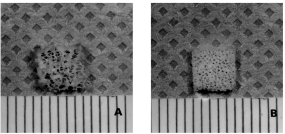
Figure 1 shows the fabricated calcium polyphos- phate matrices used in this study. The matrices exhibited a 3-dimensional interconnected porous structure. These porous matrices were composed of anastomosing network and the pore size was 450- 550㎛(CPP-45ppi) and 200-300㎛(CPP-60ppi) each.
These two porous matrices were somewhat brittle but there was no special problem in handling.
When these CPP matrices were placed in media, media was absorbed very well due to their hydrophilicity.
2. Cell attachment and proliferation in the calcium polyphosphate
The seeded cells were attached on the CPP sur- faces very well. However, the number of attached cells was not as high as expected. This is because the CPP matrices had macroporous structures, and the surface area of CPP was not large enough. The number of attached cells were counted after 1day of seeding(Table 1). The number of proliferated cells on the CPP surfaces are also written in table1. The number of attached cells at day 1 was not signifi- cantly different between that of 45ppi and that of 60ppi. The number of cells proliferated after 7, 14,
and 21days were significantly increased when com- pared with that of the first day, but there is no signif- icant difference between the two groups at each
time period. After the 7th day, the number of cells decreased over times(Figure 2).
Figure 1. Manufactured calcium polyphosphate matrices which had intercon- nected porous structure a) CPP-45pore per inch(ppi) b) CPP- 60ppi.
Table 1. Cell proliferation in CPP matrices.
Number of cells(×104/matrix)
day 45ppi 60ppi
1 0.975±0.189 1.075±0.378
7 16.400±3.940* 15.800±2.905*
14 13.567±3.465* 15.800±4.6163*
21 13.733±0.709* 14.267±3.055*
N=4, mean±S.D.,
*: P<0.01, as compared with 1 day in each group
Figure 2. Number of cells in cell-CPP complex
"No. of cells" means the Number of cells(×104/matrix) attached on the CPP surfaces.
No. of cells of day 1 is the number of initially attached cells.
No. of cells increased at day 7, but no more increased after day 7.
3. Alkaline phosphatase activity
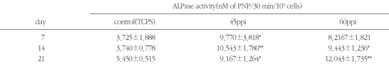
Alkaline phosphatase activities were measured in both types of CPP after 7, 14, and 21days of culture.
There was no significant difference between that of the cells cultured in 45ppi and 60ppi at all points. At day 7, alkaline phosphatase activities of cells cul- tured in 45ppi were significantly higher than that of the cells cultured in control(TCPS). At day 14, and 21, alkaline phosphatase activities of cells cultured in 45ppi and 60ppi were significantly higher than that of cells cultured in control(Table 2, Figure 3).
4. Histologic examination
SEM specimens were prepared for histologic observation. At day 1, there were many cells attached on the CPP surface. The arrow mark repre- sent attached cells on the figure4-a, & b. The num- ber of cells were increased over time(7, 14, and, 21 days) and this increasing pattern seemed somewhat different from that of cell proliferation counting. In cell counting test, the number of cells didn't increased over time. But, the number of cells were evidently increased in the SEM view. At day 7, and 14, there was many more cells proliferated from the original cells than day 1. At day 21, some of the pores of CPP were partially filled with the proliferat- ed cells(Figure 4-Figure 8).att
Table 2. Alkaline phosphatase activities of cells cultured in CPP matrices.
ALPase activity(nM of PNP/30 min/104cells)
day control(TCPS) 45ppi 60ppi
7 3.725±1.888 9.770±3.818* 8.2167±1.821
14 3.740±0.778 10.543±1.780** 9.443±1.236*
21 5.450±0.515 9.167±1.264* 12.043±1.735**
N=4, mean±S. D.,
* : P<0.05, as compared with control group in each group
** : P<0.01, as compared with control group in each group
No significant difference was found between CPP-45ppi and CPP-60ppi matrices at all periods.
Figure 3. Alkaline phosphatase activities of cultured cells in cell-CPP complex
"conc." means ALPase activity(nM of PNP/30 min/104cells) of cultured cells.
Alkaline phosphatase activities of both types of CPP was significantly increased compared with that of control group. There is no sig- nificant differences between two types of CPP.
Figure 4. SEM view of after 1 day of seeding(×400). a) CPP-45ppi, b) CPP-60ppi. Arrow marks indi- cate the attached cells on the CPP surface.
Figure 5. SEM view of after 7 days of seeding(×400). a) CPP-45ppi, b) CPP-60ppi. Arrow marks indicate the proliferated cells on the CPP surface. There were much more cells than day 1 at both two group.
Figure 6. SEM view of after 14 days of seeding(×400). a) CPP-45ppi, b) CPP-60ppi. Arrow marks indicate the proliferated cells on the CPP surface.
5. Mutagenicity of calcium polyphos- phate
To test mutagenicity of calcium polyphosphate, hypoxanthine-guanine phosphoribosyl trans-
ferase(HPRT) assay was done. The number of ini- tially seeded cells is 105/plate. Among these cells, the number of colonies of survived mutant cell were under 3. The count of colonies from survived mutants was nearly zero. It can be concluded that Figure 7. SEM view of after 21 days of seeding(×100). a) CPP-45ppi, b) CPP-60ppi. Arrow marks
indicate the attached cells on the CPP surface.
Figure 8. High magnification of Fig. 7(×400). a) CPP-45ppi, b) CPP-60ppi. Arrow marks indicate theattached cells on the CPP surface.
Table 3. Result of Mutagenicity test of Calcium PolyPhosphate(by HPRT assay).
Conc. of CPP(㎍/㎕) Mutant colony/103cell
NIH3T3 CHO-K1
1000 0.762±1.245* 1.014±1.268
100 0.539±1.070 0.660±1.080
10 1.958±2.043 0.802±1.049
1 2.842±2.260 1.117±1.197
*Means±S.D., CPP showed no mutagenicity at any concentration of CPP for both of NIH3T3 and CHO-K1 cell lines.
the CPP showed no mutagenicity at any concentra- tion of CPP for both the NIH3T3 and CHO-K1 cell lines(Table 3).
VI. Discussion
In this study, the interaction of calcium polyphos- phate(CPP) and rat bone marrow cells and the mutagenicity of CPP were investigated.
Manufactured CPP had white, interconnected porous structure. Both types of CPPs(45ppi and 60ppi) were tested for use as a scaffold of bone engineering and bone graft material. To permit tis- sue and blood vessel ingrowth, the pore must be interconnected. CPP had interconnected pore struc- ture, so this requirement is satisfied. There is some controversy about the pore size of the scaffold, but it is reasonable to make the pore size at the size of 200-600㎛. Ishaug et al36,37)also reported that in vivo transplantation of cell-scaffolds, penetration depth, mineralized tissue per surface area, and percentage of bone formation were found to be independent of pore size between 150-710㎛. In our study, there were also no significant differences between pore size of 200-300㎛ and 450-550㎛. For bone graft material, the pore size of CPP might be somewhat larger than generally accepted. But, as controlling the pore size of CPP is very easy, the pore size of CPP will make no problems for not only tissue engi- neering scaffold but also bone graft material.
Several types of cultured cells, including periosteal cells, marrow stromal cells, enzyme released fetal or neonatal rat calvarial cells and clonal osteogenic cell line, used to be employed for tissue engineered bone formation. Applications to bone engineering have been limited though by the difficulty with phe- notypic maintenance when culturing mature osteoblasts and osteocytes38).
Therefore, most studies used culturing bone mar-
row cells, and fetal or neonatal bone cells10,17,19). Present study employed the rat bone marrow cells.
Bone marrow cells are a kind of stem cells. Stem cells are capable of self-renewal, and can divide in vivo to give a daughter cell and a restored stem cell.
These hypothetical stem cells have the ability to dif- ferentiate into fibroblastic, adipogenic, osteogenic, chondrogenic, and reticular cells39). Many researchers employed this bone marrow stem cells to form bone tissue in vitro and they could achieve their objection38,40-42). In our study, whole bone mar- row cells were cultured and seeded into the CPP matrices. But, in this whole bone marrow cells, hematopoietic cells are also present besides connec- tive tissue precursor cells. If we could separate osteogenic stem cells from the whole bone marrow cells, and seed them into the matrices, the result would be more predictable. In clinical situation, the patient's own cells should be isolated and engi- neered for transplantable bone formation. Recently, Malekzadeh et al. introduced results of in vitro amplification of osteoblast-like cells isolated from fetal human calvaria43). They showed that this is invaluable source in tissue engineering approaches to restore bone in oral cavity. Ultimately, they also suggested that intraoral biopsy might be needed as an autogenous cell source for each patient.
In order for an osseous augmentation material to be successful, it should provide a matrix which is compatible with osteoblastic cell attachment and growth. Materials which are used in bone regenera- tion therapy should support attachment and prolifer- ation of the bone-forming osteoblastic cells44). The attachment of cell to the material and following pro- liferation is a very important event, because the first step of tissue formation in grafting is cell attachment to the material, and migration or proliferation of the cells is subsequent event57). Seeded cells proliferated very rapidly at the first few days in this study. In our
preliminary study, we seeded the same rat bone marrow cells, though the number of the passages were greater. In this case, the proliferation of seed- ed cells were not as great. So we seeded cells pas- saged only two times after primary culture. Then, the seeded cells proliferated very rapidly. But the proliferation stopped after day 7. At day 14 and 21, the cells proliferated in a multilayer pattern, it was impossible to detach all the cells from the scaffold.
So, the cells seen in SEM view after day 14 and day 21 were much more than that of day 7(counted number of cells). To overcome this problem, total DNA assay might be recommended.
Osteoblasts express various phenotypes such as elevated levels of ALPase activity, parathyroid hor- mone(PTH) responsiveness and osteocalcin produc- tion. These phenotypic expressions depend on the differentiation stages of osteoblasts. During osteoblast differentiation, the increase in ALPase activity and expression of PTH/PTH related protein receptor occurs earlier than does osteocalcin pro- duction. Among these phenotypes, osteocalcin pro- duction occurs preferentially in mature osteoblasts45). The expression of ALPase activities means that seeded bone marrow cells were differen- tiated into the osteoblasts. In our study, the alkaline phosphatase activities of cultured cells in CPP scaf- folds were significantly higher than that of the cells cultured on polystyrene. These phenomenons might be partly due to culturing in three-dimension- al scaffolds, and partly due to culturing on hydrox- yapatite, because three dimensional culture system permits easy and high expression of ALPase activity, and hydroxyapatite has bone conductivity. Many other researches have already shown that bone mar- row cells differentiate into osteoblasts under such conditions36,37). Generally, ALPase activities of cell- scaffolds complex seeded bone marrow were much higher than that of cell-scaffolds complex seeded
calvarial osteoblastic cells. Ishaug et al reported that they employed rat bone marrow cells and showed much higher expression of ALPase activities than that of other studies which employed calvarial osteoblastic cells10,17,19,38). In our study, we employed rat bone marrow cells, and also observed much higher expression of ALPase activities than other studies36,37). More studies are needed to eluci- date the basis of these results.
The components of culture media were also important in phenotypic expression and retention of osteoblast and matrix mineralization. In this study, the culture media was supplemented with ascorbic acid, and β-glycerophosphate. Bellows et al. report- ed that the formation of mineralized bone nodules in monolayer culture of enzymatically released rat calvaria cell population appear to be dependent upon three factors: the ability of cell to form multi- layers in vitro, the presence of ascorbic acid, and the inclusion of β-glycerophosphate in the culture medi- um46). Ascorbic acid probably stimulates the forma- tion and hydroxylation of collagen, permitting suffi- cient amount of collagenous matrix to be deposit- ed48). The organic phosphates appear to be neces- sary for mineralization. In the study of Bellows et al, nodules failed to be mineralized in absence of β- glycerophosphate while nonmineralized nodules formed in the absence of β-glycerophosphate did mineralize when β-glycerophosphate was added46). Glucocorticoids such as dexamethasone, have been shown to cause an initial increase in the activity of a number of osteoblast-like cell markers48-52). In addi- tion, data from several reports48,50,51) suggest that the immediate effects of corticosteroid on bone cell pro- liferation and ALPase activity were stimulative.
However, the results were controversial and the supplementation of glucocorticoid to media could be considered very cautiously because long-term culture application of corticosteroids might have an
opposite effect by depleting the reserves of deter- mined osteoprogenitor cells48). In our study, dexam- ethasone was not supplemented in the culture media, and there was not so much bone formation.
So, it would be better to supplement dexametha- sone into the culture media to engineer bone tissue.
After day 1, the cell-CPP complex was looked into via SEM view. The cells attached on the CPP surface very well, but the number of attached cells were not so much as expected. This might be that number of seeded cells(105cells/block) were not enough.
Because CPP had macroporous structure, most of the seeded cells passed through the blocks and attached to the bottoms of 24-well cell culture. To solve this problem, sigmacote(sigma, U.S.A.) was filmed on the bottom of 24-well cell culture, but it was in vain. Sigmacote is a substance which prevent cells from attaching on the something as culture dish. The solution of this problem might be increas- ing the seeding density. 108/block or 109/block might be necessary. At SEM view, we could find some mineralized nodules after day 21. But, it was not popular.
We employed Hypoxanthine-guanine phosphori- bosyl transferase(HPRT) assay to test whether CPP had mutagenicity or not. HPRT assay is commonly used method to test the mutagenicity of a certain material56). Mutant cells with altered, non-functional or zero levels of HPRT don't uptake 6-thioguanine (toxic purine analogues) and thus are able to sur- vive in these selective agents. Especially 6TG is the agent of choice for use with mouse fibroblasts. In our study, the count of colony from survived mutant was nearly zero level. So, the CPP showed no muta- genicity at any concentration of CPP for both the NIH3T3 and CHO-K1 cell lines.
Calcium polyphosphate have many advantages as materials for tissue engineering. It is hydrophilic, biodegradable and nontoxic1,27-29). It is available in
various forms and its degradation rate is controllable54).
Ishaug et al. suggested five prerequisites for a scaffold material for bone formation8). Concerning these criteria, CPP satisfied the first(cell attachment), the second(cell proliferation and function), and the third(tissue ingrowth) requirements, and the fourth(biodegradation), and the fifth(making 3- dimensional irregular structure) requirements are under investigation.
V. Conclusion
1. Manufactured calcium polyphosphate had inter- connected porous structure with the size of 450- 550㎛(CPP-45ppi) and 200-300㎛(CPP- 60ppi).
And its 3-dimension structure had advantage for osteoblast culture, proliferation and differentiation.
2. In cultured cell-CPP complex, cell proliferation was significantly increased after 7, 14, and 21 days than day 1. There was no significant differ- ences between two types of CPP blocks.
3. At cultured the cells, alkaline phosphatase activi- ty was significantly increased in CPP matrices than in TCPS(control) at each time period(7, 14, and 21 day). There was no significant differ- ences between two types of CPP blocks.
4. SEM view of day 1 showed well attached bone marrow cells to the CPP surfaces. And that of day 7, 14, and 21 showed increased cell popula- tion over times.
5. In HPRT assay, CPP showed no mutagenicity.
6. From these results, CPP may be good scaffolds for tissue engineering of bone tissue and may also usable as bone graft material.
VI. References
1. Mehlisch DR, Leider AS, Roberts WE. Histologic
evaluation of the bone/graft interface after mandibular augmentation with hydroxyl- apatite/purified fibrillar collagen composite implants. Oral Surg Oral Med Oral Pathol, 1990;70:685-692.
2. Wardrop RW, Wolford LM. Maxillary stability following downgraft and/or advancement pro- cedures with stabilization using rigid fixation and porous block hydroxylapatite implants. J Oral Maxillofac Surg, 1989;47:336- 342.
3. Froum SJ, Thaler R, Scopp IW, Stahl SS: Osseous autograft, I. Clinical response to bone blend or hip marrow autograft. J Periodontol 1975;46:515- 521.
4. Schallhorn RG: Present status of osseous grafting procedures. J Periodontol 1977;48:570- 576.
5. Mellonig JT: Decalcified freeze-dried bone allo- graft as an implant material in human periodon- tal defect. Int J Periodont Restorative Dent 1984;4:41-55.
6. Garrett S: Periodontal regeneration around nat- ural teeth. Ann Periodontol 1996;1:621-666.
7. Vacanti CA, Vacanti JP: Bone and cartilage reconstruction with tissue engineering approach- es. Otolaringol Clin North Am 1994;27:263- 276.
8. Ishaug SL, Crane GM, Miller MJ, Yasko AW, Yaszemski MJ, Mikos AG: Bone formation by three-dimensional stromal osteoblast culture in biodegradable polymer scaffolds. J Biomed Mater Res 1997;36:17-28.
9. Casser-Bette M, Murray AB, Closs EI, Erfle V, Schmidt J: Bone formation by osteoblast-like cells in a three-dimensional cell culture. Calcif Tissue Int 1990;46:46-56.
10. Laurencin CT, Attawia MA, Elgendy HE, Herbert KM: Tissue engineered bone-regeneration using degradable polymers: The formation of mineral- ized matrices. Bone 1996:93S-99S.
11. Goshima J, Goldberg VM, Caplan AI: The origin
of bone in composite grafts of porous calcium phosphate ceramic loaded with marrow cells.
Clin Orthop Rel Res 1991;274-283.
12. Goshima J, Victor MG, Caplan AI: The osteogenic potential of culture-expanded rat marrow mesenchymal cells assayed in vivo in calcium phosphate ceramic blocks. Clin Orthop Rel Res 1991;298-311.
13. Nakahara H, Bruder SP, Goldberg VM, Caplan AI: In vivo osteochondrogenic potential of cul- tured cells derived from periosteum. Clin Orthop 1990;259:223-232.
14. Nakahara H, Bruder SP, Haynesworth SE, Holecek JJ, Barber VM, Caplan AI: Bone and cartilage formation in diffusion chambers by subcultured cells derived from the periosteum.
Bone 1990;11:181-188.
15. Nakahara H, Goldberg VM, Caplan AI: Cultured- expanded human periosteal-derived cells exhib- ited osteochondral potential in vivo. J Orthop Res 1991;9:465-476.
16. Ohgushi H, Goldberg VM, Caplan AI:
Heterotopic osteogenesis in porous ceramic induced by marrow cells. J Orthop Res 1989;7:
568-578.
17. Vacanti CA, Kim W, Upton J, Vancanti MP, Mooney D, Schloo B, Vancanti JP: Tissue engi- neered growth of bone and cartilage. Transplant Proc 1993;25:1019-1021.
18. Puelacher WC, Vacanti JP, Ferraro NF, Schloo B, Vacanti CA: Femoral shaft reconstruction using tissue-engineered growth of bone. Int J Maxillofac Surg 1996;25:223-228.
19. Vacanti CA, Upton J: Tissue engineered mor- phogensis of cartilage and bone by means of cell transplantation using synthetic biodegrad- able polymer matrices. Clin Plast Surg 1994;:21:445-462.
20. Breitbart AS, Grande DA, Kessler R, Ryaby JT,
Fitzimmons RJ, Grant RT: Tissue engineered bone repair of calvarial defects using cultured periosteal cells. Plast Reconstr Surg 1998;101:567-576.
21. Bagambisa FB, Joos U, Shilli W. Mechanisms and structure of the bond between bone and hydroxyapatite ceramics. J Biomed Mater Res, 1993;27:1047-1055.
22. Nunes CR, Simske SJ, Sachdeva R, Wolford LM.
Long-term ingrowth and apposition of porous hydroxylapatite implants. J Biomed Mater Res, 1997;36:560-563.
23. Jarcho M. Biological aspects of calcium phos- phate. Properties and applications. Dent Clin North Am 1986;30:25-47.
24. de Groot K. Macropore tissue ingrowth: a quan- titative and qualitative study on hydroxy-apatite ceramics. Biomaterials 1986;7:137-143.
25. Muller-Mai CM, Voigt C, Gross U. Incorporation and degradation of hydroxyapatite implants of different surface roughness and surface structure in bone. Scan Microsc 1991;4:501-511.
26. Boyde A, Corsi A, Quarto R, Cancedda R, Bianco P. Osteoconduction in large macroporous hydroxyapatite ceramic implants: Evidence for a complementary integration and disintegration mechanism. Bone, 1999;24:579-589.
27. Holmes RE, Wardrop RW, Wolford.
Hydroxylapatite as a bone graft substitute in orthognathic surgery: histology and histometric findings. J Oral Maxillofac Surg, 1988;46:661- 671.
28. Jarcho M. Calcium phosphate ceramics as hard tissue prosthetics. Clin Orthop 1981;157: 259- 278.
29. Ducheyne P. Bioceramics:Material characteristics versus in vivo behavior. J Biomed Mater Res Appl Biomater. 1987;21A:219-236.
30. Ayers RA, Wolford LM, Bateman TA, Ferguson
VL, Simske SJ. Quantification of bone ingrowth into porous block hydroxyapatite in humans. J Biomed Mater Res 1999;47(1):54- 59.
31. Nunes CR, Simske SJ, Sachdeva R, Wolford LM.
Long-term ingrowth and apposition of porous hydroxyapatite implant. J Biomed Mater Res 1997;36:560-563.
32. Kim S: Bioresorbable calcium mataphos- phate ceramics: I. Preparation and preliminary in vitro study. Biomaterials Research 1998;2:48- 52.
33. Lee J, Kim S: In Transaction of 5th World Biomaterials Congress, Toronto, May 1996, University of Toronto Press, Tronto, 1966, p53.
34. Lee JS, Kaibara M, Iwaki M, Sasebe H, Suzuki YL, Kusakabe M. Selective adhesion and prolif- eration of cells on ionimplanted polymer domains. Biomaterials, 1993;14:958-960.
35. Aubin JE, Liu F, Malaval L, Gupta AK. Osteoblast and chondroblast differentiation. Bone, 1995;2:77S-83S.
36. Susan L. Ishaug, Genevieve M. Crane, Michael J.
Miller, Alan W. Yasko, Michael J. Yaszemski, and Antonios G. Mikos. Bone formation by three-dimensional stromal osteoblast culture in biodegradable polymer scaffolds. J. of Biomedical Material Research, 1997;36,17-28.
37. Susan L. Ishaug-Riley, Genevieve M. Crane, Ali Gurlek, Michael J. Millers, Alan W. Yasko, Michael J. Yaszemski, Antonios G. Mikos:
Ectopic bone formation by marrow stromal osteoblast transplantation using poly(DL-lactic- co-glycolic acid) foams implanted into the rat mesentry. J Biomed Mater Res 1997;36:1-8).
38. Yong-Moo Lee, Sang-Mook Choi, Yoon-Jeong Park, Seung-Jin Lee, Young Ku, Chong-Pyoung Chung.: Tissue Engineered Bone Formation Using Porous Chitosan and Chitosan/Tricalcium Phosphate Matrices. J Periodontol. 2000;71:410- 417.
39. Triffitt J. T. The stem cell of the osteoblast. In:
Bilizekian J. Raisz L. Rodan G. Eds. Principles of Bone Biology. San Diego, CA: Academic:
1996;39-50.
40. Bruder SP, Kraus KH, Goldberg VM, Kadiyala S.
The effect of implants loaded with autologous mesenchymal stem cells on the healing of canine segmental bone defects. J Bone Joint Surg, 1998;80-A:985-996.
41. Anselme K, Noel B, Flautre B, Blary MC, Delecourt C, Descamps M, Hardouin P.
Association of porous hydroxyapatite and bone marrow cells for bone regeneration. Bone, 1999;25:51S-54S.
42. Lamghari M, Almeida MJ, Berland S, Huet H, Laurent A, Milet C, Lopez E. Stimulation of bone marrow cells and bone formation by nacre: In vivo and In vitro studies. Bone, 1999;25:91S-94S.
43. Malekzadeh R, Hollinger JO, Buck D, Adams DF, McAllister BS. Isolation of human osteoblast- like cells and in vitro amplification for tissue engineering. J Periodontol 1998;69: 1256-1262.
44. Eugena B. Stephan, Di Jiang, Samuel Lynch, Peter Bush, and Rosemary Dziak. Anorganic bovine bone supports osteoblastic cell attach- ment and proliferation. J Periodontol 1999;70:364-369.
45. Takiguchi, T, Kobayashi M, Suzuki R, Yamaguchi A, Isatsu K, Nishihara T, Nagumo M, Hasegawa K. Recombinant human bone mor- phogenetic protein-2 stimulates osteoblast differ- entiation and suppresses matrix metallopro- teinase-1 production in human bone cells isolat- ed from mandibulae. J Periodontal Research
1998:33:476-485)
46. Bellows CG, Aubin JE, Heersche JNM, Antosz ME: Mineralization bone nodules formed in vitro enzymatically released rat calvaria cell popula- tions. Calcif Tissue Int 1986;38:143-154.
47. Barnes MJ: Function of ascorbic acid in collagen metabolism. Ann NY Acad Sci 1975; 258:264- 277.
48. Canalis R: Effect of glucocorticoid on type I col- lagen synthesis, alkaline phosphatase activity, and deoxyribonucleic acid content in cultured rat calvaria. Endocrinology 1983;112:931-939.
49. Chyun YS, Kream BE, Raisz LG: Cortisol decreas- es bone formation by inhibiting periosteal cell proliferation. Endocrinology 1984;114: 477-480.
50. Hahn TJ, Westbrook SL, Halstead LR: Cortisol modulation of osteoblast metabolic activity in cultured neonatal rat bone. Endocrinology 1984;114:1864-1870.
51. Tenebaum HC, Heerche JNM: Dexamethasone stimulates osteogenesis in vitro. Endocrnology 1985;117:2211-2217.
52. Muzzarelli RA: Biochemical significance of exogenous chitins and chitosans in animals and patients. Carbohydrate Polymers 1993;20:7-16.
53. Maniatopouls C, Sodek J, Mechler AH, Bone for- mation in vitro by stromal cells obtained from bone marrow of young adult rats. Cell Tissue Res, 1988;254:317-330.
54. Cole J, Arlett CF. Mutagenicity testing. IRL Press, Oxford. 1984:233-235.
55. Robert P Lanza, Robert Langer, William L Chick.
Principles of tissue engineering. R.G. Landes Company, Austin. Academic press. 1994.
-Abstract-
Calcium polyphosphate의 생물학적 활성도에 관한 연구
설양조1, 이재일2, 이용무1, 임윤탁3, 김석영3, 구 영1, 류인철1, 함병도1, 한수부1, 최상묵1, 정종평1
1서울대학교 치과대학 치주과학교실
2서울대학교 치과대학 구강병리학과교실
3영남대학교 공과대학 재료금속공학부
이 연구의 목적은 다공성의 CPP 내부에 쥐의 장골의 골수에서 유래된 세포를 접종하고 3차원적으로 배양하 여 CPP가 골 형성을 위한 조직공학의 지지체로 적용가능한가를 연구하는 것과 Calcium PolyPhosphate(CPP)의 돌연변이 유발성을 검사하는 것이다.
무수 Ca(H2PO4)를 condensation하여 무결정의 Ca(PO3)를 얻고 이를 용융하고 냉각시킨 후 분쇄하여 Calcium polyphosphate(CPP) powder를 얻었다. 다공성의 CPP는 5% SiO2를 첨가하여 sponge 형태로 450-550
㎛의 소공의 크기를 가지는 것과(CPP-45ppi) 200-300㎛의 소공의 크기를 가지는 것(CCP-60ppi) 2가지로 제작 하였다. 각각의 CPP matrices는 5mm×5mm×1mm의 블록 형태로 만들었다. 체중 100g 내외의 백서에서 장골 (femur, tibia)을 채취하여 백서의 장골 골수 세포를 분리하여 배양한 후 24well에 CPP block을 넣고 CPP block 당 105개의 배양한 세포를 접종하였다. 배양 1, 7, 14, 및 21 일째에 각 well에서 trypsin EDTA를 이용하여 2회 반 복하여 cell을 분리하였고, 원심분리한 후 hemacytometer로 측정하였다. 또, 45ppi와 60ppi, 그리고 Tissue Culture Polystyrene(control group)에 접종, 배양된 세포들의 염기성 인산분해효소활성도를 배양 7, 14, 및 21 일째에 각각 측정하였다. 각 기간 별로 배양된 세포-CPP 혼합체내에서 세포의 부착 및 증식과 형성된 조직의 3 차원적 형태를 관찰하기 위하여 주사전자현미경하에서의 관찰하였다. CPP의 돌연변이 유발성 검사(muta- genicity test)를 위해 hypoxanthine-guanine phosphoribosyl transferase(HPRT) assay를 하였다. NIH3T3 cell line과 CHO-K1 cell line으로 각각 1000㎍/㎖, 100㎍/㎖, 10㎍/㎖ 그리고 1㎍/㎖의 CPP 농도에서 측정하였다. 통 계적 분석을 위해서 모든 측정은 각 군당 4개체 이상 시험하였고, 각 측정값은 평균값±표준편차로 나타내었 다. 각 군간의 통계적 유의성 검정을 위해서 Analysis of variance(ANOVA)를 이용하였고 Tukey의 방법으로 사 후분석을 실시하였다. 제작된 CPP matrices는 소공들이 서로간에 연결이 잘 되어있는 형태였다. 두 가지로 제 조된 CPP(45ppi와 60ppi) 모두에서 세포의 부착이 잘 일어났고, 부착된 세포의 분열도 잘 일어났다. 2 가지의 CPP 모두에서 7, 14, 21일째의 세포 수는 1일째에 비해 유의성 있게 증가하였다(P<0.01). 3차원적 구조인 Calcium PolyPhosphate에서 배양한 세포는 24well dish(tissue culture polystyrene)에서 평면적으로 배양한 대 조군의 세포에서 보다 염기성 인산분해효소(Alkaline Phosphatase)를 유의성 있게 높게 나타냈다. 주사전자현 미경에서 세포-CPP 혼합체를 관찰한 결과, CPP block에 세포들이 잘 부착되어 있었고, 시간이 지남에 따라 세 포가 여러 층을 형성하면서 뭉치는 현상을 보였다. 또, HPRT assay 결과, Calcium PolyPhosphate는 돌연변이 유발성을 보이지 않았다. 이상의 결과로 볼 때 CPP에는 세포부착이 잘 일어나고, 지지체 상에서 세포의 분열도 활발하게 일어나므로 골조직을 위한 조직공학의 우수한 지지체가 될 수 있을 것으로 사료된다.
주요어 : calcium metaphosphate, 골전도성, 3차원적 세포배양, 세포지지체, 골수세포, 조골세포배양, 조직공학.