pISSN 1598-9992 eISSN 2233-6869
REVIEW ARTICLE
헬리코박터 파일로리 감염 진단의 최신 지견
허철웅, 김병욱
가톨릭대학교 인천성모병원 소화기내과
Diagnosis of Helicobacter pylori Infection
Cheal Wung Huh and Byung-Wook Kim
Division of Gastroenterology, Department of Internal Medicine, Incheon St. Mary's Hospital, College of Medicine, The Catholic University of Korea, Seoul, Korea
Accurate diagnosis of Helicobacter pylori (H. pylori) infection is mandatory for the effective management of many gastroduodenal diseases. Currently, various diagnostic methods are available for detecting these infections, and the choice of method should take into account the clinical condition, accessibility, advantage, disadvantage, as well as cost-effectiveness. The diagnostic methods are divided into invasive (endoscopic-based) and non-invasive methods. Non-invasive methods included urea breath test, stool antigen test, serology, and molecular methods. Invasive methods included endoscopic imaging, rapid urease test, histology, culture, and mo- lecular methods. In this article, we provide a review of the currently available options and recent advances of various diagnostic methods. (Korean J Gastroenterol 2018;72:229-236)
Key Words: Helicobacter pylori; Diagnosis; Guideline
Received September 9, 2018. Revised September 17, 2018. Accepted September 17, 2018.
CC This is an open access article distributed under the terms of the Creative Commons Attribution Non-Commercial License (http://creativecommons.org/licenses/
by-nc/4.0) which permits unrestricted non-commercial use, distribution, and reproduction in any medium, provided the original work is properly cited.
Copyright © 2018. Korean Society of Gastroenterology.
교신저자: 김병욱, 21431, 인천시 부평구 동수로 56, 가톨릭대학교 인천성모병원 소화기내과
Correspondence to: Byung-Wook Kim, Division of Gastroenterology, Department of Internal Medicine, Incheon St. Mary's Hospital, College of Medicine, The Catholic University of Korea, 56 Dongsu-ro, Bupyeong-gu, Incheon 21431, Korea. Tel: +82-32-280-5052, Fax: +82-32-280-5987, E-mail: gastro@catholic.ac.kr, ORCID:
https://orcid.org/0000-0002-2290-4954 Financial support: None. Conflict of Interest: None.
서 론
Helicobacter pylori
(H. pylori
) 감염은 한국뿐 아니라 전 세계적으로 감염률이 높고 위축성 위염, 장상피화생, 소화성 궤양, 위점막 연관 림프종(marginal zone B cell lymphoma, MALT lymphoma), 위암 등 여러 위장 질환들과 밀접한 관련 이 있다.1 근래 개정된 진료 지침에 따르면,H. pylori
제균 치료의 초기 치료는 90% 이상의 제균율을 보이는 약제를 사 용할 것을 권고하고 있다.1따라서 성공적인 제균 치료를 위해 서는H. pylori
감염 여부의 정확한 진단이 무엇보다 중요하 다고 할 수 있다. 현재까지 다양한H. pylori
진단 방법이 개 발되어 사용되고 있으며, 각 진단 방법은 민감도와 특이도가 각각 90% 이상은 되어야 실제 임상 진료에서H. pylori
감염의 정확한 진단이 가능하다.2,3
H. pylori
진단을 위한 검사 방법은 내시경 검사 여부에 따라 비침습적 검사 방법과 침습적 검사 방법으로 나누어진 다. 비침습적 검사 방법에는 요소호기 검사, 대변항원 검사, 혈청 검사 및 분자생물학적 검사가 포함되고, 침습적 검사 방법에는 급속요소분해효소 검사, 조직 검사, 배양 검사, 분 자생물학적 검사가 사용된다. 비침습적 검사 방법은 내시경 을 이용하지 않기 때문에 환자의 불편감이 적고 비용이 적게 드는 장점이 있고, 침습적 검사 방법인 조직을 이용하는 검 사는H. pylori
진단 외에도 점막의 염증, 위축성 위염 정도 및 장상피화생, 항생제 감수성 검사 등과 같은 추가적 정보 를 얻을 수 있는 장점이 있다. 각 검사 방법마다 장단점이 있으므로 검사의 정확도, 각 기관에서 검사의 사용 가능 여부, 비용-효율성, 환자 상태 등과 같은 각각의 임상 상황을 고려하여 적절한 검사의 선택이 필요하다. 본고에서는 현재
H. pylori
의 진단에 사용되는 여러 검사법의 장단점 및 제한 점을 살펴보고자 한다.본 론
1. 비침습적 검사 방법
1) 요소호기 검사(urea breath test)
요소호기 검사는
H. pylori
감염의 처음 진단뿐 아니라 제 균 치료 후 제균 여부 확인에 가장 널리 사용되는 검사 중 하나이다.1H. pylori
의 요소분해효소는 요소를 암모니아와 이산화탄소(CO2)로 분해하는데, 이 검사는 이러한H. pylori
의 요소분해효소를 이용하는 대표적인 비침습적 검사 방법이 다.H. pylori
의 요소분해효소가 구강을 통하여 섭취된 탄소 동위원소가 포함된 요소(13C-labeled urea)를 분해하여 발생 한 이산화탄소가 혈액 내로 흡수되고, 이것이 다시 폐를 통하 여 배출되는 양을 분광계(spectrometer) 또는 레이저를 이용 한 비율 분석기로 측정하는 방법이다. 가장 널리 사용되는 방 법은 시트르산(citric acid)과 75 mg의 요소를 이용하는 방법 이며, 요소 섭취 후 약 15분 뒤 호기 측정을 한다.요소호기 검사의 민감도와 특이도는 95% 정도로 매우 정 확한 검사이며 재현성이 높고 간편하다.4-6 또한, 조직 검사 및 급속요소분해효소 검사와 달리 표본 추출 오차(sampling error)가 없다는 장점이 있다. 하지만 항생제 또는 양성자펌프 억제제를 사용 중인 환자나 위장관 출혈이 있는 환자에서는 위음성이 나올 수 있고, 6세 미만 어린이는 순응도가 떨어져 검사의 민감도와 정확도가 떨어질 수 있다.7,8 아직까지는 표 준화된 방법이 없어 요소량, 시약의 종류, 검사 장비 등이 검 사 결과에 영향을 줄 수 있고 환자의 호흡 정도 및 금식 시간 에 따라서도 검사 결과가 달라질 수 있다. 또한, 드물지만
H.
pylori
이외에 요소분해효소를 생성하는 균주에 의한 위양성 이 나올 수 있다. 무엇보다 근래 항생제 저항성 균주가 증가하 는 점을 감안하면 항생제 감수성 여부를 확인할 수 없다는 것은 요소호기 검사의 중요한 한계점 중 하나이다.위음성을 줄이기 위하여 요소호기 검사 전 양성자펌프억제 제는 최소 2주, 항생제는 4주를 중단할 것을 추천한다.1따라 서, 제균 치료 후 효과를 판정하는 시기도 제균 치료 종료 후 최소 4주 이상 지나서 측정하는 것이 좋다.1,9
2) 대변항원 검사(stool antigen test)
대변항원 검사는 요소호기 검사와 같이 초기 진단 및 제균 여부 확인 검사로 추천되는 방법으로 비침습적이고, 호기 조
절이 어려운 영유아에서도 쉽게 검사를 시행할 수 있으며 금 식이 필요 없다.1,10 다클론항체(polyclonal antibody) 검사법, 단클론항체(monoclonal antibody) 검사법 모두 사용되고 있 으나 단클론항체 검사법이 다클론항체 검사법에 비하여 민감 도, 특이도, 정확도가 높다고 알려져 있다.11,12 대변항원 검사 방법으로는 효소면역분석법(enzyme immunoassay)과 면역 크로마토그래피법(immunochromatographic assay)이 이용 되고 있으며, 면역크로마토그래피법은 검사 시간이 짧고 사용 이 간편하지만 효소면역분석법에 비하여 검사의 정확도가 낮
다.13,14 대변항원 검사는 다른 검사들과 달리 집에서 대변을
채취하여 병원으로 운반 후 검사가 가능하다는 장점이 있다.
하지만 대변의 보관 온도, 보관 기간 등에 따라 검사의 정확도 가 저하되기 때문에, 냉동 보관 후 빠른 시간 내에 검사를 시 행하는 것이 좋다.1,15 대변이 묽거나 형태가 없는 경우에도 검사의 정확도가 떨어지므로 채취 시 주의해야 한다.15 또한, 대변항원 검사는 위장관 출혈, 항생제나 양성자펌프억제제와 같은 약제 사용, 장운동 기능에 따라 검사의 정확도가 달라질 수 있어 결과 해석에 주의가 필요하다.15 요소호기 검사와 마 찬가지로, 위음성을 줄이기 위하여 대변항원 검사 전 양성자 펌프억제제는 최소 2주, 항생제는 4주를 중단해야 한다.1
3) 혈청 검사(serology)
혈청 검사는 환자 혈청 내의 항헬리코박터 IgG 항체(IgG anti-
H. pylori
antibody)의 역가를 측정하는 방법이다. 비침 습적이며 경제적이고 빠르며 쉽게 검사할 수 있는 장점이 있 어H. pylori
의 감염 여부를 확인하는 선별 검사(screening teset)로 이용할 수 있다.1 주로 효소결합면역침강분석법(en- zyme linked immunosorbent assays)과 면역크로마토그래 피법을 이용하는데, 효소결합면역침강분석법의 정확도가 더 높다고 알려져 있다.16 위축성 위염 및 장상피화생과 같은 위 점막 변화, 항생제나 양성자펌프억제제와 같은 약제 복용 및 위장관 출혈 등의 상태에서 다른 검사 방법과 비교하였을 때 위음성의 가능성이 낮기 때문에, 이러한 상황에서H. pylori
감염의 초기 진단에 유용하다. 근래에는 혈청 검사를 통하여CagA, VacA, UreA
와 같은H. pylori
의 독성인자(virulence factor)를 확인하여 고위험H. pylori
감염을 확인할 수도 있 다.17-19하지만 항체는 제균 치료를 받더라도 수년 동안 양성으로 나타날 수 있기 때문에 현재의 활동성 감염과 과거의 감염을 구분하기 어렵다.1,20 따라서, 제균 성공 여부를 확인하는 검사 방법으로는 권고되지 않고 요소호기 검사나 대변항원 검사 결 과에 따라 추가적으로 감염 여부를 확진할 수 있는 검사로 이용이 가능하다.1또한 지역에 따라 유병률, 균주의 분포 및 구성의 이질성, 항체 역가의 기준 등이 다르기 때문에 반드시
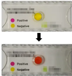
Fig. 1. Rapid urase test. When Helicobacter pylori produce ammonia then pH will increase and the color of the medium will change from yellow to red.
각 지역에서 효과가 검증된 키트를 이용하여야 한다.1,21,22
H.
pylori
의 유병률이 낮은 지역에서는 혈청 검사의 특이도가 낮 아 위양성의 가능성이 높아질 수 있어 주의해야 한다.14) 기타
근래에 대변 실시간 중합효소연쇄반응 검사(stool real-time polymerase chain reaction)를 이용하여 비침습적인 방법으로
H. pylori
진단뿐 아니라 클래리스로마이신 감수성 여부를 확인 할 수 있다는 보고가 있었다.23,24 또한 기존 진단 방법 외에 타액, 소변, 치아플라크 등의 검체를 이용하여H. pylori
를 검출 하고자 하는 노력이 있었다.25-28 이와 같이 다양한 검체를 이용하 여 항체 검사, 항원 검사, 분자생물학적인 검사 등으로H. pylori
감염을 진단하고자 하였으나 아직까지는 기존 검사에 비하여 진단의 민감도, 특이도, 정확도가 낮은 실정이다.1,22,29-33하지만 타액, 소변, 치아 플라크 등은 다른 검사에 비하여 검체를 얻기에 용이하기 때문에 추후 이러한 검체를 이용한 연구는 더욱 발전할 것으로 생각한다.2. 침습적 검사 방법
1) 내시경
조직학적 검사 없이 내시경 소견만으로
H. pylori
감염 여 부를 진단하려는 연구가 많이 있었다. 이전 연구들에서 일반 적인 백색광 내시경(conventional white-light endoscopy) 으로 관찰할 경우, 미만성 발적(diffuse redness), 점상 발적 (spotty redness), 점막 부종(mucosal swelling) 등은H. py- lori
감염을 시사하는 반면, 위점막에서 집합 소정맥의 규칙적 인 배열(regular arrangement of collecting venules)이 유지 되면H. pylori
감염이 없는 경우가 많다고 알려져 있다.34,35 하지만 이는 낮은 민감도와 특이도로 인하여 진단적 가치가 높지 않았다. 근래에는 광학기술의 발전에 따라 확대 내시경 (magnifying endoscopy), 확대 색소 내시경(magnifying chromoendoscopy; narrow band imaging, I-scan 등), 공 초점 레이저 현미경 내시경(confocal laser endomicroscopy) 등을 이용하여H. pylori
감염의 실시간 진단 정확도가 높아 지고 있다.36-38 하지만 이러한 방법은 일부 대학병원 급에서만 사용 가능한 장비이고 숙련된 기술을 필요로 하며, 관찰자 간 진단의 차이가 있을 수 있으며, 정확한 관찰에 시간이 오래 걸리기 때문에 환자에게 불편감을 줄 수 있다는 단점이 있다.2) 급속요소분해효소 검사(rapid urease test)
급속요소분해효소 검사는 내시경을 통하여 얻은 위조직을 요소 기질에 넣어
H. pylori
가 분비하는 요소분해효소로 인하 여 만들어지는 암모니아에 의하여 pH가 상승하는 것을 색조변화로 확인하는 검사이다(Fig. 1). 급속요소분해효소 검사는 비교적 싸고 빠르며 검사의 민감도는 약 90%, 특이도는 약 95-100%로, 민감도와 특이도가 모두 높아 내시경 검사가 가 능한 의료기관에서
H. pylori
감염의 진단을 위하여 가장 많 이 시행되는 검사이다.39,40겔, 종이, 액체 기반의 검사가 가능 하며, 국내에서는 겔 기반 검사인 CLO test (Delta West Limited, Bentley, Australia)가 가장 많이 사용되고 있다. 겔 기반의 검사는 24시간 뒤에, 일부 종이 기반 검사와 액체 기 반 검사는 이보다 빠른 1시간 내에 결과를 확인할 수 있다.검사 방법에 따라 위음성, 위양성이 가능하기 때문에 각 검사 키트에서 권장하는 시간에 맞게 결과를 확인하여야 한다.41 국 내에서는
H. pylori
감염의 초기 진단과 제균 여부 판정에서 모두 권고하는 검사 방법이지만, 최근 개정된 유럽 진료 지침 (Maastricht V/Florence consensus report)에서는 제균 여 부 판정 검사로 권고되지 않는다.1,42H. pylori
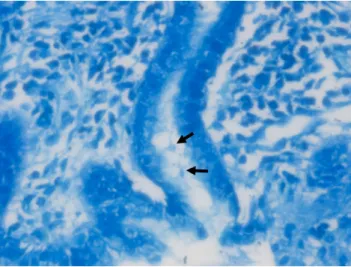
는 위점막 전체 에 동일한 밀도로 분포하고 있지 않기 때문에, 진단율을 높이 기 위하여 전정부와 체부에서 각각 조직을 채취하여 한꺼번에 같이 검사하는 것을 권유한다.1,43,44 조직 채취는 위축성 위염 이나 장상피화생이 없거나 적은 부위에서 시행하여야 위음성 을 줄일 수 있다.42 위장관 출혈이 있거나 양성자펌프억제제, 항생제 및 비스무스제제 등의 약제를 복용하는 경우, 위축성 위염이나 장상피화생이 있을 경우 위음성이 있을 수 있기 때 문에 주의해야 한다.11,45 또한 조직을 겔 등에 담는 과정에서 조직 겸자공의 포르말린이 겔에 묻을 경우 검사 민감도가 감 소할 수 있으므로 주의가 필요하다.46 요소호기 검사 및 대변 항원 검사와 마찬가지로 검사 전 양성자펌프억제제는 최소 2주,Fig. 2. Giemsa stain. The Helicobacter pylori organisms (arrows) are present in Giemsa stain (×400).
항생제 및 비스무스제제는 최소 4주 동안 중단하여야 검사의 정확도를 높일 수 있다.1,11
3) 조직 검사(histology)
조직 검사는 현재
H. pylori
감염 진단의 표준 검사법 중 하나이다.47조직 검사는H. pylori
감염 진단 외에도 위점막의 위축, 염증, 장상피화생 등의 추가적 병리정보를 얻을 수 있다.일반적인 헤마톡실린-에오진(H&E) 염색은 42-99% 민감도와 100% 특이도를 보인다고 알려져 있다.48-50 H&E 염색법만으 로는 비 나선형, 구형의
H. pylori
발견이 쉽지 않아 민감도가 낮기 때문에 Giemsa 염색법, Warthin-Starry 염색법, Genta 염색법 등의 특수 염색법을 병행해야 진단율을 높일 수 있다.이 중 Giemsa 염색법이 민감도가 높고 경제적이며, 재현성이 높아 가장 널리 사용되고 있다(Fig. 2).48,51또한 면역조직화학 (immunohistochemistry) 염색법은 민감도와 정확도가 매우 높아
H. pylori
개체수가 적어도 진단율이 높으며, 그 외에도 활동성 위염, 위축성 위염, 장상피화생이 있을 경우에서도 진 단율을 높일 수 있는 방법이다.1,52하지만 비싸고, 모든 검사실 에서 사용할 수는 없기 때문에 반드시 필요한 경우에 제한적으 로 사용해 볼 수 있다.H. pylori
의 진단율을 높이기 위해서는 전정부 중앙의 소만 과 대만, 체부 중앙의 소만과 대만에서 각각 1표본씩, 총 4표 본을 채취할 것을 권고한다.1,53 하지만 조직 검사 개수를 줄여 야 하는 경우에는 위축성 위염 및 장상피화생이 없거나 적은 부위에서 조직을 채취하기를 권장한다.1 따라서 보통 위축성 위염과 장상피화생이 비교적 심한 전정부보다는 체부 대만에 서 조직을 채취하는 것이 좋다.54 다만, 십이지장 궤양이 있는 경우에는H. pylori
집락 형성이 체부보다 전정부에서 높게나타나기 때문에 전정부에서 조직을 채취하는 것이 좋다.55,56
4) 배양 검사(culture)
위생검을 통하여 얻은 조직을 배지(Pylori agar, Columbia agar with horse blood, 등)에 도말하여 배양하면 3-5일 후에 균이 자라는 군락을 확인할 수 있다.
H. pylori
는 위를 벗어난 환경에 취약하므로 가능하면 조직 채취 직후 바로 배지에 넣 어야 한다. 조직 채취는 전정부와 체부에서 각각 얻는 것이 좋다.57 배양 검사는 특이도가 높은 검사지만 균이 자라는데 시간이 많이 소요되고 비싸며, 배양이 가능한 기술과 장비가 필요하다. 또한 균주의 채취, 운반, 보관 및 배양 기술에 따라 민감도의 차이가 커서 아직까지는 일반적으로 사용되는 진단 방법은 아니다. 하지만 근래 항생제 내성이 지속적으로 증가 하고 있으며 이는 제균 치료의 성공에 큰 걸림돌이 되고 있다 는 점을 감안할 때, 배양 검사는 항생제 감수성을 확인할 수 있는 확실한 진단 방법이므로 근래 유럽 가이드라인에서는 클 래리스로마이신 내성이 15% 이상인 지역이나 1차 제균 치료 에 실패하였을 경우에는 배양 검사를 시행할 것을 권장하고 있다.1다만, 1차 제균 치료 실패 이후 2차 제균 치료 약제로 비스무스 기반 4제를 사용할 예정이라면 2차 제균 치료 실패 이후 배양 검사를 할 것을 권장하고 있다.1 위장 출혈, 심한 위점막 염증상태, 양성자펌프억제제, 항생제 사용 등은 배양 의 성공을 저해하기 때문에 배양 검사시 고려하여야 하며 양 성자펌프억제제는 최소 2주, 항생제는 최소 4주 정도 중단하 는 것이 좋다.57,585) 분자생물학적 검사(molecular method)
중합효소연쇄반응 검사(polymerase chain reaction, PCR) 는 표적이 되는 DNA를 증폭시켜 검출의 민감도를 높이는 방 법으로 침습적으로 얻은 위 조직뿐만 아니라 대변, 타액, 소변 등의 검체를 이용해서도 검사가 가능하다. 중합효소연쇄반응 검사는 적은 양의 균만 존재하여도 양성으로 검출할 수 있기 때문에 기존 검사 방법과 비교하여 민감도와 특이도가 높으며 위장 출혈이 있는 상황에서도 진단율이 비교적 높다.2
H. py- lori
의 여러 유전자(UreA, UreC
, 16S rRNA, 23S rRNA,HSP 60
등)를 표적으로 하여 위 조직에서의 균 존재 여부를 확인 할 수 있으며 항생제 내성을 유발하는 돌연변이 및 독성인자 등을 확인할 수 있는 장점이 있다.3빠르고 정확하며, 민감도 가 높지만 비교적 비싸고 검사를 위한 기술과 장비가 필요하 다는 단점이 있다.중합효소연쇄반응 검사는 분자 역학(molecular epidemi- ology), DNA 유전자 지문법(fingerprinting)을 통한 균주의 동일성 또는
CagA, VacA, UreA
등의 독성인자의 진단, 23S rRNA,gyrA, pbp1A
등의 약제 내성과 관련된H. pylori
유Table 1. Overview of Diagnostic Methods
Characteristics Advantages Limitations
Non-invasive
UBT Sensitivity: >95%
Specificity: >95%
i) High sensitivity and specificity ii) Cheap, simple, safe, widely available iii) Useful to confirm H. pylori eradication iv) No sampling errors
i) No data about antibiotic resistance ii) Special equipment required
iii) False negative results in the case of PPI and antibiotics
SAT Sensitivity: >95%
Specificity: >95%
i) High sensitivity and specificity ii) Cheap, simple, safe iii) Practically useful for children iv) No need to skilled staffs
i) No data about antibiotic resistance ii) Patient reluctance
iii) False negative results in the case of PPI and antibiotics
iv) Variation in sensitivity and specificity over the different clinical circumstances
Serology Sensitivity: >95%
Specificity:
60-90%
i) Cheap, simple, safe
ii) Not affected by gastroduodenal bleeding
iii) No false negative result in the case of PPI and antibiotics iv) Identifies virulence factors
i) No data about antibiotic resistance
ii) Failure in distinguish between active and past infection
iii) Not useful to confirm H. pylori eradication Invasive
RUT Sensitivity: 90%
Specificity: >95%
i) High sensitivity and specificity ii) Cheap, simple, rapid
iii) Practically useful in a clinical setting
i) No data about antibiotic resistance ii) Sampling errors
iii) False negative results in the case of PPI, antibiotics and gastroduodenal bleeding
Histology Sensitivity:
60-90%
Specificity: >95%
i) Gold standard for direct H. pylori detection ii) Secondary diagnostic information iii) Cheap, simple
i) No data about antibiotic resistance ii) Sampling errors
iii) High inter-observer variability iv) Time-consuming
Culture Sensitivity:
50-90%
Specificity: 100%
i) Antibiotics sensitivity profiling ii) The most specific method
i) Limited availability, technically challenging ii) Time-consuming, expensive method
iii) False negative results in the case of PPI, antibiotics and gastroduodenal bleeding
Molecular method
Sensitivity: >95%
Specificity: >95%
i) Antibiotics sensitivity profiling ii) High sensitivity and specificity
iii) Useful to detect the mutations and virulence factors iv) Quick and accurate result
i) Expensive ii) Limited availability iii) Risk of contamination UBT, urea breath test; H. pylori, Helicobacter pylori; PPI, proton pump inhibitor; SAT, stool antigen test; RUT, rapid urease test.
Table 2. Recommended Diagnostic Method for Helicobacter pylori in Recent Guidelines
Initial diagnosis Follow up after eradication
Non-invasive Invasive Non-invasive Invasive
UBT SAT Serology RUT Histology Culture UBT SAT Serology RUT Histology Culture
Maastricht V/Florence Consensus (2017)1 O O O O O O O O
Korean College of Helicobacter and Upper Gastrointestinal Research (2013)42
O O O O O O O O O
Japanese Society of Helicobacter Research (2013)69
O O O O O O O O O O O O
UBT, urea breath test; SAT, stool antigen test; RUT, rapid urease test.
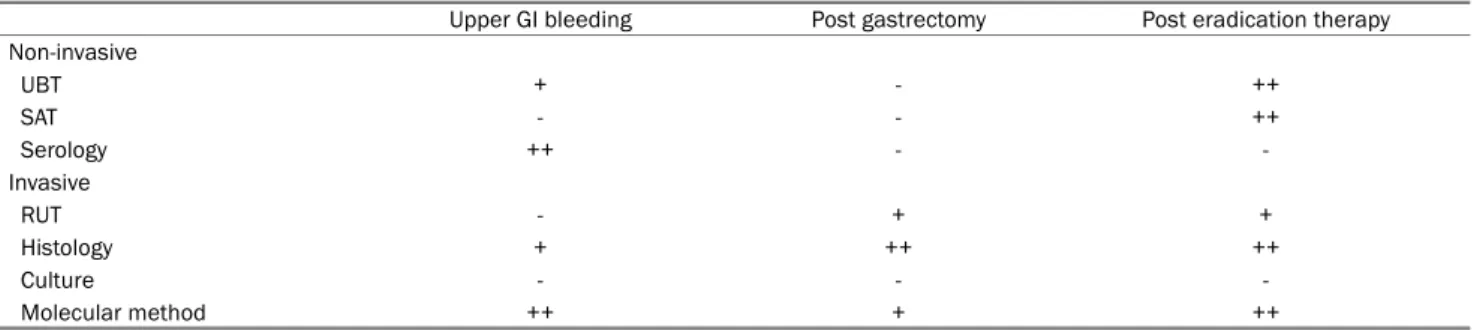
Table 3. Diagnostic Options of Helicobacter pylori Infection in Different Clinical Circumstances
Upper GI bleeding Post gastrectomy Post eradication therapy
Non-invasive
UBT + - ++
SAT - - ++
Serology ++ - -
Invasive
RUT - + +
Histology + ++ ++
Culture - - -
Molecular method ++ + ++
GI, gastrointestinal; UBT, urea breath test; SAT, stool antigen test; RUT, rapid urease test.
전자 변이 같은 영역에서 주로 이용되고 있다.59,60 또한 du- al-priming oligonucleotide-based multiplex PCR을 이용하 여
H. pylori
감염의 진단 및 클래리스로마이신 내성 돌연변 이(A2142G and A2143G mutation in 23S rRNA)를 높은 민감도와 특이도로 확인할 수 있다. 이는 기존 중합효소연쇄 반응 검사보다 간편하고 저렴하여 실제 임상에서도 이용하기 용이하다는 장점이 있다.61,62 근래에는 peptide nucleic acid fluorescence in situ hybridization (PNA-FISH) 방법을 이 용하여 더욱 간단하고 빠르게H. pylori
의 진단과 항생제 내 성과 관련된 돌연변이를 확인할 수 있는 방법이 소개되었다.63 분자생물학적인 검사는 빠르고 편리하며 정확도가 높고, 항생제 감수성을 확인할 수 있는 장점이 있어서 현재 활발한 연구가 진행되고 있는 방법으로 앞으로 더욱 기대가 되는 분야이다.3. 특별한 상황에서의 H. pylori 진단을 위한 검사 방법 위장관 출혈이 있는 경우 여러 침습적, 비침습적 검사의 진 단 정확도는 감소하게 된다. 혈청 검사는 위장관 출혈 유무에 영향을 받지 않지만
H. pylori
활동성 감염 여부를 확인하지 못하기 때문에 한계가 있다. 여러 문헌보고에 의하면, 침습적 인 검사 중에는 조직 검사와 중합효소연쇄반응 검사 방법이 급속요소분해효소 검사나 배양 검사보다 진단의 정확도가 높 다고 알려져 있다. 비침습적인 검사 방법 중에서는 대변항원 검사보다는 요소호기 검사가 더 정확도가 높다.64-66 또한, 위 장관 출혈의 경우H. pylori
감염의 확인은 매우 중요하기 때 문에 첫 검사가 음성이더라도 출혈이 멈춘 이후 최소 4주 정 도 뒤에는H. pylori
의 감염 여부를 확인하기 위한 재검사가 필요하다.67위 부분절제를 받은 환자에서
H. pylori
진단을 위해서는 조직 검사가 제일 정확한 검사이며 급속요소분해효소 검사 또 한 시행할 수 있다.68급속요소분해효소 검사를 시행할 경우에 는 잔위의 기저부 혹은 상부 체부에서 조직을 얻는 것을 추천 한다. 반면에 요소호기 검사나 대변항원 검사는 비교적 정확 도가 떨어져 추천되지 않는다.결 론
H. pylori
진단을 위한 검사 방법은 검사 목적에 따라 비침 습적 방법과 침습적 방법으로 크게 나뉘며 각 방법의 장점, 단점 및 민감도, 특이도에 차이가 있다(Table 1). 또한 국내외 주요 진료 지침들의 초기 진단 및 제균 여부 판정을 위한 검사 의 권고안도 조금씩 차이가 있다(Table 2). 따라서 각 의료기 관 및 여러 임상 상황(Table 3)을 고려하여 적절한 진단 방법 을 선택하려는 노력이 필요하다.REFERENCES
1. Malfertheiner P, Megraud F, O'Morain CA, et al. Management of Helicobacter pylori infection-the Maastricht V/Florence consensus report. Gut 2017;66:6-30.
2. Wang YK, Kuo FC, Liu CJ, et al. Diagnosis of Helicobacter pylori infection: current options and developments. World J Gastroenterol 2015;21:11221-11235.
3. Lopes AI, Vale FF, Oleastro M. Helicobacter pylori infection - re- cent developments in diagnosis. World J Gastroenterol 2014;
20:9299-9313.
4. Gisbert JP, Calvet X. Helicobacter pylori "test-and-treat" strategy for management of dyspepsia: a comprehensive review. Clin Transl Gastroenterol 2013;4:e32.
5. Nocon M, Kuhlmann A, Leodolter A, et al. Efficacy and cost-effec- tiveness of the 13C-urea breath test as the primary diagnostic in- vestigation for the detection of Helicobacter pylori infection com- pared to invasive and non-invasive diagnostic tests. GMS Health Technol Assess 2009;5:Doc14.
6. Gisbert JP, Pajares JM. Review article: 13C-urea breath test in the diagnosis of Helicobacter pylori infection -- a critical review.
Aliment Pharmacol Ther 2004;20:1001-1017.
7. Guarner J, Kalach N, Elitsur Y, Koletzko S. Helicobacter pylori di- agnostic tests in children: review of the literature from 1999 to 2009. Eur J Pediatr 2010;169:15-25.
8. Machado RS, Patrício FR, Kawakami E. 13C-urea breath test to diagnose Helicobacter pylori infection in children aged up to 6 years. Helicobacter 2004;9:39-45.
9. McColl KE. Clinical practice. Helicobacter pylori infection. N Engl J Med 2010;362:1597-1604.
10. Zhou X, Su J, Xu G, Zhang G. Accuracy of stool antigen test for the diagnosis of Helicobacter pylori infection in children: a meta- analysis. Clin Res Hepatol Gastroenterol 2014;38:629-638.
11. Gisbert JP, de la Morena F, Abraira V. Accuracy of monoclonal stool antigen test for the diagnosis of H. pylori infection: a system- atic review and meta-analysis. Am J Gastroenterol 2006;101:
1921-1930.
12. Leodolter A, Domínguez-Muñoz JE, von Arnim U, Kahl S, Peitz U, Malfertheiner P. Validity of a modified 13C-urea breath test for pre- and posttreatment diagnosis of Helicobacter pylori infection in the routine clinical setting. Am J Gastroenterol 1999;94:
2100-2104.
13. Korkmaz H, Kesli R, Karabagli P, Terzi Y. Comparison of the diag- nostic accuracy of five different stool antigen tests for the diag- nosis of Helicobacter pylori infection. Helicobacter 2013;18:
384-391.
14. Kesli R, Gokturk HS, Erbayrak M, Karabagli P, Terzi Y. Comparison of the diagnostic values of the 3 different stool antigen tests for the noninvasive diagnosis of Helicobacter pylori infection. J Investig Med 2010;58:982-986.
15. Shimoyama T. Stool antigen tests for the management of Helicobacter pylori infection. World J Gastroenterol 2013;19:
8188-8191.
16. Burucoa C, Delchier JC, Courillon-Mallet A, et al. Comparative evaluation of 29 commercial Helicobacter pylori serological kits.
Helicobacter 2013;18:169-179.
17. Pan KF, Formichella L, Zhang L, et al. Helicobacter pylori antibody responses and evolution of precancerous gastric lesions in a Chinese population. Int J Cancer 2014;134:2118-2125.
18. Khalifeh Gholi M, Kalali B, Formichella L, et al. Helicobacter pylori FliD protein is a highly sensitive and specific marker for serologic diagnosis of H. pylori infection. Int J Med Microbiol 2013;303:
618-623.
19. Formichella L, Romberg L, Bolz C, et al. A novel line immunoassay based on recombinant virulence factors enables highly specific and sensitive serologic diagnosis of Helicobacter pylori infection.
Clin Vaccine Immunol 2013;20:1703-1710.
20. Ho B, Marshall BJ. Accurate diagnosis of Helicobacter pylori.
Serologic testing. Gastroenterol Clin North Am 2000;29: 853-862.
21. Yamada K, Sugiyama T, Mihara H, et al. Fragmented CagA protein is highly immunoreactive in Japanese patients. Helicobacter 2012;17:187-192.
22. Leodolter A, Vaira D, Bazzoli F, et al. European multicentre vali- dation trial of two new non-invasive tests for the detection of Helicobacter pylori antibodies: urine-based ELISA and rapid urine test. Aliment Pharmacol Ther 2003;18:927-931.
23. Giorgio F, Ierardi E, Sorrentino C, et al. Helicobacter pylori DNA isolation in the stool: an essential pre-requisite for bacterial non- invasive molecular analysis. Scand J Gastroenterol 2016;51:
1429-1432.
24. George S, Mamani N, Lucero Y, et al. Detection of Helicobacter pylori by real-time PCR for 16s rRNA in stools of noninfected healthy children, using ELISA antigen stool test as the gold standard. Helicobacter 2016;21:606-612.
25. Wongphutorn P, Chomvarin C, Sripa B, Namwat W, Faksri K.
Detection and genotyping of Helicobacter pylori in saliva versus stool samples from asymptomatic individuals in Northeastern Thailand reveals intra-host tissue-specific H. pylori subtypes.
BMC Microbiol 2018;18:10.
26. Chaudhry S, Idrees M, Izhar M, Butt AK, Khan AA. Simultaneous amplification of two bacterial genes: more reliable method of Helicobacter pylori detection in microbial rich dental plaque samples. Curr Microbiol 2011;62:78-83.
27. Kabir S. Detection of Helicobacter pylori DNA in feces and saliva by polymerase chain reaction: a review. Helicobacter 2004;9:
115-123.
28. Adamsson I, Edlund C, Nord CE. Microbial ecology and treatment of Helicobacter pylori infections: review. J Chemother 2000;12:
5-16.
29. Shimoyama T, Sawada Y, Sawada N, Chinda D, Fukuda S.
Accuracy of a stick-type kit and enzyme-linked immunosorbent assay in detecting Helicobacter pylori antibodies in urine of peo- ple living in the Japan sea region of northern Japan. Jpn J Infect Dis 2017;70:207-209.
30. Mabe K, Kikuchi S, Okuda M, Takamasa M, Kato M, Asaka M.
Diagnostic accuracy of urine Helicobacter pylori antibody test in junior and senior high school students in Japan. Helicobacter 2017;22:e12329.
31. El Khadir M, Alaoui Boukhris S, Benajah DA, et al. Detection of Helicobacter pylori urease antigen in saliva in patients with dif- ferent gastric H. pylori status. J Chin Med Assoc 2016;79:
363-367.
32. Ogaya Y, Nomura R, Watanabe Y, Nakano K. Detection of Helicobacter pylori DNA in inflamed dental pulp specimens from Japanese children and adolescents. J Med Microbiol 2015;64(Pt 1):117-123.
33. Anand PS, Kamath KP, Anil S. Role of dental plaque, saliva and periodontal disease in Helicobacter pylori infection. World J Gastroenterol 2014;20:5639-5653.
34. Kato T, Yagi N, Kamada T, et al. Diagnosis of Helicobacter pylori infection in gastric mucosa by endoscopic features: a multi- center prospective study. Dig Endosc 2013;25:508-518.
35. Yagi K, Nakamura A, Sekine A. Characteristic endoscopic and magnified endoscopic findings in the normal stomach without Helicobacter pylori infection. J Gastroenterol Hepatol 2002;17:
39-45.
36. Qi Q, Guo C, Ji R, Li Z, Zuo X, Li Y. Diagnostic performance of magni- fying endoscopy for Helicobacter pylori infection: a meta-analysis.
PLoS One 2016;11:e0168201.
37. Dohi O, Yagi N, Onozawa Y, et al. Linked color imaging improves endoscopic diagnosis of active Helicobacter pylori infection.
Endosc Int Open 2016;4:E800-E805.
38. Ji R, Li YQ, Gu XM, Yu T, Zuo XL, Zhou CJ. Confocal laser endomicro- scopy for diagnosis of Helicobacter pylori infection: a prospective study. J Gastroenterol Hepatol 2010;25:700-705.
39. Woo JS, el-Zimaity HM, Genta RM, Yousfi MM, Graham DY. The best gastric site for obtaining a positive rapid ureas test.
Helicobacter 1996;1:256-259.
40. el-Zimaity HM, al-Assi MT, Genta RM, Graham DY. Confirmation of successful therapy of Helicobacter pylori infection: number and site of biopsies or a rapid urease test. Am J Gastroenterol 1995;90:1962-1964.
41. Vaira D, Vakil N, Gatta L, et al. Accuracy of a new ultrafast rapid urease test to diagnose Helicobacter pylori infection in 1000 con- secutive dyspeptic patients. Aliment Pharmacol Ther 2010;31:
331-338.
42. Kim SG, Jung HK, Lee HL, et al. Guidelines for the diagnosis and treatment of Helicobacter pylori infection in Korea, 2013 revised edition. J Gastroenterol Hepatol 2014;29:1371-1386.
43. Moon SW, Kim TH, Kim HS, et al. United rapid urease test is superi- or than separate test in detecting Helicobacter pylori at the gastric antrum and body specimens. Clin Endosc 2012;45:392-396.
44. Lan HC, Chen TS, Li AF, Chang FY, Lin HC. Additional corpus biopsy enhances the detection of Helicobacter pylori infection in a back- ground of gastritis with atrophy. BMC Gastroenterol 2012;12:
182.
45. Attumi TA, Graham DY. Follow-up testing after treatment of Helicobacter pylori infections: cautions, caveats, and recommendations. Clin Gastroenterol Hepatol 2011;9:373-375.
46. Ozaslan E, Koseoglu T, Purnak T, Yildiz A. A forgotten cause of false negative rapid urease test: formalin contamination of the sample. Hepatogastroenterology 2010;57:2 p. preceding table of contents.
47. Braden B. Diagnosis of Helicobacter pylori infection. BMJ 2012;344:e828.
48. Hartman DJ, Owens SR. Are routine ancillary stains required to diagnose Helicobacter infection in gastric biopsy specimens? An institutional quality assurance review. Am J Clin Pathol 2012;
137:255-260.
49. Wang XI, Zhang S, Abreo F, Thomas J. The role of routine im- munohistochemistry for Helicobacter pylori in gastric biopsy. Ann Diagn Pathol 2010;14:256-259.
50. Doglioni C, Turrin M, Macrì E, Chiarelli C, Germanà B, Barbareschi M. HpSS: a new silver staining method for Helicobacter pylori. J Clin Pathol 1997;50:461-464.
51. El-Zimaity HM, Segura AM, Genta RM, Graham DY. Histologic as- sessment of Helicobacter pylori status after therapy: compar- ison of Giemsa, Diff-Quik, and Genta stains. Mod Pathol 1998;
11:288-291.
52. Toulaymat M, Marconi S, Garb J, Otis C, Nash S. Endoscopic biop- sy pathology of Helicobacter pylori gastritis. Comparison of bac- terial detection by immunohistochemistry and Genta stain. Arch Pathol Lab Med 1999;123:778-781.
53. Satoh K, Kimura K, Taniguchi Y, et al. Biopsy sites suitable for the diagnosis of Helicobacter pylori infection and the assessment of the extent of atrophic gastritis. Am J Gastroenterol 1998;93:
569-573.
54. Lee JH, Park YS, Choi KS, et al. Optimal biopsy site for Helicobacter pylori detection during endoscopic mucosectomy in patients with extensive gastric atrophy. Helicobacter 2012;17:405-410.
55. Khulusi S, Mendall MA, Patel P, Levy J, Badve S, Northfield TC.
Helicobacter pylori infection density and gastric inflammation in duodenal ulcer and non-ulcer subjects. Gut 1995;37:319-324.
56. Bayerdörffer E, Lehn N, Hatz R, et al. Difference in expression of Helicobacter pylori gastritis in antrum and body. Gastroenterology 1992;102:1575-1582.
57. Mégraud F, Lehours P. Helicobacter pylori detection and anti- microbial susceptibility testing. Clin Microbiol Rev 2007;20:
280-322.
58. Leszczyńska K, Namiot A, Namiot Z, et al. Patient factors affect- ing culture of Helicobacter pylori isolated from gastric mucosal specimens. Adv Med Sci 2010;55:161-166.
59. Choi KD, Kim N, Lee DH, et al. Analysis of the 3' variable region of the cagA gene of Helicobacter pylori isolated in Koreans. Dig Dis Sci 2007;52:960-966.
60. Kim JM, Kim JS, Jung HC, Kim N, Kim YJ, Song IS. Distribution of antibiotic MICs for Helicobacter pylori strains over a 16-year peri- od in patients from Seoul, South Korea. Antimicrob Agents Chemother 2004;48:4843-4847.
61. Lehours P, Siffré E, Mégraud F. DPO multiplex PCR as an alter- native to culture and susceptibility testing to detect Helicobacter pylori and its resistance to clarithromycin. BMC Gastroenterol 2011;11:112.
62. Woo HY, Park DI, Park H, et al. Dual-priming oligonucleo- tide-based multiplex PCR for the detection of Helicobacter pylori and determination of clarithromycin resistance with gastric biop- sy specimens. Helicobacter 2009;14:22-28.
63. Cerqueira L, Fernandes RM, Ferreira RM, et al. Validation of a flu- orescence in situ hybridization method using peptide nucleic acid probes for detection of Helicobacter pylori clarithromycin re- sistance in gastric biopsy specimens. J Clin Microbiol 2013;51:
1887-1893.
64. Gisbert JP, Abraira V. Accuracy of Helicobacter pylori diagnostic tests in patients with bleeding peptic ulcer: a systematic review and meta-analysis. Am J Gastroenterol 2006;101:848-863.
65. Choi YJ, Kim N, Lim J, et al. Accuracy of diagnostic tests for Helicobacter pylori in patients with peptic ulcer bleeding.
Helicobacter 2012;17:77-85.
66. Ramírez-Lázaro MJ, Lario S, Casalots A, et al. Real-time PCR im- proves Helicobacter pylori detection in patients with peptic ulcer bleeding. PLoS One 2011;6:e20009.
67. Chey WD, Wong BC, Practice Parameters Committee of the American College of Gastroenterology. American college of gas- troenterology guideline on the management of Helicobacter py- lori infection. Am J Gastroenterol 2007;102:1808-1825.
68. Tian XY, Zhu H, Zhao J, She Q, Zhang GX. Diagnostic performance of urea breath test, rapid urea test, and histology for Helicobacter pylori infection in patients with partial gastrectomy: a meta- analysis. J Clin Gastroenterol 2012;46:285-292.
69. Lee SY. New guidelines for Helicobacter pylori treatment: com- parisons between Korea and Japan. Korean J Gastroenterol 2014;63:151-157.