대흉외지 2005;38:76-79 □ 증례보고 □
- 76 - 증 례
76세 환자가 내원 3개월 전부터 음식 섭취시 연하통을 주소로 입원하였다. 환자는 약 50년간의 과도한 음주력이 있었으며 입원 전까지 매일 소주 1병 정도의 음주를 하였 다. 최근 30년 동안 흡연은 하지 않았다. 폐결핵 치료의 병력이 있으며 당뇨병, 고혈압과 알콜성 간질환으로 치료 를 받고 있었다.
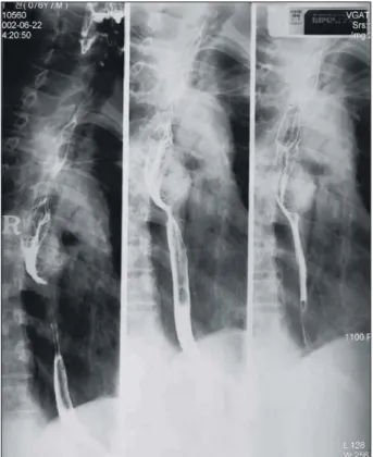
식도조영술에서 기도분기부 부근의 흉부 식도에 약 7 cm 정도 길이의 석회화된 점막하종양의 소견을 볼 수 있었다 (Fig. 1). 식도 내시경검사에서는 절치로부터 20∼27 cm 사 이에 점막이 불규칙하였으며, 점막하종양이 식도내강으로 돌출해 있었고 혈관이 충혈되어 있으며 루골 염색 결과 병 변의 경계가 명확하였다. 조직생검 결과 편평상피세포암이
었다. 흉부 전산화단층촬영에서 흉부 식도 중앙부에 석화 화를 동반한 종괴를 볼 수 있었으나 임파절 비대는 없었다.
이상의 진단으로 점막하종양과 공존하는 표재성 편평상피 암으로 진단하고 식도암절제수술을 계획하였다.
수술은 먼저 앙와위에서 상‧정중 개복술로 위장과 십이 지장을 유리하고 유문성형술을 하였다. 체위를 좌측 와위 로 변경하고 우측 제5 늑간으로 개흉하여 흉부 식도절제 술과 흉부 임파절적출술을 시행하고 흉강내에서 식도-위 문합술을 하였다.
절제된 식도의 길이는 17.5 cm였고 근위부 위장은 4.5 cm이었다. 병리 육안 소견에서 식도내강으로 돌출된 종괴 의 중앙부에 3×2 cm 크기의 점막이 불규칙한 과립성 병 변을 보였고 그 하부에 5.0×4.5×2.5 cm와 4.0×4.0×1.5 cm 크기의 두 개의 고립된 매우 단단한 종괴가 돌출되어
식도의 평활근종과 공존하는 표재성 식도암
박지권**․이철범*․전순호*․김영학**․정원상**․김 혁**
Superficial Esophageal Carcinoma Coexisting with Esophageal Leiomyoma
Ji Kwon Park, M.D.**, Chul Burm Lee, M.D.*, Soon-Ho Chon, M.D.*
Young Hak Kim, M.D.**, Won Sang Chung, M.D.**, Hyuck Kim, M.D.**
The coexistence of mesenchymal tumor and carcinoma in the esophagus is extremely rare. We report a case of squamous cell carcinoma located at the mucosal surface over leiomyoma of the esophagus. A 76-year-old man with complaints of 3 months onset of odynophagia was diagnosed preoperatively as squamous cell carcinoma over submucosal tumor with calcification. Esophagectomy and esophagogastrostomy were performed through the right thoracotomy and upper median laparotomy. The patient is doing well without evidence of recurrence in the 25 months after resection. We discuss the pathogenesis and possible relations between the two tumors.
(Korean J Thorac Cardiovasc Surg 2004;38:76-79) ꠏꠏꠏꠏꠏꠏꠏꠏꠏꠏꠏꠏꠏꠏꠏꠏꠏꠏꠏꠏꠏꠏꠏꠏꠏꠏꠏꠏꠏꠏꠏꠏꠏꠏꠏꠏꠏꠏꠏꠏꠏꠏꠏꠏꠏꠏꠏꠏꠏꠏꠏꠏꠏꠏꠏꠏꠏꠏꠏꠏꠏꠏꠏꠏꠏꠏꠏꠏꠏꠏꠏꠏꠏꠏꠏꠏꠏꠏꠏꠏꠏꠏꠏꠏꠏꠏꠏꠏꠏꠏꠏꠏ Key words: 1. Esophageal neoplasms
2. Leiomyoma
*한양대학교 구리병원 흉부외과학교실
Department of Thoracic and Cardiovascular Surgery, Hanyang University Guri Hospital
**한양대학교 의과대학 흉부외과학교실
Department of Thoracic and Cardiovascular Surgery, College of Medicine, Hanyang University 논문접수일:2004년 8월 6일, 심사통과일:2004년 10월 13일
책임저자 : 이철범 (471-701) 경기도 구리시 교문동 249-1, 한양대학교 구리병원 흉부외과 (Tel) 031-560-2301, (Fax) 031-568-9948, E-mail: cblee@hanyang.ac.kr
본 논문의 저작권 및 전자매체의 지적소유권은 대한흉부외과학회에 있다.
박지권 외 식도의 평활근종과 공존하는 식도암
- 77 - 있었다(Fig. 2).
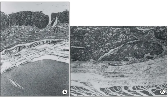
현미경 소견에서 병변 점막은 중등도로 분화된 편평상피 세포암이었으며 일부는 점막하층까지 침습되어 있었다(Fig.
3). 점막하종양은 광범위한 석회화를 포함한 전형적인 평활 근종이었다. 흉‧복부 주변 임파절의 침습은 없었다.
수술 후 회복에 특별한 문제는 없었으며 방사선 치료와 항암 치료는 하지 않았다. 수술 후 문합부 협착의 합병증 으로 2차례 풍선화장술을 시행하고 호전되었으며 25개월 된 현재 재발의 소견 없이 정상 생활을 하고 있다.
고 찰
동일 식도에 양성종양과 암종이 공존하는 경우는 흔하지 않으며, 더욱이 식도의 양성 종양을 덮는 상피세포에서 암종이 발생하는 경우는 매우 드물고 국내문헌에서는 2예 가 보고되어 있다[1,2]. Kuwano 등[3]은 식도암으로 식도 절제술을 시행한 587예 중 3예에서 편평상피세포암이 양 성종양의 표면에서 발생하였는데 2예는 평활근종이고 1 예는 지방종이라고 하였다. 편평상피세포암은 대부분 점
막층에만 국한된 조기 진단된 경우였다.
식도의 편평상피세포암의 병인에 대해서는 아직 논란 의 여지가 있다. 식도암 발생 위험 인자로 음주, 흡연과 음식물의 정체를 일으키는 조건들 즉 부식성 식도협착, 식도무이완증, 위‧식도 역류에 의한 협착, 식도계실 등이 알려져 있고 식도 상피세포가 기계적 또는 화학적으로 발 암성의 물질에 지속적으로 노출되기 때문이다.
두 종양의 공존에 대하여 두 종양의 관계는 명확하지 않다. 식도에 이미 존재하고 있던 양성 종양이 식도에서 협착과 자극으로 편평상피세포암의 발생을 유발할 수 있 다는 가정은 매우 설득력이 있다[3,4]. 또한 동기에 공존하 는 두 종양이 조직학적으로 다른 이웃의 종양을 서로 침 범하는 충돌종양(collision tumor)이라는 예도 있다[5].
식도의 점막하종양이 식도암의 발암현상과 관계가 있 다면 수술을 시행하지 않은 식도 양성종양 환자의 철저한 추적 검사가 필요하며 장기간 추적 검사로 조기 식도암을 발견한 보고도 있다[6,7].
Nagashima 등[8]은 식도 내시경검사와 조직생검으로 점 막하 종양과 공존하는 표재성 편평상피세포암을 진단한 후 내시경절제술로 두 종양을 제거하였고 단기 추적 검사 에서 재발이 없었다고 보고하였으나 수술은 식도암의 수 술에 준하는 광범위한 절제가 원칙이라고 생각한다.
Fig. 1. Esophagogram shows mucosal irregularity at the mid- esophagus, 7 cm in length. Submucosal calcified tumor is seen along the left side.
Fig. 2. (A) The mid-portion of the esophagus revealed an ill- defined granular lesion in the mucosa, measuring at 3.0×
2.0 cm, and two discrete submucosal tumors protruding into the lumen, measuring at 5.0×4.5×2.5 cm and 4.0×4.0×
1.5 cm in dimension. (B) The cut surface revealed a well demarcated submucosal mass with firm consistency and gray- ish white to pale brown appearance with severe calcification.
대흉외지 2005;38:76-79
- 78 - 참 고 문 헌
1. Son HS, Lee SH, Kim KT. Synchronous squamous cell car- cinoma and leiomyoma in the esophagus. Korean J Thorac Cardiovasc Surg 1995;28:942-5.
2. Kim JH, Lee YK. Surgical treatment of the leiomyoma of the esophagus. Korean J Thorac Cardiovasc Surg 1987;20:
156-60.
3. Kuwano H, Sadanaga N, Watanabe M, Yasuda M, Nozoe T, Sugimachi K. Esophageal squamous cell carcinoma occur- ring in the surface epithelium over a benign tumor. J Surg Oncol 1995;59:268-72.
4. Iizuka T, Kato H, Watanabe H, Itabishi M, Hirota T. Super- ficial carcinoma of the esophagus coexisting with esopha-
geal leiomyoma: A Case Report and Review of the Japanese Literature. Jpn J Clin Oncol 1984;14:115-22.
5. Sarbia M, Katoh E, Borchard F. Collision tumor of squa- mous cell carcinoma and leiomyoma in the esophagus.
Pathol Res Pract 1993;189:360-2.
6. Yoshikane H, Tsukamoto Y, Niwa Y, et al. The coexistence of esophageal submucosal tumor and carcinoma. Endoscopy 1995;27:119-23.
7. 八板朗, 東儀公哲, 金森弘明, 中村輝久. 5年間 經過觀察した 平滑筋腫 合倂の 早期食道癌の 1例. 癌の 臨床 1984;30:510-4.
8. Nagashima R, Takeda H, Motoyama T, Tsukamoto O, Taka- hashi T. Coexistence of superficial esophageal carcinoma and leiomyoma: Case report of an endoscopic resection.
Endoscopy 1997;29:683-4.
Fig. 3. (A) The underlying submucosal mass shows typical leiomyoma with well-demarcated margins and overlying mucosa shows in situ squamous cell carcinoma (×100). (B) The mucosal lesion shows in situ squamous cell carcinoma with invasion into the submucosa.
A B
박지권 외 식도의 평활근종과 공존하는 식도암
- 79 -
=국문 초록=
식도에서 간엽세포 종양과 상피성세포 종양의 공존은 매우 드물다. 저자들은 식도 평활근종의 점막 표면에 위치한 편평상피세포암 수술 치험 1예를 보고한다. 내원 3개월 전부터 음식 섭취시 연하통을 주소로 내원한 76세 남자 환자에서 수술 전 검사로 석회화를 동반한 식도 점막하종양을 의심하였고, 종양을 덮고 있는 불규칙한 점막의 식도내시경 조직검사로 편평상피세포암의 공존을 수술 전에 진단 하였다. 우측 개흉과 개복으로 흉부 식도절제술 후 식도-위 문합을 시행하였다. 수술 25개월이 경과 한 현재까지 재발의 소견 없이 정상 생활을 하고 있다. 두 종양 사이의 관계의 가능성과 빈도에 대해 문헌 고찰을 한다.
중심 단어:1. 식도 종양 2. 평활근종