Introduction
Since its introduction in the 1990s, immediate im- plant placement in aesthetic areas has been widely used as a predictable treatment option to replace tooth loss.1,2 Immediate implant placement have the advantage of preserving tissue architecture, reducing treatment time, and improving patients’ convenience through provisionalization.3,4 Also, immediate im- plant placement has been reported to have a high success rates when clinical guidelines are followed.5,6
The implant position is one of the most impor- tant factors to maintain the aesthetic and function.7 Implant should be based on prosthetic restoration plans, but are often limited by the morphology of the residual alveolar ridge. When there is a lack of residual bone in extraction socket, various factors should be considered to ensure the primary stability of implant. Factors such as root length, sagittal root position (SRP), and the morphology of the osseous housing are important in determining the feasibil- ity of immediate implant placement, and should be
*Correspondence to: Hyun-Jun Kong
Clinical Fellow, Department of Prosthodontics, College of Dentistry, Wonkwang University, 895 Muwang-ro, Iksan, 54538, Republic of Korea
Tel: +82-63-859-2929, Fax: +82-63-857-4002, E-mail: [email protected] Received: April 7, 2020/Last Revision: April 21, 2020/Accepted: April 21, 2020
a study on sagittal root position of maxillary anterior teeth in Korean
Hyun-Jun Kong*
Department of Prosthodontics, College of Dentistry, Wonkwang University, Iksan, Republic of Korea
Purpose: The purpose of this study was to analyze the sagittal root position of maxillary anterior teeth and report the frequency of each classification in Korean for immediate implant placement. Materials and Methods: A retrospective review of cone-beam computed tomography (cone-beam CT) images was conducted on 120 patients (60 male and 60 female) who fulfilled the inclusion criteria. After reorientation of the axis, cone-beam CT images were evaluated and the relationship of the sagittal root position (SRP) of the maxillary anterior teeth to its associated osseous housing was recorded. Class I, II, and III were classified respectively when the root was positioned on the labial, central, and palatal aspect of the alveolar bone. Class IV was the position that at least two thirds of the root is engaging both the labial and palatal cortical plates. Then, the angulation of the root axis and the alveolar bone axis was measured. Descriptive statistics and Kruskal-Wallis test were used to compare the angulation according to the root position and SRP class. Results: The frequency distribution of sagittal root position of maxillary anterior teeth indicated that 81.1%, 10.3%, 1.9%, and 6.7% were classified as Class I, II, III, and IV, respectively. The sagittal angulation at approximately 77.5% of central incisor, lateral incisor, and canine was < 20 degrees, but the angle at more than 42.7% of canine was ≥ 20 degrees. Within the class, the angulation was statistically significantly greater in Class I (16.19) compared to Class II (8.72) and Class III (9.93), and smaller in Class IV (3.79). Conclusion: Within the limitation of this study, a majority of the maxillary anterior roots were positioned close to the buccal cortical plate. However, some roots have very thin alveolar bone and sagittal angulation larger than 30 degrees.
Therefore, cone-beam CT analyses of the sagittal root position and the sagittal angulation are recommended for the selection of the appropriate dental implant treatment approach. (J Dent Rehabil Appl Sci 2020;36(2):88-94)
Key words: cone-beam CT; maxillary anterior teeth; sagittal root position; dental implant
Copyright© 2020 The Korean Academy of Stomatognathic Function and Occlusion.
It is identical to Creative Commons Non-Commercial License.
cc
evaluated with cone-beam computerized tomography (cone-beam CT).
The root position is critical for implant treatment planning in the maxillary anterior region, especially in immediate implant placement. Kan et al.8 presented a classification of SRP to aid implant treatment plan- ning, in which the relationship between the root position and its osseous housing is categorized as Class I, II, III, and IV. In the study, 81.1%, 6.5%, 0.7%, and 11.7% of the 600 samples were classified as Class I, II, III, and IV, respectively. However, since Caucasians and Koreans have differences in size and shape of dental arch and soft tissue profiles, it could be assumed that there is a difference in root shape and position.9-12
In a study on maxillary central incisor and lateral incisor, Jung et al.13 reported that 92.2% of central incisor and 94.0% of lateral incisor are positioned buccally.However, very few studies evaluated SRP class IV, in which the root engaged both the labial and palatal cortical plates. SRP class IV was reported to be about 10% which is considered to be contrain- dication for immediate implant placement.8,14 Also, angulation between the long axis of the tooth and the long axis of the corresponding alveolar bone was crucial importance in the selection of the appropri- ate implant approach.15
Therefore, the purpose of this study was to evalu- ate and compare sagittal root position and sagittal angulation of maxillary anterior teeth for immediate implant placement. The first null hypothesis was that there is no difference in sagittal angulation between central incisor, lateral incisor, and canine. The second null hypothesis was that there is no difference in sag- ittal angulation between SRP classes.
Materials and Methods
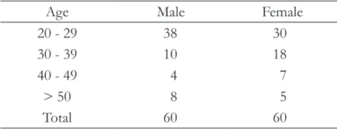
This study was approved by the local institutional review board (protocol no. IRB202003-01). A retro- spective review of cone-beam CT images was con- ducted on 120 patients (60 male and 60 female) who met the criteria of this study. The gender and age distribution of patients is in Table 1. The following inclusion criteria were applied as reported by previ-
ous studies8,13: at least 18 years of age at the time of the cone-beam CT scan; all maxillary and mandibular teeth were present; Angle class I occlusion; no rota- tion or malposition of anterior teeth; no radiographic evidence of infection, root resorption, or trauma to maxillary anterior dentition; and no history of surgi- cal treatment in the maxillary anterior dentition.
Cone-beam CT images were analyzed using soft- ware (Invivo 5.1, Anatomage, San Jose, USA). The orientation of axis was conducted as reported by previous study.16 First, the vertical line was set to the line passing through Nasion and Anterior Nasal Spine (ANS) in the sagittal view. Next, the horizontal line was set to the line passing through the Orbitale on both sides in coronal view. Finally, the vertical line was set to the line passing through the ANS and Pos- terior Nasal Spine in axial view. It was reported that there were no significant differences between the measurements on the right and left sides or between sexes.17,18 Therefore, in this study, only one side of each subject’s cone-beam CT images was selected for evaluation. The Image analysis was conducted in left or right view mode for central incisor and lateral incisor, and in right 3/4 or left 3/4 view mode for canine considering arch form and canine position.
Images were clipped by 5 mm and teeth mode was selected for confirming root position.
Each tooth images was classified according to the classification reported by Kan et al.8 (Fig. 1).
Class I: The root is positioned against the labial cortical plate
Class II: The root is centered in the middle of the alveolar housing without engaging either the labial or the palatal cortical plate at the apical third of the root
Table 1. Patient age and gender information
Age Male Female
20 - 29 38 30
30 - 39 10 18
40 - 49 4 7
> 50 8 5
Total 60 60
Class III: The root is positioned against the palatal cortical plate
Class IV: at least two thirds of the root is engaging both the labial and palatal cortical plates The angulation between the long axis of the tooth and the long axis of the corresponding alveolar bone was determined in accordance with the procedure described by Lau et al. (Fig. 2).19 Line A shows the al- veolar bone axis which bisects the angle of the buccal and palatal line. Line B shows the tooth axis which was defined as the line through the lowest point of the crown to the highest point on the apex.
The normality of the data was determined using the Kolmogorov-Smirnov test. Descriptive statistics were presented, including means values, frequencies, and percentages. The Kruskal-Wallis test was used to compare the angulation between teeth and SRP classes because the samples did not follow normal distributions. After that, Mann-Whitney U test with Bonferroni correction was used to verify differences between groups. Statistical analysis was performed with software (SPSS 23.0, IBM Inc., Armonk, USA) and statistical significance was set at P < 0.05.
Results
In present study, the sagittal root position in rela- tionship to the osseous housing was examined. The frequency distribution was categorized according to tooth position and SRP class (Table 2). A majority of the maxillary anterior roots were positioned more buccally within the alveolar bone. Only seven ante- rior roots were positioned more palatally.
Fig. 1. Classification of sagittal root position. (A) Schematic diagram, (B) Cone-beam CT image.
Fig. 2. The angulation between the alveolar bone axis and the tooth axis. Line A: long axis of the alveolar bone, Line B: long axis of the tooth.
A
B
Class I Class II Class III Class IV
Class I Class II Class III Class IV
The sagittal angulation of the maxillary anterior teeth within the alveolar bone was determined (Table 3). The angulation of the canine showed the larg- est angulation, followed by lateral incisor, and finally central incisor showed the smallest angulation (P <
0.05, Table 4). Within the class, Class I showed the largest angulation and Class III showed the smallest one.
Discussion
The present study evaluated the sagittal root posi- tion and sagittal angulation of maxillary anterior teeth in Korean using cone-beam CT. According to the results of the study, the first null hypothesis that there is no difference in sagittal angulation be-
tween central incisor, lateral incisor, and canine was rejected. The second null hypothesis that there is no difference in sagittal angulation between SRP classes was partially rejected.
In this study, 81.1%, 10.3%, 1.9%, and 6.7% of the 360 samples were classified as Class I, II, III, and IV, respectively. SRP Class I, in which the root is in contact with the labial cortical bone, has a consider- able amount of bone in palatal aspect generally. This provides the primary stability for immediate implant placement. Also, implant-socket gap on labial side allows an aesthetic tissue profile by using bone graft materials. Thus, SRP Class I was categorized as a po- sition that is favorable for immediate implant place- ment. It means that, if guidelines are followed, the majority of maxillary anterior region could be indi- Table 2. Frequency distribution of the sagittal root position classification
Sagittal root position Percentage (no.)
Central incisor Lateral incisor Canine Overall
Class I 74.2 (89) 81.7 (98) 87.5 (105) 81.1 (292)
Class II 18.3 (22) 5.8 (7) 6.7 (8) 10.3 (37)
Class III 1.7 (2) 3.3 (3) 0.8 (1) 1.9 (7)
Class IV 5.8 (7) 9.2 (11) 5.0 (6) 6.7 (24)
Total 100 (120) 100 (120) 100 (120) 100 (360)
Table 4. Sagittal angulation (in degrees) of the maxillary anterior teeth according to SRP class
Sagittal root position Central incisor Lateral incisor Canine Overall
Class I 11.78 16.25 19.88 16.19A
Class II 6.30 8.67 15.40 8.72B
Class III 3.90 3.98 2.80 3.79C
Class IV 5.93 12.25 10.33 9.93B
Total 10.31a 15.03b 18.96c 14.77
The different lower case letters in rows and the different capital letters in columns indicate significant difference (P < 0.05).
Table 3. Frequency distribution of the sagittal angulation (in degrees)
Angulation Percentage (no.)
Central incisor Lateral incisor Canine Overall
< 10 46.7 (56) 20 (24) 6.7 (8) 24.4 (88)
≥ 10, < 20 50.0 (60) 57.50 (69) 51.7 (62) 53.1 (191)
≥ 20, < 30 3.3 (4) 20.83 (25) 35 (42) 19.7 (71)
≥ 30 0 (0) 1.67 (2) 6.7 (8) 2.8 (10)
Total 100 (120) 100 (120) 100 (120) 100 (360)
cation for immediate implant placement. Especially, Class I was the most common in canine and this re- sult is consistent with the result of previous study.14 However, clinicians should evaluate the implant insertion angles, because Class I showed the greatest sagittal angulation between the tooth axis and the al- veolar axis.
Overall, SRP Class II was 10.3% and generally, the volume of alveolar bone on both the labial and pala- tal aspects is less than what is encountered in Class I or III.8 This amount of bone could be inadequate to ensure implant primary stability. Therefore, when considering immediate implant placement in Class II, the available bone beyond the apex of the extraction socket should be evaluated. Next, similar to previous studies, Class III was rarely found. The frequency of Class III has been reported to vary from 0.2% to 1.8%.19,20
Class IV was 6.7% and especially 9.2% for lateral incisors in present study. In this case, the root oc- cupies the majority of the alveolar volume, and after tooth extraction, there is a limited alveolar bone for primary stability. Thus, bone grafting procedures are often necessary prior to implant placement. For this reason, Class IV is considered to be a contraindica- tion for immediate implant placement. Frequency of Class IV in this study was less than that of Cauca- sians, and more than that of Thai people.8,14 This ap- pears to be due to differences in race and the average age of the subjects studied.
Sagittal angulation between the long axis of the tooth and the alveolar bone was below 10 degrees at 24.4% of maxillary anterior teeth. It is relatively sim- ple to insert implant in this case. The implant can be placed in the same direction along the root, however, slightly palatal to ensure that the labial bone wall is sufficiently thick and to ensure primary stability.21 In present study, 53.1% of the samples featured a sagittal angulation between about 10 and 20 degrees.
More technical placement is needed considering sagittal angulation and may require narrow-diameter implant, angulated abutment, or cement-type pros- thesis.
However, about 20% of anterior maxillary teeth showed a sagittal angulation that was greater than 20
degrees and 8% of canine was greater than 30 de- grees. In this situation, the tooth root is close to the buccal cortical bone, and difficulties are encountered in setting the implant placement angles. Therefore, socket preservation procedure with late implant placement or narrow-diameter implant with palatal position should be considered for primary stability.
It is also recommended to use customized abutment for the aesthetic prosthesis.
The limitation of this study is that cone-beam CT images of normal teeth and alveolar bone is different from the situation weakened by periodontal disease or other reasons. However, the present study could be used as data on the position and angulation of the root that should be considered when planning imme- diate implant placement for Koreans.
Conclusion
Within the limitation of this study, a majority of the maxillary anterior roots were positioned close to the buccal cortical plate and showed a sagittal angulation that was smaller than 20 degrees. How- ever, some roots have very thin alveolar bone and sagittal angulation larger than 30 degrees, which is inappropriate for immediate implant placement.
Consequently, cone-beam CT analysis before tooth extraction is recommended so that the clinicians can choose the appropriate implant treatment approach to achieve aesthetic and functional prosthesis.
ORCID
Hyun-Jun Kong https://orcid.org/0000-0001-9331-3572
References
1. Buser D, Martin W, Belser UC. Optimizing esthet- ics for implant restorations in the anterior maxilla:
anatomic and surgical considerations. Int J Oral Maxillofac Implants 2004;19 Suppl:43-61.
2. Koh RU, Rudek I, Wang HL. Immediate implant placement: positives and negatives. Implant Dent 2010;19:98-108.
3. Chen ST, Darby IB, Reynolds EC, Clement JG. Im-
mediate implant placement postextraction without flap elevation. J Periodontol 2009;80:163-72.
4. Kois JC, Kan JY. Predictable peri-implant gingival aesthetics: surgical and prosthodontic rationales.
Pract Proced Aesthet Dent 2001;13:691-8.
5. Rosenquist B, Grenthe B. Immediate placement of implants into extraction sockets: implant survival.
Int J Oral Maxillofac Implants 1996;11:205-9.
6. Grunder U, Polizzi G, Goené R, Hatano N, Henry P, Jackson WJ, Kawamura K, Köhler S, Renouard F, Rosenberg R, Triplett G, Werbitt M, Lithner B.
A 3-year prospective multicenter follow-up report on the immediate and delayed-immediate place- ment of implants. Int J Oral Maxillofac Implants 1999;14:210-6.
7. Chan HL, Garaicoa-Pazmino C, Suarez F, Monje A, Benavides E, Oh TJ, Wang HL. Incidence of im- plant buccal plate fenestration in the esthetic zone:
a cone beam computed tomography study. Int J Oral Maxillofac Implants 2014;29:171-7.
8. Kan JY, Roe P, Rungcharassaeng K, Patel RD, Waki T, Lozada JL, Zimmerman G. Classification of sag- ittal root position in relation to the anterior maxil- lary osseous housing for immediate implant place- ment: a cone beam computed tomography study.
Int J Oral Maxillofac Implants 2011;26:873-6.
9. Lee KY, Lee DJ. A comparative study of teeth and dental arch of Korean and Caucasian. Oral Biol Res 1993;71:1-15.
10. Song WC, Yun KH, Koh KS. Facial flatness of Ko- rean: using facial depth. Korean J Anat 2003;36:499- 506.
11. Kook YA, Nojima K, Moon HB, McLaughlin RP, Sinclair PM. Comparison of arch forms between Korean and north American white populations. Am J Orthod Dentofacial Orthop 2004;126:680-6.
12. Hwang HS, Kim WS, McNamara Jr JA. Ethnic dif- ferences in the soft tissue profile of Korean and European-American adults with normal occlusions and well-balanced faces. Angle Orthod 2002;72:72- 80.
13. Jung YH, Cho BH, Hwang JJ. Analysis of the root position of the maxillary incisors in the alveolar bone using cone-beam computed tomography. Im- aging Sci Dent 2017;47:181-7.
14. Petaibunlue S, Serichetaphongse P, Pimkhaokham A. Influence of the anterior arch shape and root position on root angulation in the maxillary esthetic area. Imaging Sci Dent 2019;49:123-30.
15. Kim JH, Lee JG, Han DH, Kim HJ. Morphomet- ric analysis of the anterior region of the maxil- lary bone for immediate implant placement using micro-CT. Clin Anat 2011;24:462-8.
16. Bayome M, Park JH, Kook YA. New three-dimen- sional cephalometric analyses among adults with a skeletal Class I pattern and normal occlusion. Ko- rean J Orthod 2013;43:62-73.
17. Braut V, Bornstein MM, Belser U, Buser D. Thick- ness of the anterior maxillary facial bone wall - a retrospective radiographic study using cone beam computed tomography. Int J Periodontics Restor- ative Dent 2011;31:125-31.
18. Zekry A, Wang R, Chau AC, Lang NP. Facial alveo- lar bone wall width - a cone-beam computed to- mography study in Asians. Clin Oral Implants Res 2014;25:194-206.
19. Lau SL, Chow J, Li W, Chow LK. Classification of maxillary central incisors-implications for immedi- ate implant in the esthetic zone. J Oral Maxillofac Surg 2011;69:142-53.
20. Chung SH, Park YS, Chung SH, Shon WJ. Deter- mination of implant position for immediate im- plant placement in maxillary central incisors using palatal soft tissue landmarks. Int J Oral Maxillofac Implants 2014;29:627-33.
21. Wang HM, Shen JW, Yu MF, Chen XY, Jiang QH, He FM. Analysis of facial bone wall dimensions and sagittal root position in the maxillary esthetic zone: a retrospective study using cone beam com- puted tomography. Int J Oral Maxillofac Implants 2014;29:1123-9.
*교신저자: 공현준
(54538)전라북도 익산시 무왕로 895 원광대학교 치과대학병원 치과보철과 Tel: 063-859-2929|Fax: 063-857-4002|E-mail: [email protected] 접수일: 2020년 4월 7일|수정일: 2020년 4월 21일|채택일: 2020년 4월 21일
한국인에서 상악 전치의 시상 치근 위치에 대한 연구
공현준* 전임의
원광대학교 치과대학 치과보철학교실
목적: 본 연구는 한국인에서 상악 전치의 치근 위치를 시상면에서 분석하고, 분류에 따른 빈도를 보고함으로써 즉시 식 립 임플란트를 위한 방사선학적 자료를 수집하기 위함이다.
연구 재료 및 방법: 콘빔형 전산화단층영상(cone-beam CT)을 촬영한 환자 중 연구 기준에 적합한 120명(남성 60명, 여성 60명)을 대상으로 후향적 분석을 시행하였다. 축의 방향설정을 시행한 후에, 상악 전치부 치아와 치조골 사이의 관계에 대한 시상 치근 위치를 분석하였다. 치근이 치조골의 협측, 중앙, 구개측으로 위치한 경우 각각 Class I, II, III로 분류하였 으며, 치근이 협측과 구개측 모두에서 피질골판에 2/3 이상 닿아있는 경우에는 Class IV로 하였다. 다음으로, 치아의 장 축과 치조골의 장축 사이의 각도를 측정하였다. 기술적 분석 및 Kruskal-Wallis 분석을 시행하였으며, 치아의 위치 및 분 류에 따른 시상각을 비교하였다.
결과: 상악 전치부의 시상 치근 위치에 대한 빈도분석 결과, Class I은 81.1% , Class II는 10.3%, Class III는 1.9%, 그리 고 Class IV는 6.7%로 나타났다. 상악 전치부의 77.5%에서 시상각이 20도 이하로 나타났다. 그러나 견치의 경우, 42.7%
에서 20도 이상의 시상각을 보였다. 분류에 따라서는 Class I (16.19)에서 Class II (8.72) 및 Class III (9.93)에 비해 통계 학적으로 유의하게 높은 시상각을 보였으며, Class IV (3.79)에서 낮았다.
결론: 본 제한된 연구의 결과를 근거로, 상악 전치의 치근은 일반적으로 협측 치조골에 가깝게 위치하고 있으나, 일부 치
아는 매우 얇은 치조골을 가지고 있으며, 30도 이상의 큰 시상각을 보였다. 따라서 적절한 치과 임플란트 치료 계획 수립 을 위해 시상 치근 위치 및 시상각에 대한 cone-beam CT 분석이 필요할 것이다.
(구강회복응용과학지 2020;36(2):88-94)
주요어: 콘빔형 전산화단층영상; 상악 전치; 시상 치근 위치; 치과 임플란트