• 검색 결과가 없습니다.

Assessment of antero-posterior skeletal relationships in adult Korean patients in the natural head position and centric relation

N/A
N/A
Protected

Academic year: 2022

Share "Assessment of antero-posterior skeletal relationships in adult Korean patients in the natural head position and centric relation"

Copied!
11
0
0

로드 중.... (전체 텍스트 보기)

전체 글

(1)

aAssociate Professor, Department of Orthodontics, Kangnam Sa- cred Heart Hospital, Hallym University.

bAssistant Professor, Department of Preventive Dentistry, School of Dentistry, Seoul National University.

cProfessor, eAssistant Professor, Department of Oral and Maxillofacial Surgery, Kangnam Sacred Heart Hospital, Hallym University.

dChairman, Department of Orthodontics, Chong-A Dental Hospital.

fAssistant Professor, Department of Orthodontics, Hangang Sacred Heart Hospital, Hallym University.

Corresponding author:

Mi-Ja Kim.

Department of Orthodontics, Hangang Sacred Heart Hospital, Hallym University, 94-200 Yeongdeungpo-dong, Yeongdeungpo- gu, Seoul 150-719, Korea.

+82 2 2639 5580; e-mail, [email protected].

Received February 10, 2010; Last Revision June 11, 2010;

Accepted June 14, 2010.

DOI:10.4041/kjod.2010.40.6.421 / pISSN 1225-5610

Assessment of antero-posterior skeletal relationships in adult Korean patients in the natural head position and centric relation

Jang-Hoon Ahn, DDS, MSD, PhD,

a

Kwang-Hak Bae, DDS, MSD, PhD,

b

Young-Ju Park, DDS, MSD, PhD,

c

Ryoon-Ki Hong, DDS, PhD,

d

Joeng-Hun Nam, DDS, MSD, PhD

e

, Mi-Ja Kim, DDS, MSD, PhD

f

Objective: This study aimed to verify the intra-individual reproducibility of the natural head position (NHP) in adult Korean patients in the centric relation (CR) position and to prove the inter-individual variability of the Frankfurt horizontal (FH) plane and sella-nasion (SN) line compared to the true horizontal line (THL).

In addition, the study aimed to investigate the correlations between linear measurements from A-point and B-point to the nasion true vertical line (NTVL) and angular measurements from A-point and B-point to the SN line. Methods: Two lateral cephalograms were taken of 116 subjects (23 males, 93 females) with CR wax bites in a NHP at a one-week interval. Results: Method errors of three variables and intraclass correla- tion coefficients of six parameters proved the intra-individual reproducibility of NHP (p < 0.001). The angle of the FH to the THL was not significantly different from 0

o

(p > 0.05), but it was clinically variable (SD 3.89

o

) on the inter-individual level. Conversely, the angle of the SN line to the THL was significantly differ- ent from 7

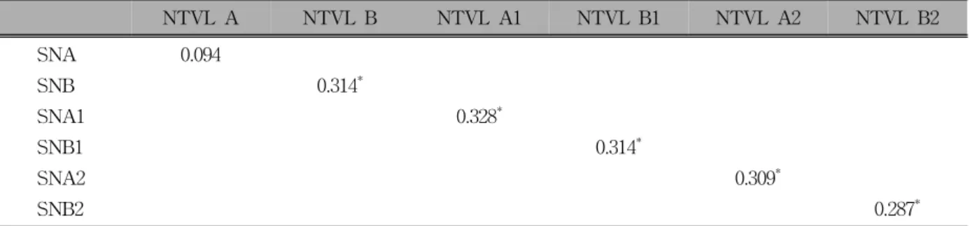
o

(p < 0.05). Very low correlation was found between the linear measurements and angular measurements of A-point and B-point (p < 0.01). Conclusions: The NTVL could be a useful reference line for assessing the antero-posterior position of the maxilla and mandibleof Korean adult patients in NHP and CR. (Korean J Orthod 2010;40(6):421-431)

Key words: Natural head position, Centric relation, Frankfurt horizontal plane, Sella-nasion line, Nasion true vertical line

INTRODUCTION

Many cephalometric measurements have been used

to assess the antero-posterior position of the skeleton

in patients since Broadbent

1

and Hofrath

2

described

roentgenographic cephalometry. Downs

3

classified skel-

etal patterns and skeleton-to-denture relationships using

the Frankfurt horizontal (FH) plane. Steiner

4

utilized

sella-nasion (SN)-A and SN-B angles to assess jaw re-

lationships in patients. Riedel

5

suggested the angle of

SN to point-A for measuring the relative ante-

ro-posterior position of the maxilla. Tweed

6,7

measured

the FH plane to the mandibular plane angle and the

mandibular plane to the lower incisor angle focusing

on mandibular position and mandibular incisor posi-

tion. Ricketts

8

used A-NPog for maxillary convexity

(2)

Fig 1. Registration of the centric relation wax bite. A, Guiding the mandible using the tripoding method; B, centric relation wax bite (anterior and posterior portion).

(in millimeters) as an indicator of horizontal jaw rela- tionships. Jenkins

9

and Jacobson

10

described Wits’ ap- praisal for measuring antero-posterior jaw disharmony.

McNamara

11

suggested the McNamara line, which passes through a nasion point perpendicular to the FH plane, as a critical plane for measuring the distance from A-point or the pogonion point.

Two common intracranial reference planes exist for orthodontic diagnosis. The representative line is the SN line, which is reliable and biologically meaningful, but it has been shown to have large inter-individual variability.

12,13

The SN reference line is thought to be less valid due to this high inter-individual variability.

The other reference line is the FH plane, which was originally introduced at an anthropological conference in 1884. The FH plane was defined as extending from the orbitale to both porion points. The orbitale is de- fined as the lowest point of the infraorbital margin and the porion as the outer upper margin of the porus acousticus externus. The FH plane is widely used as a reference, and it may produce the most acceptable esti- mation of a true horizontal line. However, this refer- ence plane also has inter-individual variability due to the positions of orbitale and porion.

The desire for facial aesthetics has been increasing.

Thus, havinga reproducible and reliable reference posi- tion and plane is important for the diagnosis and treat- ment of orthodontic patients and two-jaw surgery pa- tients. Orthodontists and oral surgeons do not have a common reference line for assessing the position of the maxilla and mandible with regard to facial aesthetics.

As early as the 1860s, craniologists realized the natural

head position (NHP) in human beings. Broca

14

defined this head position as “when man is standing and his visual axis is horizontal, he is in the natural position”.

The NHP was introduced into orthodontics in the late 1950s

15

and has been advocated as a craniofacial refer- ence system because of its good intra-individual re- producibility

15-17

to a true vertical plumb line. Another characteristic of the NHP is the representation of the true-to-life appearance and ease of registration. There- fore, setting up a reliable reference line with the NHP is necessary. Orthodontists also need to have a reliable and repeatable position of the mandible to diagnose maxillo-mandibular relationships and occlusion in pa- tients. Centric relation (CR) is known as the single most repeatable position, but few reports have ad- dressed the reproducibility of NHP in adult Korean pa- tients in CR or the clinical application of the NHP and CR position.

The purpose of this study was to verify the intra-in- dividual reproducibility of the NHP in CR position and to prove the inter-individual differences in the FH plane and SN line compared to the true horizontal line (THL) in adult Korean patients. In addition, the study aimed to investigate the correlations between linear measurements from A-point and B-point to the nasion true vertical line (NTVL) and angular measurements from A-point and B-point to the SN line on the basis of the first two studies.

MATERIAL AND METHODS

Ethical approval for the present investigation was

(3)

Fig 2. Registration in the natural head position and lat- eral cephalogram. A, Subjects looked into their eyes in the mirror (orthoposition); B, standing position with

arms and chin relaxed. Fig 3. Reference points used in this study.

obtained from the Kangnam Sacred Heart Hospital eth- ics committee, Hallym University Medical Center, and written informed consent was obtained for all subjects.

Samples

A total of 116 subjects (23 males, age range 19 - 41 years and mean age 27.9 years; 93 females, age range 19 - 47 years and mean age 23.6 years) were in- vestigated. The patients visited the Department of Orthodontics at the university hospital to receive treat- ment for malocclusions and skeletal discrepancies. No orthodontic treatment, orthognathic surgery, or pros- thetic treatment more than a 3-unit bridge was pro- vided in advance. Subjects with more than two missing teeth were excluded from the study.

Centric relation bite registration

The CR was recorded according to the Roth techni- que (power centric registration method) described by Choi

18

before taking lateral cephalograms (Fig 1).

NHP registration and lateral cephalograms

Solow and Tallgren’s method

19

was used to achieve the NHP (Fig 2). The subjects were asked to stand in front of a mirror with their feet a shoulder width apart and arms relaxed, wearing eyeglasses attached to a flu- id leveler. The subjects were placed 120 cm from a 30

× 40 cm mirror and asked to stare at their own eyes after exercising their head up and down. All of the

subjects were asked to bite their CR wax records and to relax their lips and arms during X-ray.

An auto IIIN CM (Asahi Corp., Japan) cephalo- metric X-ray machine was used to take the lateral cephalograms. X-ray (69 KV, 12 mA) was irradiated for 2 seconds. Subjects were positioned in the cephalo- stat without ear rod insertion. A metal chain was drooped in front of the subject to draw the NTVL on the lateral cephalometric tracing. Two lateral cephalo- grams were taken for each subject at a one-week interval.

Tracing technique and statistical analysis

V-ceph orthodontic diagnosis program version 6.0

(Osstem Corp., Korea) was used to digitize the lateral

cephalograms. The initial (T1) and second (T2) lateral

cephalograms were taken with the same NHP and CR,

and both were digitized and measured. Fourteen land-

marks and the soft tissue profile line were digitized

with the software. The NTVL was made by moving

the metal chain line parallel and drawing a line

through the nasion point. The data were transferred to

SPSS for windows version 11.5 (SPSS Inc., Chicago,

IL, USA) for analysis using intraclass correlation co-

efficients, one sample t-test, and Pearson correlation

coefficients. The mean, standard deviation, minimum,

and maximum was calculated for each variable. The

(4)

Fig 4. Reference planes used in this study. 1, Nasion true vertical line (NTVL) passing through the nasion point perpendicular to the floor; 2, E-line, tip of nose- soft tissue pogonion; 3, cervical vertebrae tangent- plane the posterior tangent to the odontoid process through cv4ip; 4, odontoid process tangent plane, the posterior tangent to the odontoid process through cv2ip; 5, palatal plane, ANS-PNS; 6, true horizontal line passing through nasion point perpendicular to the NTVL; 7, sella-nasion plane; 8, Frankfurt horizontal plane, porion-orbitale.

Fig 5. Angular measurements. A, 1, Nasion true vertical line (NTVL) to palatal plane; 2, NTVL to E-Line; 3, NTVL to cervical vertebrae tangent; 4, NTVL to sella-nasion (SN); 5, true horizontal line (THL) to odontoid process tangent (OPT); 6, SN to OPT. B, 1, THL to Frankfurt horizontal plane; 2, THL to SN.

reference points and reference planes used in this study are illustrated in Figs 3 and 4. Six angular parameters were calculated (Fig 5A). The THL to the FH plane and SN line angles were measured (Fig 5B). The above angle compared to the THL was positive and the below angle was negative. The data for this analy- sis were the mean T1 and T2 measurements. In addi- tion to linear measurements from A-point and B-point to the NTVL (Fig 6A), SN-A and SN-B angles (Fig 6B) were measured and compared by Pearson correla- tion coefficients using T1, T2, and mean data.

Method errors of digitizing and positioning

A method error study was performed to determine the errors in digitizing landmarks and measurements.

Method errors in digitizing were calculated in 15 ran- domly selected subjects. Each T1 lateral cephalogram was retraced by the same observer 1 week after the in- itial tracing. Double determinations of three variables (SN/NTVL, SN/OPT, and OPT/THL) were calculated using Dahlberg’s formula (Eq. 1).

20

Method errors in positioning were evaluated in 116

subjects using Dahlberg’s formula. Three variables

(SN/NTVL, SN/OPT, and OPT/THL) were also cal-

culated. The T1 and T2 data were used.

(5)

Fig 6. Linear measurements. A, 1, Nasion true vertical line (NTVL to A-point); 2, NTVL to B-point. B, 1, Sella nasion A-point (SNA) angle; 2, sella nasion B-point (SNB) angle.

Variables This study (n = 116)

Huggare

33

(n = 33)

Solow &

Tallgren

19

(n = 120)

SN/NTVL 1.06 1.60 1.43

SN/OPT 1.28 1.35 1.43

OPT/THL 0.77 2.10 1.95

Date are in degrees. NTVL, Nasion true vertical line;

OPT, odontoid process tangent; SN, sella-nasion; THL, true horizontal line.

Table 2. Positioning method errors (n = 116) Table 1. Digitizing method errors (n = 15)

Variables Degrees

SN/NTVL 0.22

SN/OPT 0.52

OPT/THL 0.46

NTVL, Nasion true vertical line; OPT, odontoid process tangent; SN, sella-nasion; THL, true horizontal line.

Table 3. Reproducibility of natural head position (NHP) by intraclass correlation coefficients (n = 116)

Variables Average of

raters (α) p-value

NTVL/E-line 0.9925 < 0.001

NTVL/palatal plane 0.6102 < 0.001

NTVL/CVT 0.9883 < 0.001

NTVL/SN 0.9703 < 0.001

SN/OPT 0.9815 < 0.001

NTVL/OPT 0.9917 < 0.001



(Eq. 1)

Where d is the difference between two measurements of a pair and n is the number of paired measurements.

RESULTS

The method errors in digitizing and positioning are summarized in Tables 1 and 2, respectively.

Six parameters were assessed to prove the reliability and reproducibility of the NHP according to intraclass correlation coefficients (Table 3, p < 0.001). The cor- relation coefficient for NTVL and the palatal plane was relatively low.

The differences between FH to THL and SN to THL compared to norms are summarized in Table 4. No significant differences were found in the angles of FH to THL according to one sample t-test (p > 0.05).

The prerequisite for the one sample t-test was a normal

FH to THL angle of 0°. In contrast, we identified sig-

(6)

Table 4. Differences in Frankfurt horizontal line (FHL) to true horizontal line (THL) and SN to true horizontal line compared to norms (n = 116)

Variables N Mean SD Min Max p-value

THL/FH 116 −0.16 3.89 −12.43 7.65 < 0.05

THL/SN 116 9.73 4.60 −2.11 17.50 < 0.05

Norms: THL/FH= 0

o

, THL/SN=7

o

. SD, Standard deviation; Min, minimum; Max, maximum.

Table 5. Correlations between linear measurements of A-point to nasion true vertical line (NTVL) and sella-naison (SN)-A angles, and linear measurements of B-point to NTVL and SN-B angles using Pearson correlation coefficients (n = 116)

NTVL A NTVL B NTVL A1 NTVL B1 NTVL A2 NTVL B2

SNA 0.094

SNB 0.314

*

SNA1 0.328

*

SNB1 0.314

*

SNA2 0.309

*

SNB2 0.287

*

A and B, Mean of T1 and T2 data; A1 and B1, T1 data; A2 and B2, T2 data.

*

p < 0.01.

nificant differences in the angles of SN to THL when they were compared to the normal angle of 7° (p < 0.05).

The correlations between the linear measurements of A-point to the NTVL and SN-A angles, and B-point to the NTVL and SN-B angles are summarized in Table 5. Significant correlations were identified but the corre- lation coefficients were very low.

DISCUSSION

Intracranial reference lines, such as the SN and FH plane, have been used by orthodontists to set the ante- ro-posterior position of the maxilla and mandible in or- thodontic and orthognathic surgical cases. However, these reference lines do not always coincide with facial aesthetics. Therefore, adopting the NHP and CR is necessary to include the real appearance of patients in the diagnostic position. Some studies

21,22

have eval- uated the relationships between craniocervical posture and craniofacial morphology in Korea. Kim et al.

23

showed the reproducibility of NHP irrespective of sex and type of malocclusion, but these studies were car-

ried out in centric occlusion (CO). Studies in centric relation have not been reported. Before the clinical ap- plication of NHP and CR, lateral cephalograms should be taken with the same NHP and CR. Even if there is no CO-CR discrepancy, the operator should register the CR bite because any CO-CR discrepancy cannot be found without a guiding CR at the initial diagnostic stage.

The CR is the most important factor in reproducibly determining not only antero-posterior, but also vertical and transverse skeletal relationships. If the treatment of patients is planned without considering the CR posi- tion, relapse and temporomandibular disorder may sometimes be confronted after treatment. However, the CR determined in this study is not the real one, but if a CO-CR discrepancy can be found, finding the real CR position is possible using a CR splint.

The concept of the CR has changed since Hanau

24

defined it as the mandible position in which the con-

dylar heads are resting on the menisci in the sockets

of the glenoid fossae, regardless of the opening of the

jaws. Before 1987, CR was considered to be a retruded

(posterior-superior) condylar position. The latest edition

(7)

of the Glossary of Prosthodontic Terms

25

defines CR as a maxillo-mandibular relationship in which the con- dyles articulate with the thinnest avascular portion of their respective disks with the complex in the ante- ro-superior position against the slopes of the articular eminences. The Roth technique used in the present study relies on the latest concept of CR using masseter muscle force.

Many methods and devices have been used to pre- cisely locate and record the CR of the mandible. For example, a pantographic recording of mandibular bor- der movement,

26

an acrylic resin jig (Lucia jig),

27

cot- ton roll,

28

popsicle stick,

29

and leaf gauge

30

have all- been used for a long time. Dawson’s bimanual techni- que

31

is also a very accurate method of recording the CR bite, but it is difficult to hold the wax bite in the mouth firmly. On the other hand, handling the registra- tion wax and rechecking CR with the wax bite is easy with the Roth technique because of the two-step method.

Reproducibility of NHP in CR

Proving the intra-individual reproducibility of NHP is the most important step for applying it as a diag- nostic position in patients. Digitizing errors must be verified before proving the intra-individual reproduci- bility because positioning errors include digitizing errors. The digitizing of landmarks seemed to be rela- tively precise as the method error was less than 1

o

. The data for calculating method errors in digitizing were from randomly selected subjects and digitized by one orthodontist with a 1-week interval. Random errors can arise as a result of variations in positioning of the patients in cephalostat and the greatest source of ran- dom errors is difficulty in identifying a particular land- mark or imprecision in its definition.

32

The reason for selecting three variables (SN to NTVL, SN to OPT, and OPT to THL) was to compare the results of Huggare

33

and Solow and Tallgrens’

19

studies.

Method errors were relatively smaller in this study compared to the previous studies.

19,33

According to Solow and Tallgren,

19

NHP can be obtained in two positions. One position is the self-balance position de- termined by the subject’s own feeling of natural head

balance. The other position is the mirror position, so called orthoposition used in this study, in which the subject looks straight into a mirror and stares at their eyes. Solow and Tallgren carefully inserted ear rods, but ear rods were not inserted in this study because they might inhibit natural head positioning. In contrast, Cooke and Wei

34

reported no significant differences in reproducibility between NHP recorded with or without ear rods.

The smaller positioning errors were likely due to the fluid leveler on the eyeglasses. The fluid leveler showed the head position during manipulation and while taking the lateral cephalograms. Showfety et al.

35

showed that the fluid level method can be used to re- producibly register the NHP.

Many methods have been developed to record the NHP since Moorrees and Kean

15

and Bjerin

36

in- troduced the NHP in orthodontics. Ferrario et al.

37

sug- gested that the NHP can be captured on a photograph with a plumb line and superimposed onto a conven- tional cephalogram. Raju et al.

38

reported that the true vertical can be captured on the patient’s face with light and then the cephalogram taken in a conventional manner. Uşümez and Orhan

39

used the inclinometer method to record and transfer the NHP to cepha- lometrics. Among the many methods, the mirror posi- tion with fluid leveler used in this study is simple and easy to determine the NHP, as well as very reprodu- cible and repeatable.

The reproducibility of NHP is commonly reported as a Dahlberg’s value. However, Bister et al.

40

suggested that this formula has a tendency to camouflage the true variability of the results and advocated the use of re- producibility coefficients. Thus, the reproducibility of NHP was also verified by intraclass correlation co- efficients for six parameters. The criteria for selecting the six parameters were as follows: NTVL/E-line for reproducibility of facial aesthetics, NTVL/palatal plane as anintracranial reference line, NTVL/CVT and OPT for the cervical vertebrae, NTVL/SN for the anterior cranial base (SN), and SN/OPT for the anterior cranial base to cervical vertebrae.

Five parameters had very high correlation coeffi-

cients, but the coefficient for NTVL to the palatal

plane was relatively low due to not being able to re-

(8)

producibly digitize the posterior nasal spine (PNS).

The PNS is hard to discriminate because it is located at the junction area between the bony hard palate and soft palate. Madsen et al.

41

also showed the intra-in- dividual reproducibility of THL to many reference lines, including the Krogman-Walker line and the pala- tal plane, using paired t-test and coefficients of reli- ability for 39 subjects.

Variability of FH to THL and SN to THL an- gles

Marcotte

42

suggested that the FH line should be equal to THL and the FH to SN angle should be 7

o

. The results of this study showed that the FH to THL angles were not significantly different from 0°, but they were clinically variable due to a high standard de- viation (3.89

o

) and wide range (12.43

o

- 7.65

o

). The angle above the THL was positive and the angle below was negative. The angles used in this study were the mean of the T1 and T2 measurements. Bjerin’s

36

FH to THL angle range was −11.7

o

to +15.3

o

in the standing position and −13.8

o

to +13

o

in the sitting position. If orthodontists use McNamara’s line

11

in the diagnosis of orthodontic cases, the FH line must co- incide with the THL, otherwise many errors may occur in the antero-posterior measurements of the A-point, B-point, and Pog to nasion perpendicular.

The angle of SN to THL was significantly different from 7

o

, both statistically and clinically, in this study.

Lundström and Lundström

43

reported a SN to THL an- gle of 3.8

o

(SD 5.6

o

) for 27 boys and 4.1

o

(SD 5.0

o

) for 25 girls. In regard to SN variation, Taylor

12

re- ported that the ANB angle is influenced by changes in the positions of its three points relative to nasion.

Additionally, many reports

9,12,13

have shown that the ANB angle can differ because of variance in the length of the cranial base and/or jaw rotation. Therefore, the SN-A and SN-B angles may be a cause of misdiag- nosis in antero-posterior skeletal relationships when SN severely deviates from the normal range. Bjerin

36

also reported that the range of variation in the SN to THL angle was +7.7

o

to −16.3

o

in the standing position and +7.0

o

to −9.4

o

in the sitting position. The SN to THL angle ranged from −2.11

o

to 17.5

o

in this study.

The results of this study also showed large inter-in- dividual variability in the SN to THL angle. Lund- ström and Lundström

44

concluded that the large varia- tion in both intracranial reference lines (FH and SN) compared to the NHP, as well as NHO, confirmed their relative unsuitability as cephalometric references for clinical purposes.

Correlations between linear and angular measurements

The above two studies were prerequisites to inves- tigating these correlations in terms of the NTVL. That is, the intra-individual reproducibility of NHP is in- evitable and the differences in the FH plane and SN line compared to THL were also required for this study. Although statistical correlations were identified according to Pearson correlation coefficients, very low correlation coefficients were found between the linear measurements of A-point to the NTVL and SN-A an- gle, and B-point to the NTVL and SN-B angle. These results explain that linear measurements using the NTVL rarely coincide with angular measurements, such as the SN-A and SN-B angles.

To overcome variations in the SN-A and SN-B an- gles, many methods comparing angular measurements to THL have been used to determine the skeletal rela- tionships in patients. Cooke and Wei

45

used the angle of the AB line to the THL and the antero-inferior an- gle between the Y-axis and THL, which is an im- proved method for assessing the antero-posterior skel- etal pattern. Viazis

46

also suggested the angle of the nasion-point A line to the THL and nasion-pogonion line to the THL to determine the skeletal pattern. How- ever, linear measurements are more comfortable and easy for assessing the skeleton antero-posteriorly in clinical applications. In most cases, when deciding the amount of teeth and jaw movement on the sagittal plane, orthodontists and oral surgeons want to know the linear measurements. Thus, the NTVL perpendicu- lar to the THL could be a guide line for assessing the- antero-posterior linear relationships of A-point, B-point, and Pog.

Kang et al.

47

compared the soft tissue profiles of 27

males and 20 females selected for a normal facial pro-

(9)

file with the true vertical line (TVL). This study showed thicker lower facial tissue, longer facial length, and prominent lower face in men, but the TVL should be used in the normal antero-posterior position of the maxilla. If the TVL is not used in this way, errors may occur in determining the facial profile because the TVL can move relative to the sagittal position of the maxilla. Thus, orthodontists should assess the position of the maxilla on the basis of the NTVL in order to use the TVL as a reference line in advance.

McNamara

11

reported that the normal distance from the nasion perpendicular to the FH plane to A-point was 0.4 mm (SD 2.3) in 73 females and 1.1 mm (SD 2.7) in 38 males, and from Pog to nasion perpendicular was −1.8 mm (SD 4.5) in females and −0.3 mm (SD 3.8) in males. McNamara’s samples were from 111 untreated adults with well-balanced faces and good occlusions (Ann Arbor sample). However, the nasion perpendicular line to the THL is more reliable because of the variations in the FH plane compared to the THL. Michiels and Tourne

48

reported that the A- NTVL was 0.4 mm (SD 3.3), B-NTVL −5.0 mm (SD 5.2), and Pog-NTVL −4.4 mm (SD 6.4) in 27 se- lected women in the NHP. In that study, the subjects were asked to sit comfortably, relax on a chair, and look into the reflection of their own eyes in the mirror during the lateral cephalogram.

However, the subjects’ bites were in the CO position. Linear measurements from the NTVL to the B-point and pogonion point should be measured in the CR position because they can change according to the position of the mandible. In future studies, the linear norms from A-point, B- point, and Pog to the NTVL will need to be established in normal Korean adults in the NHP and CR. These norms could be used as guidelines for orthodontic tooth movements and or- thognathic surgical movements on the sagittal plane.

Taken together, the results of the present study show that the antero-posterior position of the maxilla and mandible in patientscan be determined by linear meas- urements using the NTVL for orthodontic and orthog- nathic surgical cases in the NHP and CR instead of the SN line and FH plane.

CONCLUSION

Orthodontic and orthognathic surgical cases are dif- ficult to diagnose reproducibly and reliably because the FH plane and SN line do not always coincide with fa- cial aesthetics. The present study found that the NHP can be a useful position for diagnosing orthodontic and orthognathic surgical cases reproducibly and reliably.

In addition, the NTVL can be used to assess the ante- ro-posterior skeletal relationships of adult Korean pa- tients in the NHP and CR instead of the SN line and FH plane.

- 국문초록 -

자연 두부 위치 및 안정위에서 한국 성인 환자 골격의 전ㆍ후 관계 결정

안장훈ㆍ배광학ㆍ박영주ㆍ홍윤기ㆍ남정훈ㆍ김미자

SN 기준선과 FH 기준선이 안모의 심미에 종종 맞지 않아 서 모든 치열 교정 및 악교정 수술 증례를 이 기준선을 이용 하여 반복적으로 정확히 진단하기가 매우 어렵다. 따라서, 이 연구의 목적은 한국 성인 환자를 대상으로 안정위에서 자 연 두부 위치의 개인내 재현성을 확인하고 진성 수평선에 대 해 SN 기준선과 FH 기준선의 개인간 다양성을 증명하는 것 이다. 그리고, 상기 두 연구를 바탕으로 Na 기준 진수선에서 A점 및 B점까지의 거리가 SNA 및 SNB 각도와 상관 관계가 있는지 여부를 조사하는 것이다. 부정교합 및 악골의 부조화 를 치료받기 위해 강남성심병원과 한강성심병원 교정과에 내원한 116명의 환자(남자 23명, 여자 93명)를 조사하였다.

중심위 왁스 바이트는 모든 대상들로부터 채득하였고 1주일 간격으로 자연 두부 위치와 중심위 상태에서 모든 대상자들 의 측모두부방사선사진을 2번 촬영하였다. 3개의 변수에서 위치 선정의 방법 오류는 다른 연구에 비해 낮은 수치를 보 였다. 그리고 6개의 변수에서 군내 상관계수를 이용하여 자 연두부 위치의 개인간 재현성을 증명하였다 (p < 0.001).

각 환자에서 진성 수평선에 대한 FH 기준선간의 각도는 통

계적으로 기준치 0도와 다르지 않았다 (p > 0.05). 하지만

임상적으로 다양한 수치를 보였다 (SD: 3.89

o

). 반면 각 환자

에서 진성 수평선에 대한 SN 기준선간의 각도는 기준치 7도

와 매우 다르게 나타났다 (p < 0.05). Na 기준 진수선에서

A점 및 B점까지의 거리와 SNA 및 SNB 각도와의 피어슨 상

관 계수에서 유의하지만 매우 낮은 수치를 보였다 (p <

(10)

0.01). 이에 자연 두부 위치는 치열 교정 환자 및 악교정 수 술 환자의 진단을 위해 재현적이고 믿을 만한 위치이며 자연 두부 위치 및 중심위 상태에서 Na 기준 진수선은 SN과 FH 기준선을 대신하여 한국인 성인 환자에서 상악골과 하악골 의 전후 관계를 결정하는데 매우 유용한 기준선으로 이용될 수 있을 것으로 생각한다.

주요 단어: 자연 두부 위치, 중심위, FH 기준선, SN 기준선, Na 기준 진수선

REFERENCES

1. Broadbent BH. A new X-ray technique and its application to orthodontia. Angle Orthod 1931;1:45-66.

2. Hofrath H. Die Bedeutung der Röntgenfern – und Abstand- saufnahme für die Diagnostik der kieferanomalien. Fortschritte der kieferorthopädie 1931;1:232-58.

3. Downs WB. Variations in facial relationships; their sig- nificance in treatment and prognosis. Am J Orthod 1948;34:

812-40.

4. Steiner CC. Cephalometrics for you and me. Am J Orthod 1953;39:729-55.

5. Riedel RA. Relation of maxillary structures to cranium in mal- occlusion and in normal occlusion. Angle Orthod 1952;22:

142-45.

6. Tweed CH. The Frankfort-mandibular plane angle in ortho- dontic diagnosis classification, treatment planning, and prog- nosis. Am J Orthod Oral Surg 1946;32:175-230.

7. Tweed CH. The Frankfort-mandibular incisor angle (FMIA) in orthodontic diagnosis, treatment planning and prognosis. Angle Orthod 1954;24:121-69.

8. Ricketts RM. Cephalometric synthesis: An exercise in stating objectives and planning treatment with tracings of the head roentgenogram. Am J Orthod 1960;46:647-73.

9. Jenkins DH. Analysis of orthodontic deformity employing lat- eral cephalostatic radiography. Am J Orthod 1955;41:442-52.

10. Jacobson A. The “Wits” appraisal of jaw disharmony. Am J Orthod 1975;67:125-38.

11. McNamara JA Jr. A method of cephalometric evaluation. Am J Orthod 1984;86:449-69.

12. Taylor CM. Changes in the relationship of nasion, point A, and point B and the effect upon ANB. Am J Orthod 1969;

56:143-63.

13. Beatty EJ. A modified technique for evaluating apical base relationships. Am J Orthod 1975;68:303-15.

14. Broca M. Sur les projections de la tète, et sur un nouveau pro- cède de cephalometrié. Bull de la Société D’Anthropologie de Paris 1862;3:514-44.

15. Moorrees CFA, Kean MR. Natural head position: a basic con- sideration in the interpretation of cephalometric radiographs.

Am J Phys Anthrop 1958;16:213-34.

16. Leitão P, Nanda RS. Relationship of natural head position to craniofacial morphology. Am J Orthod Dentofacial Orthop 2000;117:406-17.

17. Peng L, Cooke MS. Fifteen-year reproducibility of natural

head posture: A longitudinal study. Am J Orthod Dentofacial Orthop 1999;116:82-5.

18. Choi BT. Articulator mounting. In: Choi BT editor. Steps of preparation for orthognathic surgery. Seoul: Jee sung; 2004. p.

113-32.

19. Solow B, Tallgren A. Natural head position in standing subjects. Acta Odontol Scand 1971;29:591-607.

20. Dahlberg G. Statistical methods for medical and biological students. New York: Interscience Publications; 1940.

21. Park EJ, Suhr CH. Study of craniocervical posture and cranio- facial morphology in Korean young adults. Korean J Orthod 1995;25:129-42.

22. Lee CM, Cha KS. A study on the adaptation of the craniofa- cial structure to the variations of head postures. Korean J Orthod 1992;22:169-77.

23. Kim HR, Lee DY, Kim KW, Yoon YJ. A study on the re- producibility of the natural head position according to the skel- etal malocclusion types and sex. Korean J Orthod 2000;30:

307-15.

24. Hanau RL. Occlusal changes in centric relation. J Am Dent Assoc 1929;16:1903-15.

25. The glossary of prosthodontic terms. The Academy of Prosthodontics. J Prosthet Dent 1994;71:41-112.

26. Lundeen HC, Shryock EF, Gibbs CH. An evaluation of man- dibular border movements: their character and significance. J Prosthet Dent 1978;40:442-52.

27. Lucia VO. A technique for recording centric relation. J Prosthet Dent 1964;14:492-505.

28. Dawson PE. Centric relation. In: Dawson PE editor.

Evaluation, diagnosis, and treatment of occlusal problem. St Loius: CV Mosby; 1974. p. 48-70.

29. Strohaver RA. A comparison of articulator mountings made with centric relation and myocentric position records. J Prosthet Dent 1972;28:379-90.

30. Long JH. Locating centric relation with a leaf gauge. J Prosthet Dent 1973;29:608-10.

31. Dawson PE. Optimum TMJ condyle position in clinical practice. Int J Periodontics Restorative Dent 1985;5:10-31.

32. Houston WJ. The analysis of errors in orthodontic measurements. Am J Orthod 1983;83:382-90.

33. Huggare JA. A natural head position technique for radio- graphic cephalometry. Dentomaxillofac Radiol 1993;22:74-6.

34. Cooke MS, Wei SH. The reproducibility of natural head pos- ture: a methodological study. Am J Orthod Dentofacial Orthop 1988;93:280-8.

35. Showfety KJ, Vig PS, Matteson S. A single method for taking natural head position cephalograms. Am J Orthod 1983;83:

495-500.

36. Bjerin R. A comparison between the Frankfort Horizontal and the sella turcica-nasion line as reference planes in cephalo- metric analysis. Acta Odontologica Scandinavica 1957;15:1-12.

37. Ferrario VF, Sforza C, Germanò D, Dalloca LL, Miani A Jr.

Head posture and cephalometric analyses: an integrated photo- graphic/radiographic technique. Am J Orthod Dentofacial Orthop 1994;106:257-64.

38. Raju NS, Prasad KG, Jayade VP. A modified approach for ob-

taining cephalograms in the natural head position. J Orthod

2001;28:25-8.

(11)

39. Uşümez S, Orhan M. Inclinometer method for recording and transferring natural head position in cephalometrics. Am J Orthod Dentofacial Orthop 2001;120:664-70.

40. Bister D, Edler RJ, Tom BD, Prevost AT. Natural head pos- ture-considerations of reproducibility. Eur J Orthod 2002;24:

457-70.

41. Madsen DP, Sampson WJ, Townsend GC. Craniofacial refer- ence plane variation and natural head position. Eur J Orthod 2008;30:532-40.

42. Marcotte MR. Head posture and dentofacial proportions. Angle Orthod 1981;51:208-13.

43. Lundström F, Lundström A. Natural head position as a basis for cephalometric analysis. Am J Orthod Dentofacial Orthop 1992;101:244-7.

44. Lundström A, Lundström F. The Frankfort horizontal as a ba-

sis for cephalometric analysis. Am J Orthod Dentofacial Orthop 1995;107:537-40.

45. Cooke MS, Wei SH. A summary five-factor cephalometric analysis based on natural head posture and the true horizontal.

Am J Orthod Dentofacial Orthop 1988;93:213-23.

46. Viazis AD. A cephalometric analysis based on natural head position. J Clin Orthod 1991;25:172-81.

47. Kang SG, Lee YJ, Park YG. A comparative study of soft tis- sue profile between Korean and Caucasian young adults under NHP. Korean J Orthod 2003;33:323-37.

48. Michiels LY, Tourne LP. Nasion true vertical: a proposed

method for testing the clinical validity of cephalometric meas-

urements applied to a new cephalometric reference line. Int J

Adult Orthodon Orthognath Surg 1990;5:43-52.

수치

Fig 1. Registration of the centric relation wax bite. A, Guiding the mandible using the tripoding method; B, centric relation wax bite (anterior and posterior portion).
Fig 2. Registration in the natural head position and lat- lat-eral cephalogram. A, Subjects looked into their eyes in the mirror (orthoposition); B, standing position with
Fig 5. Angular measurements. A, 1, Nasion true vertical line (NTVL) to palatal plane; 2, NTVL to E-Line; 3, NTVL to cervical vertebrae tangent; 4, NTVL to sella-nasion (SN); 5, true horizontal line (THL) to odontoid process tangent (OPT); 6, SN to OPT
Fig 6. Linear measurements. A, 1, Nasion true vertical line (NTVL to A-point); 2, NTVL to B-point
+2

참조

관련 문서