J. Appl. Biol. Chem. 50(4), 293-295 (2007)
Short Communication
Antiproliferative Effect of Silkworm Extract
Haeyoung Lim
1, Yoong-Eui Lim
2, Sungwon Hong
3, Chul-Hoon Lee
1, and Yoongho Lim
3,*
1Department of Medical Genetics & Institute of Biomedical Science, College of Medicine, Hanyang University, Seoul 133-791, Korea
2HyeSung Hospital, Seoul 121-200, Korea
3Department of Bioscience and Biotechnology, Bio/
Molecular Informatics Center, Konkuk University, Seoul 143-701, Korea
Received November 6, 2007; Accepted November 26, 2007
Key words: apoptosis, ethylacetate, extract, HeLa cell, proliferative, silkworm
In normal cell growth, the balance between cellular proliferation and apoptosis is maintained; however, in the cancer cells, this balance gets out of control [Khosraviani et al., 1996]. Abnormal proliferation increases in the cancer cells, thereby inducing the tumor growth. Therefore, the induction of apoptosis could be important to the cancer chemotherapy. Apoptotic inducers are screened to discover anticancer drugs [Fotedar et al., 1996]. While apoptosis preserves the cell membrane, necrosis destructs it, enabling the differentiation of the two systems based on their effects on the cell morphology [Darzynkiewicz et al., 1992]. A flow cytometry is used for the observation of apoptosis, and MTT assay for the observation of the antiproliferative effect of the cell growth. In searching for novel anticancer agents, natural products became the center of interest. The silkworm, the larva of Bombyx mori, is important as the producer of silk. The silk culture has been practiced for over 5,000 years. In addition, the silkworm has been used as an oriental medicine for spasm relief, dispelling flatulence, and dissolving phlegm.
Recently, to obtain information on the silk proteins, the genetics of the silkworm and its genome sequence have been studied [Mita et al., 2004]. The biological activities of the silkworm, such as its anti-diabetic effects on α- glycosidase, antiviral effects, inhibitory effect on monoamine oxide, and lectin-like properties, have been reported [Hiraki et al., 1997; Kang et al., 2005; Hirayama et al., 1993]. In particular, studies on its effects on diabetes are currently in progress by many research groups [Ryu et al., 2002; Zhaohui et al., 2007]. However, no reports have yet been made on the induction of apoptosis and the growth inhibition of the HeLa cells by the silkworm. Because the silkworm peptides and proteins are currently under study by many research groups, in this study the authors focused on the non- peptidic compounds using the ethyl acetate extract of the silkworm powder (S-EA).
The human cervical adenocarcinoma cell line HeLa was obtained from the American Type Culture Collection (Rockville, MD). The cells were cultured in MEM (GIBCO, Carlsbad, CA) containing 10% heat-inactivated FBS and 1% penicillin-streptomycin at 37oC in a humidified atmosphere of 5% CO2. The cell density in the culture did not exceed 1×10⁶ cells/mL. One kg each of the silkworm powder (Dongsung-Pharm, Seoul, Korea) and methanol were mixed, shaken for 24 h at room temperature, and filtered through a 0.45-µm filter unit.
The filtrate was concentrated in vacuo on a rotary evaporator at 60oC. The remnant was dissolved in 100 mL hexane, mixed with equal amount of water, and the mixture was separated using a separation funnel. The aqueous layer was concentrated in vacuo using a rotary evaporator. Likewise, the same procedures were applied for chloroform and ethylacetate. Finally, methanol, hexane, chloroform, ethyl acetate, and aqueous fractions were collected and concentrated.
The antiproliferative effects of the silkworm fractions were determined by the MTT assay [Mosmann, 1983]
using an MTT reagent kit purchased from Sigma (St.
Louis, MO) for counting of the viable cells. The cells (1.4
×105 cells per well) were seeded in a 96-well plate in 100
µL of the cell culture medium. After incubation for 24 h, 1µL each of various concentrations (0-120µg/mL) of the fractions were added to the HeLa cells. After incubation at 37oC for 48 h, the culture medium was carefully removed without disturbing the cells, and replaced with 100µL of the fresh cell medium. Subsequently, 15µL of the MTT reagent was added to each well, and the plates were incubated again in a CO2 incubator at 37oC for 3 h. The supernatant was then removed from each well, and 100
*Corresponding author
Phone: 82-2-450-3760; Fax: 82-2-456-3761 E-mail: [email protected]
Abbreviations: ELISA, Enzyme-Linked ImmunoSorbent Assay;
FBS, fetal bovine serum; MEM, Minimal Essential Medium;
MTT, 3-(4,5-Dimethylthiazol-2-yl)-2,5-diphenyltetrazolium bro- mide; PBS, Phosphate Buffered Saline; PI, propidium iodine
294 Haeyoung Lim et al.
µL DMSO was added to dissolve the colored formazan crystals produced by the MTT. Subsequently, the optical density was measured at 570 nm using an ELISA Reader (Molecular Devices Corp., Sunnyvale, CA)
The HeLa cells (1-2×10⁶ cells/mL) were incubated in an MEM medium with 10% FBS for 24 h after the treatments with various concentrations (0-100µg/mL) of the fractions. The cells were then washed twice with ice- cold PBS, harvested, fixed with ice-cold PBS in 70%
ethanol, and stored at 4oC. For the flow cytometric analysis, the cells were incubated with 0.1 mg/mL RNase A at 37oC for 30 min, stained with 50µg/mL PI for 30 min on ice, and measured with a FASTAR flow cytometer (Becton Dickinson, San Diego, CA) using the Cell Quest software. Because the ethyl acetate fraction (S-EA, 700.6 mg) showed the highest effect among the silkworm fractions as revealed by MTT assay, further experiments were carried out using S-EA.
The detection of apoptotic cells was performed according to the manufacturer’s protocol using an Apoptosis Detection System, Fluorescein (Promega, Madison, WI).
After the treatment with 60µg/mL of S-EA, the HeLa cells (1-2×10⁶ /mL) were incubated in MEM with 10%
FBS for 48 h. The cells were then washed in PBS and fixed in a 1% formaldehyde solution for 20 min on ice.
The fixed cells were washed with cold 70% ethanol, dehydrated, and incubated for 4 h at −20oC. Subsequently, the cells were washed again in PBS and re-suspended in an equilibration buffer for 5 min at room temperature, then in a TdT reaction buffer (50µL) at 37oC for 60 min.
After the termination of the TdT reaction, the cells were
incubated in 1 mL PBS containing 25µg/mL PI and 250
µg/mL RNase A at room temperature for 30 min in the dark. The fluorescein-12-dUTP-labeled DNA was quantitated using a FASTAR flow cytometer (Becton Dickinson, San Diego, CA) with the Cell Quest software. The data reported are the means ± standard deviation of three independent experiments and were evaluated using the Student’s t-test.
Values of p< 0.05 were considered to be statistically significant.
To determine the S-EA-induced cell growth inhibition of the human cancer cell line, the effect of S-EA on the viability of the human cervical adenocarcinoma HeLa cell line was assessed using the MTT assay. The growth of the HeLa cells was inhibited in a dose-dependent manner when treated with 60 to 120µg/mL S-EA for 48 h, (IC50: 59µg/mL), with 60µg/mL S-EA significantly inhibiting (p< 0.05) the HeLa cell growth when compared Fig. 1. Antiproliferative effects of S-EA on the cell
growth of HeLa cells as determined by MTT assay. The cells were treated with various concentrations of S-EA for 48 h as described in the text. The results represent the means ± SD of three independent experiments, and a sig- nificant difference was established at p< 0.05. *p< 0.01, and **p< 0.05 compared with the control group (0.1%
DMSO) for the indicated concentration. IC50= 59µg/mL.
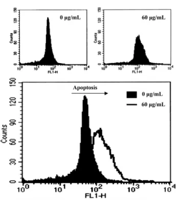
Fig. 2. Quantification of apoptosis by flow cytometry with PI staining. The HeLa cells were treated with vari- ous concentrations of S-EA for 24 h. The cells were then stained with PI, and the nuclei analyzed for their DNA contents using a flow cytometry with Cell Quest software.
A total of 10,000 nuclei were analyzed from each sample.
Data are means ± SD of three separate experiments.
Antiproliferative Effect of Silkworm Extract 295
with the control (Fig. 1). The growth of the HeLa cells was also significantly inhibited (p< 0.01) by 120µg/mL S-EA.
To evaluate whether the growth inhibition of the HeLa cells by S-EA was related to apoptosis, a flow cytometry analysis was first performed. The HeLa cells were treated with various concentrations (0-100µg/mL) of S-EA for 24 h, and subjected to the flow cytometric analysis after the DNA staining by PI. The apoptotic cell population increased gradually from 9% without S-EA to 15% at 50
µg/mL, to 34.2% at 75µg/mL, and to 41.9% at 100µg/
mL after exposure to S-EA for 24 h (Fig. 2). Moreover, the DNA fragmentation of the HeLa cells due to apoptosis was measured directly by the terminal deoxynucleotidyl transferase-mediated dUTP nick-end labeling (TUNEL) assay. When the HeLa cells were incubated with 60µg/
mL of S-EA for 48 h, the apoptotic DNA fragmentation was observed in the HeLa cells (Fig. 3).
The ethyl acetate fraction used in this experiment did not contain peptides and proteins, because the probability that the ethyl acetate layer can contain them was very
low; the extract thus was treated with ammonium sulfate.
The effects of the non-peptidic compounds on the biological activities of silkworm have not yet been reported. Further study should be performed on the identification of the active compound contained in the extract.
Acknowledgments. This work was supported by the grants from Biogreen 21 (Korea Ministry of Agriculture and Forestry) and the second Brain Korea 21 (Korea Ministry of Education).
References
Darzynkiewicz Z, Bruno S, Del Bino G, Gorczyca W, Hotz MA, Lassota P, and Traganos F (1992) Features of apop- totic cells measured by flow cytometry. Cytometry 13, 795-808.
Fotedar R, Diederich L, and Fotedar A (1996) Apoptosis and the cell cycle. Prog Cell Cycle Res 2, 147-163.
Hiraki A, Yukawa M, Kim J, and Ueda S (1997) Antiviral substance from silkworm faeces: purification and its chemical characterization. Biol Pharm Bul. 20, 547-555.
Hirayama E, Ishikawa N, Yano-Inoue T, and Kim J (1993) Purification and characterization of a lectin-like sub- stance from silkworm faeces. Cell Struc Funct 18, 161- Kang YK, Nam SH, Sohn HO, and Lee DW (2005) Inhibi-171.
tory effect of silkworm-Extract (SE) on monoamine oxi- dase activity in vitro and in vivo. Entomol Res 35,189- Khosraviani K, Williamson K, and Hamilton P (1996) Apo-193.
ptosis and proliferation: The balance. Gastroenterology
110, 1323-1324.
Mita K, Kasahara M, Sasaki S, Nagayasu Y, Yamada T, Kanamori H,Namiki N, Kitagawa M, Yamashita H, Yasukochi Y, Kadono-Okuda K, Yamamoto K, Ajimura M, Ravikumar R, Shimomura M, Nagamura Y, Shin-I T, Abe H, Shimada, T, Morishita S, and Sasaki T (2004) The Genome Sequence of Silkworm, Bombyx mori. DNA Res 11, 27-35.
Mosmann T (1983) Rapid colorimetric assay for cellular growth and survival: Application to proliferation and cytotoxicity assays. J ImmunolMethods65, 55-63.
Ryu KS, Lee HS, and Kim IS (2002) Effects and mecha- nisms of silkworm powder as a blood glucose-lowering agent. Int J Ind Entomol4, 93-100.
Zhaohui G, Yongfeng J, and Yaozhou Z (2007) Suppression of diabetes in non-obese diabetic (NOD) mice by oral administration of a cholera toxin B subunit-insulin B chain fusion protein vaccine produced in silkworm. Vac- cine25, 1444-1451.
Fig. 3. Induction of apoptosis by S-EA in the HeLa cells as determined by TUNEL assay. The cells were incubated for 48 h in 60µg/mL of S-EA, fixed, permeabi- lized, and stained with a fluorescent TUNEL reaction (Promega, Madison, WI). The cells were analyzed using the flow cytometry.