PGHN
Original Article
Association between Elevated Alanine Aminotransferase and Urosepsis in Children with Acute Pyelonephritis
Dongwan Kim, Sung Hyun Lee, Hann Tchah, Eell Ryoo, Hye Kyung Cho, and Yun Mi Kim*
Department of Pediatrics, Gachon University Gil Medical Center, *Department of Nursing, Gachon University, Incheon, Korea
Purpose: The aim of this study is to investigate the association between elevated alanine aminotransferase (ALT) and urosepsis in children with acute pyelonephritis (APN).
Methods: We retrospectively identified all children who were managed in our hospital with APN during a decade period. In our study a diagnosis of APN was defined as having a positive urine culture and a positive (99m)Tc-di- mercaptosuccinic acid scintigraphy. We compared those with elevated ALT and those with normal ALT according to the following variables: age, gender, duration of fever prior to admission, presence of hypotension, C-reactive protein (CRP), creatinine, presence of anemia, white blood cells count, platelet count, blood culture result, and grades of vesicoureteral reflux. In addition, the correlation between elevated ALT and positive blood culture was analyzed in detail.
Results: A total of 996 children were diagnosed with APN, of which 883 were included in the study. ALT was elevated in 81 children (9.2%). In the analysis of demographic characteristics, the number of children with elevated ALT was higher in children between 0 to 3 months, boys, and in those with positive blood culture (p=0.002, 0.036, and 0.010, respectively). In multivariate analysis of variables associated with positive blood culture, age younger than 3 months, elevated ALT, elevated CRP, and elevated creatinine showed statistical significance (p=0.004, 0.030, 0.043, and 0.044, respectively).
Conclusion: Our study demonstrates the association between elevated ALT and increased prevalence of urosepsis in addition to elevated CRP, elevated creatinine, and age younger than 3 months in children with APN.
Key Words: Alanine transaminase, Pyelonephritis, Urosepsis, Infant, Child
Received:July 3, 2015, Revised:September 1, 2015, Accepted:October 19, 2015
Corresponding author: Hann Tchah, Department of Pediatrics, Gachon University Gil Medical Center, 21 Namdong-daero 774beon-gil, Namdong-gu, Incheon 21565, Korea. Tel: +82-32-460-8429, Fax: +82-32-460-2362, E-mail: [email protected]
Copyright ⓒ 2016 by The Korean Society of Pediatric Gastroenterology, Hepatology and Nutrition
This is an openaccess article distributed under the terms of the Creative Commons Attribution NonCommercial License (http://creativecommons.org/licenses/by-nc/4.0/) which permits unrestricted noncommercial use, distribution, and reproduction in any medium, provided the original work is properly cited.
INTRODUCTION
Urinary tract infection (UTI) is a common febrile illness in children, especially in infancy. Acute pyelo-
nephritis (APN) in particular is of concern because it can easily lead to bacteremia and sepsis in children, and cause renal scarring as a complication [1]. APN is suspected in children with UTI who exhibit signs
of systemic inflammatory response, hence the need for diagnostic tests such as renal scintigraphy with 99mTc-dimercaptosuccinic acid (DMSA).
APN, like many other disease entities, can cause liver injury via sepsis. In patients with sepsis, proin- flammatory cytokines derived from Kupffer cells are responsible for hepatocellular dysfunction [2-4]. Liver injury in APN patients has been studied in adults [5].
In addition, a study on children with UTI and ele- vated alanine aminotransferases (ALT) has been reported. However, it did not show direct correlation of elevated ALT with sepsis [6].
The aim of our study was to investigate the associ- ation between elevated ALT and urosepsis in chil- dren with APN.
MATERIALS AND METHODS
Children diagnosed with APN at Gachon University Gil Medical Center (Incheon, Korea) between January 2005 and December 2014 were included in our study.
The research process was approved by Gachon University Gil Medical Center Institutional Review Board (GCIRB2015-65).
In our study diagnosis of APN was defined as hav- ing a positive urine culture and a positive DMSA. The results of urine culture and DMSA were collected via a retrospective chart review of children who were clinically diagnosed with UTI during their stay.
Urine samples for urinalysis and urine culture were collected using sterile urine bags, urine catheter- ization, or suprapubic aspiration. The exact pro- portion of urine samples collected via each method is unknown, although sterile urine bags were used in most cases. DMSA was performed when a working diagnosis of UTI was made.
Clinical data and laboratory test results were re- corded for all children diagnosed with APN. This da- ta include age, gender, duration of fever prior to ad- mission, types of antibiotics used, systolic blood pressure, and laboratory tests such as ALT, total bilir- ubin, C-reactive protein (CRP), creatinine, hemoglo- bin, white blood cells (WBC) count, platelet count, and blood culture results, if performed. For the labo-
ratory test results, data from the day of admission before the initiation of treatment were used. Results of voiding cystourethrogram were also recorded if available.
Hypotension was defined as a systolic blood pres- sure of less than (2 × age in years+65) mmHg, in ac- cordance with an analysis by Hague and Zaritsky [7].
ALT levels over 50 IU/mL were defined as elevated ALT. Cut-off values of total bilirubin and CRP were 1.2 mg/dL and 0.5 mg/dL, respectively. For crea- tinine, the cut-off values were the upper reference limits calculated by Pottel et al. [8]. Patients with he- moglobin values under the 3rd percentile of Korean children were diagnosed as anemic [9] and a cut-off value of 9.5 g/dL was used for infants under the age of 6 months. Urosepsis was defined as a positive blood culture of the same agent found in the urine culture with at least two of the signs of systemic in- flammatory response syndrome [10]. Vesicoureteral reflux (VUR) of grades I and II were grouped as low grade and III and over as high grade.
We compared those with elevated ALT and those with normal ALT according to the following varia- bles: age, gender, duration of fever prior to admis- sion, presence of hypotension, CRP, creatinine, pre- sence of anemia, WBC count, platelet count, blood culture result, and grades of VUR. In addition, the correlation between elevated ALT and positive blood culture was analyzed in detail.
Exclusion criteria
Patients were excluded from the study if any of the following were true: the DMSA scan was performed as a follow-up study; there was any history of hospi- talization and administration of parenteral anti- biotics within a month of the diagnosis of UTI; there was any history of liver disease; or another etiology for ALT elevation was identified during their hospi- talization for APN. A few patients whose laboratory data was unavailable for a variety of reasons were ex- cluded from analysis.
Statistical analysis
For univariate analysis, either a chi-square (χ2)
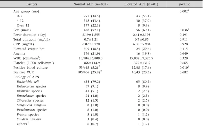
Table 1.Demographic Characteristics of Children with Normal and Elevated ALT Levels
Factors Normal ALT (n=802) Elevated ALT (n=81) p-value
Age group (mo) 0.002§
0-3 277 (34.5) 43 (53.1)
4-12 348 (43.4) 30 (37.0)
Over 12 177 (22.1) 8 (9.9)
Sex (male) 458 (57.1) 56 (69.1) 0.036§
Fever duration (day) 2.19±1.855 2.41±2.195 0.391
Total bilirubin (mg/dL) 0.7±1.21 0.7±0.85 0.911
CRP (mg/dL) 6.02±5.770 6.08±5.904 0.928
Elevated creatinine* 309 (38.5) 24 (29.6) 0.115
Anemia 176 (21.9) 16 (19.8) 0.649
WBC (cells/mm3) 15,784±6,808.0 15,002±7,323.5 0.328
Platelet (1,000 cells/mm3) 366±114.9 372±131.9 0.665
Positive blood culture 53/648 (8.2)† 12/68 (17.6) 0.010§
Positive VUR 105/406 (25.9)‡ 10/43 (23.3) 0.682
Etiology of APN
Escherichia coli 635 (79.2) 65 (80.2)
Enterococcus species 57 (7.1) 8 (9.9)
Klebsiella species 41 (5.1) 2 (2.5)
Enterobacter species 24 (3.0) 2 (2.5)
Citrobacter species 12 (1.5) 2 (2.5)
Morganella morganii 8 (1.0) 0 (0.0)
Pseudomonas species 8 (1.0) 0 (0.0)
Proteus species 8 (1.0) 1 (1.2)
Candida albicans 3 (0.4) 0 (0.0)
Others∥ 6 (0.7) 1 (1.2)
Values are presented as number (%) or mean±standard deviation.
ALT: alanine aminotransferase, CRP: C-reactive protein, WBC: white blood cells, VUR: vesicoureteral reflux, APN: acute pyelonephritis.
*Different cut-off values were used for different ages [8]. †Total among 716 children on whom at least one blood culture was performed. ‡Total among 449 children on whom VCUG was performed. §Statistically significant. ∥Streptococcus agalactiae, Serratia marcescens, Elizabethkingia meningoseptica, Raoultella planticola, Staphylococcus saprophyticus, Acinetobacter baumannii.
test or an independent-samples t-test was used. The χ2 test was used for categorical variables. For con- tinuous variables, if different cut-off values were used for different ages, the variables were catego- rized according to normal or abnormal values, and analyzed as categorical variables using a χ2 test. For other continuous variables, independent t-tests were used to compare the means between normal and ele- vated ALT groups. For analysis with blood culture re- sults, all variables were categorized according to ei- ther normal or abnormal values for logistic re- gression analysis. All statistical analyses were per- formed using SPSS ver. 12.0 (SPSS Inc., Chicago, IL, USA).
RESULTS
Demography of children with APN
The total number of children diagnosed as APN was 996. The number of patients excluded from this study by each criteria is as follows: 87 were excluded because the DMSA scan was a follow-up study; 18 were excluded due to prior use of antibiotics; three were excluded due to prior history of liver disease;
one patient was excluded because another etiology for hepatitis was identified; and four patients were excluded because laboratory data were unavailable.
A total of 883 children with APN were included in this study, 514 were boys and 369 were girls. The male to female ratio for all APN patients was 1.4:1.
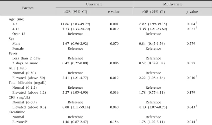
Table 2.Logistic Regression Model of Factors Associated with Urosepsis
Factors Univariate Multivariate
uOR (95% CI) p-value aOR (95% CI) p-value
Age (mo)
1-3 11.86 (2.83-49.79) 0.001 8.82 (1.99-39.15) 0.004†
4-12 5.73 (1.33-24.70) 0.019 5.35 (1.21-23.60) 0.027†
Over 12 Reference Reference
Sex
Male 1.67 (0.96-2.92) 0.070 0.84 (0.45-1.56) 0.579
Female Reference Reference
Fever
Less than 2 days Reference Reference
2 days or more 0.47 (0.27-0.80) 0.006 0.57 (0.32-1.02) 0.057
ALT (IU/L)
Normal (0-50) Reference Reference
Elevated (above 50) 2.41 (1.21-4.77) 0.012 2.22 (1.08-4.56) 0.030†
Total bilirubin (mg/dL)
Normal (0-1.2) Reference Reference
Elevated (above 1.2) 2.27 (1.05-4.90) 0.036 1.78 (0.77-4.11) 0.179
CRP (mg/dL)
Normal (0-0.5) Reference Reference
Elevated (above 0.5) 8.08 (1.11-59.14) 0.040 8.13 (1.07-60.75) 0.043† Creatinine
Normal Reference Reference
Elevated* 1.46 (0.87-2.47) 0.156 1.78 (1.02-3.11) 0.044†
OR: odds ratio, uOR: unadjusted OR, aOR: adjusted OR, CI: confidence interval, ALT: alanine aminotransferase, CRP: C-reactive protein.
*Different cut-off values were used for different ages [8]. †Statistically significant.
The number of children in each of the age groups was 320 for 0 to 3 months of age, 378 for 4 to 12 months of age, and 185 for over 12 months of age. The pro- portion of boys in ages 0 to 8 months was greater than 50%, especially during the earlier months when the proportion of boys was greater than 80%. After 8 months of age, the proportion of boys declined rapidly.
This difference in proportions was statistically sig- nificant when comparing each of the age groups de- scribed above (77.2% in ages 0 to 3 months, 55.6% in ages 4 to 12 months, and 30.8% in ages greater than 12 months, p=0.000).
Elevated ALT and its association with de- mographic and clinical variables
Of the 883 children with APN, 81 children (9.2%) had elevated ALT. The maximum level of ALT was 774 IU/L, and the median elevated ALT was 95 IU/L.
In the analysis of demographic characteristics be- tween the children with normal and elevated ALT levels, differences were observed between the num- ber of children in each age group, sex, and blood cul- ture results (Table 1).
Urosepsis and its association with demographic and clinical variables
To confirm the correlation between elevated ALT and positive blood culture, we decided that multi- variate analysis of variables related to positive blood culture was required. In the multivariate analysis of the variables, age, elevated ALT, elevated CRP, and elevated creatinine showed statistical significance (p<0.050) (Table 2).
The duration of intravenous antibiotic treatment for the children with positive blood culture was lon- ger than that of the children with negative blood cul-
ture, 8.2±2.38 days and 6.8±2.14 days, respectively (p=0.000).
Total bilirubin and hypotension
Of the 883 children with APN, 58 had elevated to- tal bilirubin levels (7 children with elevated ALT and 51 children with normal ALT), and 81% of these chil- dren were under the age of 3 months. Hypotension was observed in only three children, one with ele- vated ALT and two without. None of these children had a systemic blood pressure of less than 60 mmHg.
DISCUSSION
The results of our study demonstrate a correlation between elevated ALT levels and increased preva- lence of sepsis in children with APN. Roughly 9% of children with APN had ALT elevation, and among these children, 17.6% had a positive blood culture, double the rate in children without ALT elevation, which is 8.2%.
Younger age and elevated serum levels of CRP and creatinine had been found to be associated with uro- sepsis among children with UTI or APN. Among in- fants and children with UTI, patients with a positive blood culture were younger [1,11]. UTI has been studied most often among young infants since they were the most prone to be implicated, and as high as 85% of serious bacterial infections among infants younger than 3 months of age has been identified as UTI [12]. We found that age younger than 3 months was significantly related to increased prevalence of urosepsis among children with APN. Elevated CRP has been associated with increased risk for sepsis [13]. Our study also showed difference of blood cul- ture positivity between children with elevated and normal CRP levels, with adjusted odds ratio of 8.13.
Our study also showed difference of blood culture positivity between children with elevated and nor- mal creatinine levels, with adjusted odds ratio of 1.78. This finding is in accordance with a prior study analyzing factors associated with bacteremia in young infants with UTI which found association of increased blood creatinine values with bacteremia
[14]. Therefore elevated ALT can be another parame- ter for anticipating increased prevalence of urosepsis in children with APN, complementary to age young- er than 3 months, elevated CRP, and elevated creati- nine.
Three main mechanisms have been proposed for the liver injury during sepsis. The liver plays a central role when a sepsis occurs. Kupffer cells are the main scavengers of bacteria and endotoxins, preventing them from entering the systemic circulation [4].
However, Kupffer cells have been found to be respon- sible for the increase in proinflammatory cytokines and decrease in hepatocellular function. Decrease number of Kupffer cells have shown beneficial effect on hepatocellular function and decreased levels of the cytokines [3]. During sepsis, liver parenchymal cells are involved in the immune response, and acute phase proteins produced by hepatocytes enhances both host defense and protective functions. However, the acute phase proteins response markedly contrib- utes to the procoagulant state [4]. Lastly, neutrophils are proposed to be the main effector cells that cause septic liver injury [15]. Activated neutrophils injure hepatocytes by production of oxygen-derived radi- cals and protease that may injure hepatocytes [16].
Liver enzyme elevation among patients with UTI or APN has been studied in adults and children. In the study of liver enzyme elevation among adult pa- tients with APN by Campos et al. [5], arterial hypo- tension and older age seemed to be associated with increased prevalence of urosepsis. In the study among children [6], however, younger age was the only meaningful factor that showed difference between patients with liver enzyme elevation and those without. These studies did not consider presence of urosepsis as a factor in their analysis. Whether the effect of arterial hypotension and older age in adults and younger age in children on elevated liver en- zymes is actually caused by the presence of urosepsis remains to be studied.
UTI during the first three months of life is more common in boys, and the sex ratio reverses after the first year [17]. The results of our study are in con- cordance with the known demography, with boys
constituting more than 80% of children under age of 3 months, and dropping with increasing age.
Oddly, in univariate analysis of our study, fever durations of two or more days showed negative cor- relation with positive blood culture. A prospective cohort study in children of 3 to 36 months of age by Teach and Fleisher reported similar results [18].
Another study on fever and bacteremia in children concluded that a high fever of greater than or equal to 40.5oC was specific for bacteremia [19]. It would be logical to suppose that parents with younger chil- dren or children with high fevers would be more in- clined to visit a hospital and consent to a urinalysis for detection of UTI. Although we did not collect data on the degree of fever, an analysis of our data showed that children who had a fever for less than two days were younger than those who had a fever for two or more days (21.9±1.0 months and 25.2±1.2 months, respectively, p=0.001). A logistic regression analysis of fever duration and age showed that fever duration had no statistically significant correlation with blood culture positive rate, which confirmed the possibility of age being the confounding variable for the fever duration (adjusted odds ratio 0.62, 95% confidence interval 0.35-1.09, p=0.094).
Systemic viral, bacterial, and protozoan infections invading the liver may cause cholestasis [20]. This effect occurs predominantly in Gram-negative ba- cilli, such as Escherichia coli, Kelbsiella pneumoniae, Pseudomonas aeruginosa, and Proteus species. Proposed mechanisms are increased bilirubin load due to he- molysis that can occur in bacterial infections, and de- creased bilirubin uptake, intrahepatic processing, and canalicular excretion [21]. In our study, 58 chil- dren had elevated total bilirubin levels among 883 children with APN, but the majority (81%) was un- der the age of 3 months. Neonates with UTI may present with jaundice only [22], and sepsis is more likely to manifest with jaundice in infants and chil- dren than in adults [21]. In our study, no association was found between elevated total bilirubin and ele- vated ALT. More accurate analysis of the relationship between elevated total bilirubin with the adjusted normal values for age and the rate of sepsis would be
valuable.
Prior studies on UTI and elevated aminotrans- ferases included both aspartate aminotransferase (AST) and ALT elevation, and the prevalence of in- creased aminotransferase levels in UTI patients was approximately 20% [5,6]. If elevated AST was in- cluded in our analysis as well, approximately 15% of the children with APN had elevated aminotransfe- rases.
One of the limitations of our analysis is that blood cultures were not performed in 154 children (19.2%) among 802 with normal ALT and in 13 children (16.0%) among 81 with elevated ALT, but the pro- portions of the children without blood cultures do not differ much between the two groups, and we do not think it has affected the results of our study.
In conclusion, our study demonstrates the associ- ation between elevated ALT and increased preva- lence of urosepsis, in addition to elevated CRP, ele- vated creatinine, and age younger than 3 months in children with APN.
REFERENCES
1. Pitetti RD, Choi S. Utility of blood cultures in febrile children with UTI. Am J Emerg Med 2002;20:271-4.
2. Szabo G, Romics L Jr, Frendl G. Liver in sepsis and sys- temic inflammatory response syndrome. Clin Liver Dis 2002;6:1045-66.
3. Koo DJ, Chaudry IH, Wang P. Kupffer cells are respon- sible for producing inflammatory cytokines and hep- atocellular dysfunction during early sepsis. J Surg Res 1999;83:151-7.
4. Dhainaut JF, Marin N, Mignon A, Vinsonneau C.
Hepatic response to sepsis: interaction between coagu- lation and inflammatory processes. Crit Care Med 2001;29(7 Suppl):S42-7.
5. Campos J, Alende R, Gonzalez-Quintela A. Abnormali- ties in aminotransferase levels during acute pyelone- phritis. Eur J Intern Med 2009;20:e53-6.
6. Park JY, Ko KO, Lim JW, Cheon EJ, Yoon JM. Increase in aminotransferase levels during urinary tract in- fections in children. Pediatr Gastroenterol Hepatol Nutr 2013;16:89-94.
7. Haque IU, Zaritsky AL. Analysis of the evidence for the lower limit of systolic and mean arterial pressure in children. Pediatr Crit Care Med 2007;8:138-44.
8. Pottel H, Vrydags N, Mahieu B, Vandewynckele E, Croes K, Martens F. Establishing age/sex related se- rum creatinine reference intervals from hospital labo- ratory data based on different statistical methods. Clin Chim Acta 2008;396:49-55.
9. Kim SK, Son BK. Studies on normal values for red blood cells in Korean children. J Korean Pediatr Soc 1996;
39:673-81.
10. Wagenlehner FM, Lichtenstern C, Rolfes C, Mayer K, Uhle F, Weidner W, et al. Diagnosis and management for urosepsis. Int J Urol 2013;20:963-70.
11. Bachur R, Caputo GL. Bacteremia and meningitis among infants with urinary tract infections. Pediatr Emerg Care 1995;11:280-4.
12. Bachur RG, Harper MB. Predictive model for serious bacterial infections among infants younger than 3 months of age. Pediatrics 2001;108:311-6.
13. Wang HE, Shapiro NI, Safford MM, Griffin R, Judd S, Rodgers JB, et al. High-sensitivity C-reactive protein and risk of sepsis. PLoS One 2013;8:e69232.
14. Averbuch D, Nir-Paz R, Tenenbaum A, Stepensky P, Brooks R, Koplewitz BZ, et al. Factors associated with bacteremia in young infants with urinary tract infection.
Pediatr Infect Dis J 2014;33:571-5.
15. Holman JM Jr, Saba TM. Hepatocyte injury during post-operative sepsis: activated neutrophils as poten- tial mediators. J Leukoc Biol 1988;43:193-203.
16. Doi F, Goya T, Torisu M. Potential role of hepatic macro- phages in neutrophil-mediated liver injury in rats with sepsis. Hepatology 1993;17:1086-94.
17. Stull TL, LiPuma JJ. Epidemiology and natural history of urinary tract infections in children. Med Clin North Am 1991;75:287-97.
18. Teach SJ, Fleisher GR. Duration of fever and its rela- tionship to bacteremia in febrile outpatients three to 36 months old. The Occult Bacteremia Study Group.
Pediatr Emerg Care 1997;13:317-9.
19. McCarthy PL, Grundy GW, Spiesel SZ, Dolan TF Jr.
Bacteremia in children: an outpatient clinical review.
Pediatrics 1976;57:861-8.
20. Trauner M, Fickert P, Stauber RE. Inflammation-in- duced cholestasis. J Gastroenterol Hepatol 1999;14:
946-59.
21. Chand N, Sanyal AJ. Sepsis-induced cholestasis.
Hepatology 2007;45:230-41.
22. Afzal N, Qadir M, Qureshi S, Ali R, Ahmed S, Ahmad K. Urinary tract infection presenting as jaundice in neonates. J Pak Med Assoc 2012;62:735-7.