INTRODUCTION
Dacryocystorhinostomy (DCR), which has been performed for the past hundred years, is a surgical procedure by which lacrimal flow is diverted into the nasal cavity through an arti- ficial opening made at the level of the lacrimal sac. The oper- ation can be carried out using either an external or endonasal surgical approach. The external approach was popularized first and became the surgery of choice for most ophthalmol- ogists, until recently (1). Since its first description by McDo- nough and Meiring (2), ten years ago, endoscopic DCR has been gaining popularity, largely due to technological advances in endoscopes and other modern instruments of rhinologic surgery (3).
Despite much debate, many ophthalmologists still believe that external DCR provides a higher success rate than endo- scopic DCR, and consider external DCR to be the gold stan- dard treatment for nasolacrimal duct obstruction. Though many types of endonasal approaches have been attempted, long-term success rates have not been equivalent to that achi- eved with external DCR, which approximates 90% (4-7).
Endoscopic DCR with laser assistance is successful between 58 and 85 percent of the time (6, 8, 9), whereas endoscopic DCR procedures using other tools (eg. drill, cold knife, punch) appear to yield slightly higher success rates (10, 11).
The most common cause of a surgical failure in endoscopic DCR is obstruction of the neo-ostium by granulation tissue or synechia that forms post-operatively (5-11). Most previ- ously-described endoscopic DCR procedures involve a small opening at the medial wall of the lacrimal sac, and sacrifice nasal and lacrimal sac mucosa during the procedure. Inade- quate exposure of the lacrimal sac, due to limited resection of bone and excessive and unnecessary removal or injury of surrounding nasal and lacrimal sac mucosa, and, hence, expo- sure of bone around a small neo-ostium, appear to contribute to obstruction of the neo-ostium by granulation tissue.
This retrospective study estimates the effectiveness of a sur- gical procedure that is a modification of previously reported endoscopic DCR techniques. This technique exposes the lac- rimal sac fully after removing the maxillary bone surround- ing the sac, creates a large marsupialized lacrimal sac, and cov- ers the exposed bone with preserved nasal mucosal flaps.
MATERIALS AND METHODS
At our institution, from 2002 to 2004, 42 patients (3 male and 39 female) underwent 46 endoscopic DCR (17 left sides and 29 right sides) for chronic epiphora. Patients’ ages ranged from 7 to 71 yr, with a mean age of 47. All patients were eval-
Hong-Ryul Jin, Je-Yeob Yeon*, Mi-Young Choi�
Department of Otorhinolaryngology-Head and Neck Surgery, Seoul National University Boramae Hospital, Seoul; Departments of Otolaryngology*and Ophthalmology�, Chungbuk National University Hospital, Cheongju, Korea
Address for correspondence Hong-Ryul Jin, M.D.
Department of Otorhinolarygology-Head and Neck Surgery, Seoul National University Boramae Hospital, 425 Shindaebang 2-dong, Dongjak-gu, Seoul 156-707, Korea
Tel : +82.2-840-2412, Fax : +82.2-831-2826 E-mail : hrjin@paran.com
719
Endoscopic Dacryocystorhinostomy: Creation of a Large Marsupialized Lacrimal Sac
This retrospective study describes and evaluates the effectiveness of a modified te- chnique of conventional endoscopic dacryocystorhinostomy (DCR) that minimizes the obstruction of a neo-ostium by creating an enlarged marsupialized lacrimal sac using mucosal flaps. Forty-two patients who had undergone 46 endoscopic DCR at a tertiary medical center, from 2002 to 2004, for correction of lacrimal system obstruc- tion were investigated. The surgical technique involves elevation of a nasal mucosal flap, full sac exposure using a power drill, and shaping of the mucosal flap to cover denuded bone and juxtapose exposed sac mucosa. Postoperative symptoms and endoscopic findings of the neo-ostium were evaluated. Mean duration of follow-up was 5.9 months. An eighty-three percent primary success rate was observed, with- out any serious complications. Obstruction of the neo-ostium with granulation tissue was observed in eight cases, among which six underwent revision with success in all cases. Overall, 44 (96%) of 46 cases experienced surgical successes. Endoscop- ic DCR, a procedure in which a large marsupialized lacrimal sac is created from mu- cosal flaps, yields a very satisfactory success rate with straightforward and highly successful revision available for those in whom the primary procedure yields a sub- standard result.
Key Words : Dacryocystorhinostomy; Endoscopy; Lacrimal Duct Obstruction; Lacrimal Apparatus
Received : 25 November 2005 Accepted : 27 January 2006
uated by an ophthalmologist before surgery. Pre-operative evaluation consisted of a standard examination that included lacrimal irrigation, conventional dacryocystography, and/or dacryoscintigraphy. The nasal cavity was examined and the need for additional nasal surgery (i.e., septoplasty, middle tur- binate reduction) also was determined pre-operatively.
Of the 46 primary surgery cases, fourteen cases previously had failed insertion of a nasolacrimal polyurethane stent. Eti- ologies of epiphora were proximal nasolacrimal duct obstruc- tion in all cases and one case had associated inferior canaliculi obstruction.
Patient symptoms and endoscopic findings of the neo-osti- um were evaluated postoperatively. Irrigation through the pu- nctum was performed to evaluate the patency of the neo-osti- um post-operatively, and the neo-ostium was judged “wide”
in size if the marsupialized sac had been well maintained in shape and size. Surgery was considered unsuccessful if the pa- tient had one or more of the following postoperative out- comes: 1) no marked improvement of preoperative chronic epi-
phora, or any episode of dacryocystitis; 2) inability to irrigate the lacrimal system; and 3) nasal endoscopy revealing obstruc- tion of the neo-ostium with granulation tissue or synechia.
Operative techniques
At our institution, DCR is performed under local or gener- al anesthesia, as determined by considering a number of pa- tient factors. After shrinkage of the nasal cavity by inserting a gauze packing soaked in a mixture of 1:2,000 epinephrine and 1% lidocaine, the head of the middle turbinate and the mucosa surrounding the lacrimal sac are infiltrated with a mixture of 1:200,000 epinephrine and 2% lidocaine. The pa- tient is placed in a supine position with the head elevated 15 degrees. A zero or thirty degree, 4-mm diameter endoscope is used.
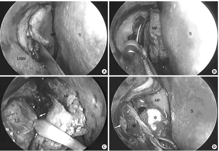
Operating steps are shown in Fig. 1. In the first step, using a slit knife (angled, 2.7 mm, Alcon Co., Cleveland, Ohio, U.S.A.), a reverse “C” shaped mucosal incision, 10×10 mm,
Fig. 1. (A) A nasal mucosal flap is being elevated after a reverse “C” shaped incision on the mucosa of the lateral nasal wall with slit knife just anterior to the insertion of middle turbinate. (B) The maxillary bone covering the lacrimal sac is drilled out with a curved diamond dacry- ocystorhinostomy bur (15°, 2.9 mm, Xomed Co., Jacksonville, Florida, U.S.A.). (C) The extent of the lacrimal sac is verified with lacrimal pro- be (arrow) and the sac wall is tented to allow incision. The vertical incision is made with a slit knife. (D) The mucosal flap covers the exposed bony portion after cutting and trimming. The edges of the exposed lacrimal sac (arrow) are everted to match the nasal mucosa and the sac lumen is filled with gelfoam (asterisk) to keep the flap anastomosis in position. A silicone bicannalicular tube is seen through the new opening. LNW, lateral nasal wall; MT, middle turbinate; S, septum; LS, lacrimal sac; MF, mucosal flap.
A B
C D
MT S
MF
S MF
LS
*
*
MF
S S
LNW
is made at the lateral nasal wall anterior and slightly superi- or to the insertion of the middle turbinate (Fig. 1A). The pos- teriorly based mucosal flap is elevated backwards off the max- illary bone, extending up to the uncinate process. The max- illary bone covering the lacrimal sac then is gently drilled (Curved diamond DCR bur, 15 degree, 2.9 mm, Xomed Co., Jacksonville, Florida, U.S.A.) until the sac is widely exposed, extending to the level of the fundus (Fig. 1B). It is important to remove all bone covering the common cannalicular open- ing. Metallic lacrimal probes are passed medially through both canaliculi, and gently pushed so as to tent the sac, thus facilitating incision through the sac while precisely localizing the position of the sac lumen (Fig. 1C). An incision then is made with a slit knife, avoiding injury to the sac lumen and, hence, minimizing hemorrhage.
The elevated nasal mucosal flap is bisected and trimmed.
The mucosal flaps are adjusted in size to cover the denuded bone surrounding the opened sac. The lacrimal sac flaps are incised, everted, and adjusted to accurately appose the nasal mucosa. A small gel foam patch is packed lightly in the ex- posed sac to keep the flap anastomosis in position through- out the initial healing period (Fig. 1D). A silicone bicanalic- ular tube (Canaliculus intubation set tube, Xomed Co., Jack- sonville, Florida, U.S.A.) is positioned, except in those ins- tances in which the sac is marsupialized widely due to prior longstanding dilatation of the sac.
Light nasal packing is required unless there has been asso- ciated nasal surgery (i.e. septoplasty). Post-operatively, each patient is prescribed antibiotics and ophthalmic drops, and followed regularly for nasal dressings. Irrigation and spray of the nasal cavity with saline are performed to prevent crust for- mation. The silicone tube generally is removed after six weeks.
RESULTS
The average follow-up period varied from three to 33 mon- ths, with an average of 5.9 months. Of 46 cases, 38 cases (83%) demonstrated primary surgical success, defined as decreased or absent epiphora and an adequately patent neo-ostium. In eight cases (17%), including one case of functional obstruc- tion, obstruction of the neo-ostium by granulation tissue or synechia was identified, all associated with persistent epiphora (Table 1). Of 14 cases with a history of previously failed inser- tion of a nasolacrimal polyurethane stent, three showed ob-
struction (21%) while five showed obstruction (16%) out of the remaining 32 cases.
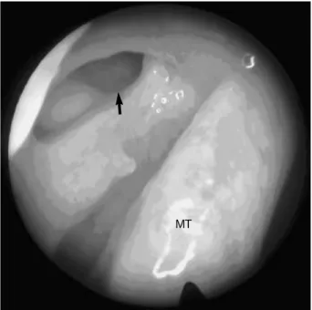
The neo-ostium was widely patent in 35 cases (76%) (Fig.
2) and “narrow but patent” in three cases (7%). Though six cases (four with wide and patent neo-ostium and two with narrow but patent neo-ostium) complained of occasional epi- phora in spite of great symptom improvement postoperatively, these cases were considered surgical successes, in accordance with our prior criteria.
Among the eight patients with persistent obstruction of the neo-ostium, six subsequently underwent revision proce- dures. Two patients refused revision surgery and are being followed for symptoms. All six patients who underwent revi- sion became free of epiphora and ultimately had an adequately patent ostium that has persisted throughout the mean follow- up of six months post revision surgery. Overall, 44 of 46 cases (96%) ultimately had a successful surgical outcome.
All five patients without placement of silicone tube stent after sac opening had widely patent neo-ostium without epi- phora. In addition to the DCR procedure, septoplasty was performed in three cases (7%) and anterior middle turbin- ectomy in one case.
There were no serious complications, beyond obstruction of the neo-ostium causing surgical failure. In one case, orbital fat was mildly exposed during the operation but had no influ- ence on the postoperative surgical outcome.
DISCUSSION
For DCR, the endoscopic approach has several advantages
Wide and patent Narrow but patent Obstructed Neo-osteum
Total Epiphora
Absent 31 1 0 32 (70%)
Improved 4 2 0 6 (13%)
Persistent 0 0 8 8 (17%)
Total 35 (76%) 3 (7%) 8 (17%) 46 (100%)
Table 1.Surgical results in 46 cases of primary endoscopic dacry- ocystorhinostomy
Fig. 2.A wide neo-ostium made by marsupialization of the lacrimal sac (arrow) at six months after surgery. MT indicates the middle turbinate.
MT
over the external approach: 1) it is less traumatic and, thus, shortens the hospital stay; 2) a facial scar is avoided; 3) there is no disruption of the medial canthal tendon, which conse- quently enables preservation of lacrimal pump function; 4) access to the sac is direct through the lacrimal bone, avoid- ing double-side dissection of the sac; and 5) it is excellent in controlling tissue and, thus, results in less trauma to the nasal mucosa. Conversely, disadvantages are: 1) the surgical field may be limited because of bleeding; 2) there is an occasional need for septoplasty or removal of the middle turbinate; and 3) there appears to be increased likelihood of granulation tis- sue formation, resulting in stenosis and, thereby, obstruction of the opening. Previously described, conventional endoscop- ic DCR techniques generally involved limited opening of the sac, yielding frequent obstruction of the neo-ostium by granulation tissue, an outcome which explains the higher failure rates.
To avoid or prevent obstruction of the neo-ostium, many modified techniques have been attempted. These include complete separation of the sac from the nasolacrimal duct to divert lacrimal flow to the neo-ostium (12), use of steroids or mitomycin-C (13, 14), and use of mucosal flaps after wide resection of bone surrounding the sac (15-17); this last appro- ach is technically similar to our method.
Our surgical technique modified the conventional endo- scopic techniques in order to solve previously reported sources of surgical failure. Our modifications included the following:
1) A nasal mucosal flap was elevated to avoid unnecessary injury or removal of the mucosa; 2) We exposed the lacrimal sac as wide and as high as possible, up to the level of the fun- dus, by removing the bone surrounding the sac using a power drill; 3) Instead of cutting off the sac wall, it was incised and everted to meet the nasal mucosa; 4) Tailored mucosal flaps were created to cover all exposed bone and appose the evert- ed lacrimal sac flap. With these techniques, we intended to make a large, epithelialized fistula, thereby potentially min- imizing the formation of granulation tissue and synechia, which represent the most common causes of failure in endo- scopic DCR.
Our primary success rate of 83% is similar to the rates (80- 88%) reported using other endoscopic techniques performed without the use of a mucosal flap (10, 11). Other investiga- tors who have reported using techniques similar to ours have noted slightly higher primary success rates, which may not be significantly different from ours (15-17). Nonetheless, con- sidering our original intention to reduce granulation tissue formation and synechia and, thus, increase primary success by making a large marsupialized opening, our results are somewhat contrary to our expectations. There are a few pos- sible explanations for this. First, among our 46 patients who underwent surgery, 14 previously had failed insertion of a nasolacrimal polyurethane stent; many of these 14 had devel- oped a constricted sac with thick walls due to chronic inflam- mation likely caused by long-standing placement of the stent
(18). In patients with a constricted sac with thick walls, structur- ing a large marsupialized cavity is technically more difficult.
Though statistically not significant, these patients showed a slightly increased primary failure rate compared to other pat- ients. Second, proper postoperative care was not delivered in all of our cases, due to inadequate follow-up. Good postoper- ative care is a necessity and increases the likelihood of a suc- cessful outcome. With more favorable case selection and im- proved postoperative care, a higher success rate might have been accomplished at the time of primary surgery.
Though our primary success rate was not high enough to demonstrate advantages of our technique over others, our overall success rate after revision surgery was very high, at 96%. During revision surgery performed under local anesthe- sia, removal of granulation tissue or lysis of synechia which obstructed the ostium was facilitated by the previously insert- ed bicannalicular silicone tube exposing a wide ostium. This can be attributed to the wide opening of the sac from exten- sive removal of surrounding bone at the time of primary sur- gery. These results imply that our modified technique, though it failed to achieve a higher primary success rate compared to conventional endoscopic DCR techniques, nonetheless suc- ceeded in achieving an excellent ultimate success rate, with technically-simpler and more highly-successful revision pro- cedures.
The purposes of using silicone tubing are: 1) to maintain the opening of the neo-ostium; 2) to prevent or correct syne- chia of the canaliculus; and 3) to facilitate postoperative dress- ings (19, 20). There are debates over the use of silicone tub- ing in endoscopic DCR. Some investigators have reported 81-87% success rates without using silicone tubes after endo- scopic DCR, and recommend not using silicone tubing or removing it early because of granulation formation stimulat- ed by the tubing itself (19-21). As suggested by the results of our study, in which widely-patent neo-ostium was achieved in all five cases without silicone tube stenting, a silicone tube can be used, as determined by the pre-operative status of the lacrimal sac and canaliculus.
Complications of endoscopic DCR include re-stenosis of the opening, bleeding from the nasal cavity, orbital injury, CSF leakage through a fractured ethmoid, and corneal abra- sion or canaliculi erosion due to the overly-tight silicone tube placement (22). A lacrimal sump syndrome and associated recurrent infections can occur if the lower portion of the bone surrounding the sac is removed inadequately (5, 22). Our marsupialization technique opened the sac inferior to the pro- ximal nasolacrimal duct after bone removal, thereby prevent- ing lacrimal sump syndrome.
In conclusion, the authors obtained a large marsupialized lacrimal sac with wide removal of the covering bone and use of mucosal flaps. This technique yields a good surgical result that is comparable to the results of conventional endoscopic DCR techniques. Advantages of this procedure over the con- ventional endoscopic DCR techniques are two. First, primary
surgical failures are amenable to technically simple and high- ly successful surgical revisions. Second, there is a possibility for obtaining wide ostium without silicone tube stenting.
REFERENCES
1. Jones LT. The cure of epiphora due to canalicular disorders, trauma and surgical failures on the lacrimal passages. Trans Am Acad Oph- thalmol Otolaryngol 1962; 66: 506.
2. McDonough M, Meiring JH. Endoscopic transnasal dacryocystorhi- nostomy. J Laryngol Otol 1989; 103: 585-7.
3. Watkins LM, Janfaza P, Rubin PA. The evolution of endonasal dacry- ocystorhinostomy. Surv Ophthalmol 2003; 48: 73-84.
4. Hartikainen J, Antila J, Varpula M, Puukka P, Seppa H, Grenman R.
Prospective randomized comparison of endonasal endoscopic dacryo- cystorhinostomy and external dacryocystorhinostomy. Laryngoscope 1998; 108: 1861-6.
5. Ben Simon GJ, Joseph J, Lee S, Schwarcz RM, McCann JD, Gold- berg RA. External versus endoscopic dacryocystorhinostomy for acquired nasolacrimal duct obstruction in a tertiary referral center.
Ophthalmology 2005; 112: 1463-8.
6. Ibrahim HA, Batterbury M, Banhegyi G, McGalliard J. Endonasal laser dacryocystorhinostomy and external dacryocystorhinostomy outcome profile in a general ophthalmic service unit: a comparative retrospective study. Ophthalmic Surg Lasers 2001; 32: 220-7.
7. Tsirbas A, Davis G, Wormald PJ. Revision dacryocystorhinostomy:
a comparison of endoscopic and external techniques. Am J Rhinol 2005; 19: 322-5.
8. Massaro BM, Gonnering RS, Harris GJ. Endonasal laser dacryocys- torhinostomy: A new approach to nasolacrimal duct obstruction. Arch Ophthalmol 1990; 108: 1172-6.
9. Metson R, Woog JJ, Puliafito CA. Endoscopic laser dacryocystorhi- nostomy. Laryngoscope 1994; 104: 269-74.
10. Zilelioglu G, Tekeli O, Ugurba SH, Akiner M, Akturk T, Anadolu Y.
Results of endoscopic endonasal non-laser dacryocystorhinostomy.
Doc Ophthalmol 2002; 105: 57-62.
11. Gurler B, San I. Long-term follow-up outcomes of nonlaser intranasal endoscopic dacryocystorhinostomy: how suitable and useful are con- ventional surgical instruments? Eur J Ophthalmol 2004; 14: 453-60.
12. Sham CL, van Hasselt CA. Endoscopic terminal dacryocystorhinos- tomy. Laryngoscope 2000; 110: 1045-9.
13. Selig YK, Biesman BS, Rebeiz EE. Topical application of mitomycin- C in endoscopic dacryocystorhinostomy. Am J Rhinol 2000; 14: 205-7.
14. Park DJ, Kwak MS. The effect of mitomycin-C on the success rate of endoscopic dacryocystorhinostomy. J Korean Ophthalmol Soc 2000;
41: 1674-9.
15. Wormald PJ. Powered endoscopic dacryocystorhinostomy. Laryn- goscope 2002; 112: 69-72.
16. Tsirbas A, Wormald PJ. Mechanical endonasal dacryocystorhino- stomy with mucosal flaps. Br J Ophthalmol 2003; 87: 43-7.
17. Massegur H, Trias E, Adema JM. Endoscopic dacryocystorhino- stomy: modified technique. Otolaryngol Head Neck Surg 2004; 130:
39-46.
18. Ozturk S, Konuk O, Ilgit ET, Unal M, Erdem O. Outcome of patients with nasolacrimal polyurethane stent implantation: do they keep tear- ing? Ophthal Plast Reconstr Surg 2004; 20: 130-5.
19. Unlu HH, Toprak B, Aslan A, Guler C. Comparison of surgical out- comes in primary endoscopic dacryocystorhinostomy with and with- out silicone intubation. Ann Otol Rhinol Laryngol 2002; 111: 704-9.
20. Mortimore S, Banhegy GY, Lancaster JL, Karkanevatos A. Endo- scopic dacryocystorhinostomy without silicone stenting. J R Coll Surg Edinb 1999; 44: 371-3.
21. Lee TS, Kim SW. The effects of placement of bicanalicular silicone tube and silicone stent on granuloma formation in endoscopic intra- nasal dacryocystorhinostomy. J Korean Ophthalmol Soc 1999; 40:
16-22.
22. Fayet B, Racy E, Assouline M. Complications of standardized en- donasal dacryocystorhinostomy with unciformectomy. Ophthalmol- ogy 2004; 111: 837-45.