INTRODUCTION
Epstein-Barr virus (EBV) is an oncogenic virus associated with various lymphoproliferative disorders (LPDs) (1-4). In immunocompetent hosts, the relative frequency of the occur- rence, its clinical presentation, and EBV positivity of differ- ent histological subtypes of LPDs vary in different geograph- ic areas, and this may be ascribed to genetic and environmen- tal etiologic factors. In Western countries, Hodgkin’s lym- phoma (HD) and infectious mononucleosis are the most com- mon EBV-associated diseases, whereas some EBV-associated LPDs, especially those associated with T lymphocytes or nat- ural killer (NK) cells, are more prevalent in Asian and Latin American countries (5, 6).
T or NK cell LPDs associated with EBV have been differ- ently named based on their clinical findings as well as the immunophenotype of proliferating cells. EBV-associated hemophagocytic lymphohistiocytosis (EBV-HLH) is an unusu- al syndrome characterized by fever, organomegaly, pancytope- nia, and disseminated intravascular coagulation, and com- monly occurs in children and adolescents (7). Chronic active EBV (CAEBV) infection has been reported mainly in Japan
and is characterized by long-lasting infectious mononucleo- sis-like symptoms in children and young adults with no appar- ent immune deficiency (7). Fulminant T cell lymphoma fol- lowing acute EBV infections, as described by Quintanilla- Martinez et al. (8), is characterized by hepatosplenomegaly - often without significant lymphadenopathy and by fever, liver failure, and hemophagocytic syndrome following recent viral-like upper respiratory illnesses. Aggressive NK cell leu- kemia (ANKL) is a neoplasm of NK cells that causes systemic illness and pursues an aggressive clinical course (9). Nasal- type NK/T cell lymphomas are distinct from other EBV-asso- ciated T or NK cell LPDs in their localization to the upper aerodigestive tract at the initial presentation, but once dis- seminated, they follow a similar fatal clinical course to other types of EBV-associated T or NK LPDs (10-12).
Previous studies on patients with EBV-associated T or NK LPDs mainly focused on the clinicopathological and virolog- ical aspects of a specific category of these diseases, but knowl- edge on the overall distribution among lymphoproliferative disorders and relationships between these diseases is lacking.
This might be because such EBV-associated T or NK LPDs are uncommon, even in Asia.
185
Eun-Yoon Cho, Ki-Hyun Kim*, Won-Seog Kim*, Keon Hee Yoo�, Hong-Hoe Koo�, and Young-Hyeh Ko
Departments of Pathology, Division of Hematology- oncology of Internal Medicine*, and Pediatrics�, Samsung Medical Center, Sungkyunkwan University School of Medicine, Seoul, Korea
Address for correspondence Young-Hyeh Ko, M.D.
Department of Pathology, Samsung Medical Center, Sungkyunkwan University School of Medicine, 50 Irwon-dong, Gangnam-gu, Seoul 135-710, Korea Tel : +82.2-3410-2800, Fax : +82.2-3410-0025 E-mail : [email protected]
*This study was supported by SBRI grants C-A3-201- 1&2, and C-A3-202-3.
DOI: 10.3346/jkms.2008.23.2.185
The Spectrum of Epstein-Barr Virus-Associated Lymphoproliferative Disease in Korea: Incidence of Disease Entities by Age Groups
This study is to identify the spectrum of Epstein-Barr virus (EBV)-positive lympho- proliferative diseases (LPD) and relationships between these diseases in Korea.
The EBV status and clinicopathology of 764 patients, including acute EBV-associ- ated hemophagocytic lymphohistiocytosis (EBV-HLH), chronic active EBV (CAEBV) infections, B-LPD arising in chronic latent EBV infection, T & natural killer (NK) cell non-Hodgkin’s lymphomas (NHL), B-NHLs, and Hodgkin’s lymphomas (HD), were analyzed. T or NK cell NHLs were the most common forms of EBV-positive NHLs (107/167, 64%); among these, nasal-type NK/T cell lymphomas were the most common (89/107, 83%). According to the age, Burkitt’s lymphoma was the most common in early childhood; in teenagers, chronic (active) EBV infection-associat- ed LPD was the most common type. The incidence of NK/T cell lymphoma began to increase from the twenties and formed the major type of EBV-associated tumor throughout life. Diffuse large B cell lymphoma formed the major type in the sixties and seventies. In conclusion, primary infections in early childhood are complicated by the development of CAEBV infections that are main predisposing factors for EBV-associated T or NK cell malignancies in young adults. In old patients, decreased immunity associated with old age and environmental cofactors may provoke the development of peripheral T cell lymphoma, unspecified, and diffuse large B cell lymphoma.
Key Words : Lymphoma; Epstein-Barr Virus; Lymphohistiocytosis, Hemophagocytic
Received : 12 April 2007 Accepted : 11 September 2007
Korea is an endemic area for EBV infection and shows a higher frequency of peripheral T or NK cell lymphomas than Western countries. There is a high prevalence of EBV infec- tion in early childhood and nasal-type NK/T cell lymphomas are common, accounting for 8.7% of non-Hodgkin’s lym- phomas (NHL) in Korea (13). Thus, it is conceivable that many LPDs in Korea are associated pathogenetically with EBV and an analysis of their distribution may provide an insight into the relationships between each category of EBV- associated LPDs. Here, we analyzed the distribution and clini- cal findings for all patients with EBV-associated LPDs, with a special emphasis on the relationship among these diseases.
MATERIALS AND METHODS Case selection
A total of 764 patients, including 67 children, were enroll- ed for the retrospective study. Among all lymphoprolifera- tive disorders diagnosed in the Samsung Medical Center from 1994 to 2005, all patients with HD (n=52), T&NK cell lymphomas (n=226), and acute or chronic EBV-infec- tions with or without subsequent development of LPD (n=
21) were included for the study. To identify the frequency of EBV-positive B-cell NHL, 465 cases of B-NHLs for which paraffin blocks were available for study were retrieved from surgical pathology files. LPDs arising in immunocompro- mised hosts such as those receiving organ transplantation, or patients with congenital immune deficiency syndrome were excluded. Information for the EBV-encoded small nuclear RNAs (EBER) in situ hybridization study was avail- able for all patients, either from previous studies performed during routine diagnostic work or from retrospective analy- sis. Clinical information, including age, sex, site of involve- ment, stage, and outcome, was obtained from the medical records.
Immunohistochemistry
Hematoxylin and eosin-stained slides were reviewed for all patients. Immunohistochemical analyses were performed on paraffin sections using monoclonal and polyclonal anti- bodies for the detection of lineage-specific or lineage-char- acteristic antigens. These included antibodies for CD3, CD4, CD8, CD20 (Novocastra, Newcastle upon Tyne, U.K.), CD10, CD15, CD21, CD23, CD30, cyclinD1, TIA-1, and granzyme B (DAKO, Glostrup, Denmark), and the anti-CD56 antibody (Novocastra). Sections of formalin-fixed tissue were stained with the avidin-biotin peroxidase procedure using diami- nobenzidine as a chromogen. Immunohistochemical stain- ing for LMP-1 and EBNA-2 (DAKO) was performed for selected EBER-positive tissues. Depending on the required protocol, the paraffin sections were pretreated in a microwave
oven or with proteolytic enzymes for antigen retrieval.
Case definition
Acute EBV-associated hemophagocytic lymphohistiocy- tosis was defined as an acute EBV infection with fulminant manifestations such as persistent fever, severe hepatospleno- megaly, severe cytopenia, coagulopathy, hypertriglyceridemia, and/or hypofibrinogenemia, and histiocytic erythrophagocy- tosis in the bone marrow and secondary lymphoid organs (14).
CAEBV infection was defined according to the following cri- teria suggested by Straus: 1) severe illness that had lasted more than 6 months and began as a primary EBV infection; 2) his- tological evidence of major organ involvement; 3) increased quantities of EBV in affected tissues detected by EBER in situ hybridization; and 4) no evidence of previous immunolog- ical abnormalities or other recent infections that might explain the observed condition (15). The lymphomas were classified according to the World Health Organization scheme (9).
EBER in situ hybridization
EBER in situ hybridization was performed for all speci- mens using paraffin-embedded tissue and fluorescein isoth- iocyanate-labeled oligonucleotide probe directed to EBV encoded RNA (EBER-1) (Novocastra). To identify cases with strong pathogenetic association with EBV in NHL, a positive reaction was defined only when more than 20% of the exam- ined cells showed a nuclear signal. For HD, a positive case was defined when Hodgkin’s cells showed a nuclear signal.
RESULTS
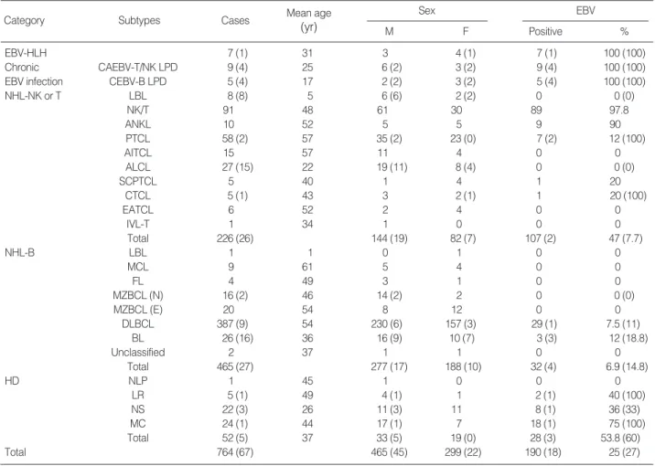
Demographic findings(Table 1)
The patients were all Korean, consisting of 299 women and 465 men, and had a mean age of 49 yr (range, 1-90).
Histological subtypes included HD (n=52), B-cell NHL (n=465), T&NK cell lymphoma (n=226), acute EBV-HLH (n=7), CAEBV infection (n=9), and B-cell LPD arising in chronic EBV infection (n=5).
Malignant lymphomas(Table 1)
Histological type of EBV-positive malignant lymphoma (Table 1) Among 743 cases of malignant lymphomas analyzed by in situ hybridization, 169 (23%) were EBER-positive. EBV was positive in 47% of T&NK cell lymphomas, 6.9% of B cell lymphomas, and 53.8% of HD. EBV was positive in almost all cases of NK/T cell lymphoma and aggressive NK cell leukemia. Compared with NK/T cell lymphoma or ANKL, less association was observed in other types of NHLs including diffuse large B cell lymphoma (DLBCL) (29/387,
7.5%), peripheral T cell lymphoma, unspecified (PTCL) (7/58, 12%), and Burkitt’s lymphoma (3/26, 12%). In HD, EBER positivity was relatively high in those tumors with mixed cellularity (75%, 18/24), but low in lymphocyte-rich forms (40%, 2/5), and in nodular sclerosis (36%, 8/22).
Among all EBV-positive T and NK cell type NHL cases, NK/T cell lymphoma was the most common subtype, accoun- ting for 89 of 107 EBV-positive T or NK type NHL cases (83%). Other T or NK NHLs showed similar incidences:
8.4% of ANKL (9/107) and 6.5% of PTCL (7/107). EBV- positive B-NHL cases comprised with DLBCL (29/32, 91%) and Burkitt’s lymphoma (3/32, 9%) (Fig. 1).
Age distribution of patients with EBV-positive malignant lymphoma
The ages of the patients with EBV-positive malignant lymphomas ranged from 5 to 88 yr with a median of 49.
EBV-negative malignant lymphomas showed an age peak at the sixties whereas EBV-positive malignant lymphomas showed a broad peak from the forties to sixties.
The age distribution of patients with EBV-negative HD showed a major peak in the twenties, whereas the occurrence of EBV-positive HD showed a large peak from the forties to sixties and a smaller peak in the twenties. DLBCLs occurred in old patients, with an age peak in the sixties.
NK/T cell lymphoma showed the highest incidence in the forties. A few cases of EBV-associated PTCL and ANKL developed in the patients in the twenties, but the major peak was in the forties for ANKL and in the sixties for PTCL .
Clinicopathology of EBV-positive malignant lymphomas
The patients with EBV-positive NK/T cell lymphomas were 89 patients. Primary sites of presentation were the nasal cavity and nasopharynx in 62 patients and 27 in extranasal-
Category Subtypes Cases Mean age
(yr)
EBV
Positive %
Sex
M F
EBV-HLH 7 (1) 31 3 4 (1) 7 (1) 100 (100)
Chronic CAEBV-T/NK LPD 9 (4) 25 6 (2) 3 (2) 9 (4) 100 (100)
EBV infection CEBV-B LPD 5 (4) 17 2 (2) 3 (2) 5 (4) 100 (100)
NHL-NK or T LBL 8 (8) 5 6 (6) 2 (2) 0 0 (0)
NK/T 91 48 61 30 89 97.8
ANKL 10 52 5 5 9 90
PTCL 58 (2) 57 35 (2) 23 (0) 7 (2) 12 (100)
AITCL 15 57 11 4 0 0
ALCL 27 (15) 22 19 (11) 8 (4) 0 0 (0)
SCPTCL 5 40 1 4 1 20
CTCL 5 (1) 43 3 2 (1) 1 20 (100)
EATCL 6 52 2 4 0 0
IVL-T 1 34 1 0 0 0
Total 226 (26) 144 (19) 82 (7) 107 (2) 47 (7.7)
NHL-B LBL 1 1 0 1 0 0
MCL 9 61 5 4 0 0
FL 4 49 3 1 0 0
MZBCL (N) 16 (2) 46 14 (2) 2 0 0 (0)
MZBCL (E) 20 54 8 12 0 0
DLBCL 387 (9) 54 230 (6) 157 (3) 29 (1) 7.5 (11)
BL 26 (16) 36 16 (9) 10 (7) 3 (3) 12 (18.8)
Unclassified 2 37 1 1 0 0
Total 465 (27) 277 (17) 188 (10) 32 (4) 6.9 (14.8)
HD NLP 1 45 1 0 0 0
LR 5 (1) 49 4 (1) 1 2 (1) 40 (100)
NS 22 (3) 26 11 (3) 11 8 (1) 36 (33)
MC 24 (1) 44 17 (1) 7 18 (1) 75 (100)
Total 52 (5) 37 33 (5) 19 (0) 28 (3) 53.8 (60)
Total 764 (67) 465 (45) 299 (22) 190 (18) 25 (27)
Table 1. Summary of disease entities enrolled for the study
Children (0-18 yr old) are shown in parentheses.
EBV-HLH, acute EBV-associated hemophagocytic lymphohistiocytosis; CAEBV, chronic active EBV-infection; CEBV, chronic EBV infection; NHL, non- Hodgkin’s lymphoma; LBL, lymphoblastic lymphoma; ANKL, aggressive NK cell leukemia; AITCL, angioimmunoblastic T cell lymphoma; ALCL, anaplas- tic large cell lymphoma; SCPTCL, subcutaneous panniculitis-like T cell lymphoma; CTCL, cutaneous T cell lymphoma other than mycosis fungoides, SCPTCL, and ALCL; EATCL, enteropathy-type T cell lymphoma; IVL-T, intravascular large cell lymphoma of T-lineage; MCL, mantle cell lymphoma;
FL, follicular lymphoma; MZBCL (E), marginal zone B cell lymphoma, extranodal; MZBCL (N), marginal zone B cell lymphoma, nodal; BL, Burkitt’s lym- phoma; HD, Hodgkin’s lymphoma; NLP, nodular lymphocyte predominance; LR, lymphocyte-rich; NS, nodular sclerosis; MC, mixed cellularity.
sites. The clinicopathology of these patients has been report- ed previously (10, 11, 16). Most patients presented with local symptoms such as nasal obstruction, testicular enlargement, or abdominal pain, without any previous history suggesting chronic EBV infection or immune deficiency. Only one patient had a history of mosquito-bite hypersensitivity in childhood.
The nine patients with EBV-positive ANKL presented with acute syndromes of fever, pancytopenia, coagulopathy, and hepatosplenomegaly. These patients had no previous history suggestive of chronic EBV infection. Three of seven patients with EBV-positive PTCL showed a past history such as recurrent upper respiratory infection-like symptoms, hep- atosplenomegaly, and increased liver enzymes with a duration shorter than 6 months, which suggested acute or subacute EBV infection. Other 3 patients with EBV-positive PTCL had a past or present illness of hepatitis C virus (HCV) infec- tion, prostate cancer, or medication for rheumatoid arthritis.
There were 29 patients with EBV-positive DLBCL: 17 men and 12 women. Primary sites of presentation were lymph node in 12 patients and extranodal sites in 17 patients such as brain, peritoneum, and gastrointestinal tract. Most patients had no history suggestive of chronic EBV infection. Two
patients had a concurrent illness such as hepatitis C and hep- atitis B, and three patients had previous diseases such as tuber- culosis, angioimmunoblastic T cell lymphoma, and HD.
Acute hemophagocytic lymphohistiocytosis and chronic EBV infection
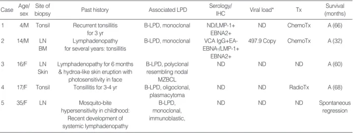
The clinicopathology of patients with acute HLH, chron- ic active EBV infection associated with NK or T LPD, and B-LPD of various spectra arising in the background of chron- ic EBV infection was summarized in Table 2-4.
Among the patients with CAEBV infection, ANKL develop- ed in two patients, hydroa-like T cell lymphoma in one, PTCL, unspecified in one, and monoclonal T-LPD in one patient.
Common EBV-positive LPD type by age groups
In early childhood, Burkitt’s lymphoma was the most com-
Fig. 1. Cell lineage and histological types among EBV-positive lymphomas.
17%
9%
DLBCL 8.4%
6.5% 2.1%
19%
64%
91% 83%
NK/T cell lymphoma Others NK or T-NHL HD
B-NHL
PTCL ANKL
Burkitt’s
Case Age/
sex Site of biopsy Past history Serology Viral load* Tx Survival
(months)
1 2/F Lymph node, BM N-C VCA IgG+IgM-EBNA+ ND ND D (1.3)
2 21/F BM N-C ND ND PBSCT A (60)
3 28/M LN Ankylosing spondylitis for 5 yr VCA IgM+EBNA+ ND PBSCT A (45)
4 29/F Skin, BM Postpartum ND ND ND D (2)
5 48/M BM Arthritis: dsDNA+(weak) VCA IgG+IgM-EA-EBNA+ 16,720 copy ChemoTX D (2)
6 61/M BM N-C ND ND ChemoTX D (2.3)
7 61/F Spleen, BM Hepatitis C ND ND ChemoTX D (2)
Table 2. Acute EBV-associated hemophagocytic lymphohistiocytosis
*, The viral load in peripheral mononuclear cells were examined by real-time PCR analysis.
Tx, treatment; N-C, not contributory; ND, not done; LN, lymph node; BM, bone marrow; PBSCT, peripheral blood stem cell transplantation; A, alive; D, died.
Fig. 2. The change of common histological types of EBV-positive lymphoproliferative disease according to age.
CEBV-B, chronic EBV infection associted B-LPD; CAEBV-T/NK, chronic active EBV infection-associated T or NK cell LPD.
%
100
80
60
40
20
0
0-9 10-19 20-29 30-39 40-49 50-59 60-69 70- Age
HD Burkitt’s DLBCL Acute HLH CAEBV-T/NK PTCL ANKL NK/T
CEBV-B
mon; in teenagers, chronic (active) EBV infection-associated LPD was the most common type. The incidence of NK/T cell lymphoma began to increase from the twenties and form- ed the major type of EBV-associated tumor throughout life.
DLBCL increased from the fifties and tended to form the major type in the sixties and seventies (Fig. 2).
Comparison of the overall survival of patients with EBV- associated LPDs
Among 190 EBV-positive samples of LPDs, survival data were available for 177 patients (Fig. 3). The follow-up period ranged from a few days to 111.6 months, with a median of nine months and a mean of 19 months. Median patient sur-
Case Age/
sex
Site of
biopsy Past history Associated LPD Serology/IHC Viral load* Tx Survival
(months)
1 16/F BM Nasal lymphoma (7 yr ago) ANKL ND ND N-C D (0.6)
with spontaneous regression
2 29/M BM Splenomegaly for 10 yr; ANKL ND ND ChemoTx D (2)
fever, cytopenia
3 11/M LN Hepatitis Sx for 2 yr; T-LPD, monoclonal VCA IgG+ IgM- 14,680 copy ChemoTx D (8) BM fever, lymphadenopathy
4 17/F Skin IgA nephropathy (5 yr ago): Hydroa-like T cell lymphoma ND ND ChemoTx D (6) Larynx hydroa-like skin eruption in skin/monoclonal
with photosensitivity
5 18/M BM Fever, fatigue for 6 months; PTCL, monoclonal ND ND ChemoTx D (10)
hepatosplenomegaly
6 22/M LN Mosquito-bite hypersensitivity T and NK LPD, VCA IgG+ ND ChemoTx D (10)
BM since childhood: fever, polyclonal EBNA+
Liver lymphadenopathy, cytopenia
7 29/F BM Recurrent tonsillitis since T and NK LPD, ND 6,936 copy N-C A (6)
Lung childhood: fever, polyclonal Wax and wane
LN respiratory Sx
8 35/M BM Hepatitis Sx for 1 yr T-LPD, polyclonal ND 17,230 copy N-C A (12)
Wax and wane
9 45/M LN Hepatitis Sx and T-LPD, polyclonal EA+VCA IgG+ ND Acyclovir IFN D (6)
Liver lymphadenopathy for 8 months EBNA+
Table 3. Chronic active EBV infection
*, The viral load in peripheral mononuclear cells examined by real-time PCR analysis.
LPD, lymphoproliferative disease; IHC, immunohistochemical staining on paraffin tissue; Tx, treatment; Sx, symptoms; LN, lymph node; BM, bone marrow; IFN, interferon; A, alive; D, died.
Case Age/
sex Site of
biopsy Past history Associated LPD Serology/
IHC Viral load* Tx Survival
(months)
1 4/M Tonsil Recurrent tonsillitis B-LPD, monoclonal ND/LMP-1+ ND ChemoTx A (66)
for 3 yr EBNA2+
2 14/M LN Lymphadenopathy B-LPD, monoclonal VCA IgG+EA- 497.9 Copy ChemoTx A (32)
BM for several years: tonsillitis EBNA-/LMP-1+
EBNA2+
3 16/F LN Lymphadenopathy for 6 months B-LPD, polyclonal ND ND ND A (60)
Skin & hydroa-like skin eruption with resembling nodal photosensitivity in face MZBCL
4 17/F Tonsil Tonsillitis for 3-4 yr B-LPD, oligoclonal, ND ND RadioTx A (68)
plasmacytoma
5 35/F LN Mosquito-bite B-LPD, ND ND ND Spontaneous
hypersensitivity in childhood: monoclonal, regression
Recent development of immunoblastic, systemic lymphadenopathy
Table 4. B-lymphoproliferative disease associated with chronic EBV infection
*, The viral load in peripheral mononuclear cells examined by real-time PCR analysis.
LPD, lymphoproliferative disease; IHC, immunohistochemical staining on paraffin tissue; Tx, Treatment; N-C, not contributory; ND, not done; LN, lymph node; BM, bone marrow; IFN, interferon; A, alive; D, died.
vivals for each category of T or NK EBV-positive LPDs were 2 months for acute HLH, 1.6 months for ANKL, 5.5 months for PTCL, 8 months for T/NK LPD associated with CAEBV, and 13.2 months for NK/T cell lymphomas. In contrast to patients with T and NK LPD, those with B-lineage LPD and HD showed excellent prognosis; all five patients with B-LPD arising in chronic EBV infection were alive at follow- up. The median survival for patients with DLBCL and HD were 65 months and longer than 69.5 months, respectively.
DISCUSSION
Populations from different ethnic groups have variable susceptibility to EBV infection, as is demonstrated by geo- graphical variations in the prevalence of EBV-related cancers.
The relative incidence of T or NK/T cell lymphomas, espe- cially EBV-associated nasal or nasal type NK/T cell lym- phoma, is much higher in Asians than in Western popula- tions (6, 13, 17). In Korea, T or NK/T cell lymphomas com- prise 25% of NHLs (13), which is in line with reports from other Far Eastern countries. In the present study, T and NK cell lymphoproliferative diseases accounted for 64% of EBV- associated disease and 47.5% of T or NK cell lymphomas were associated with EBV. If we exclude those EBV-positive T and NK cell lymphomas, the incidence of NK and T cell lymphoma in Korea is similar to that of Western countries, accounting for 10-15% of NHLs (17), which again empha- sizes the role of EBV in the pathogenesis of NK and T cell lymphomas in Asia.
B-cell tropism of EBV through the EBV-specific receptor CD21 (CR2), which is expressed on B cells and developing T cells but not on mature peripheral T cells, is well known
(18). In individuals with a normal immune system, sustained T-cell infection by EBV occurs only rarely, raising the possi- bility that the infection of T lymphocytes and their subse- quent unregulated growth is caused, at least in part, by a defect in immune surveillance (7). It has been suggested that a genetically determined susceptibility, possibly based on certain HLA types, results in an abnormal response to pri- mary EBV infection in certain parts of Asia. A study of Chi- nese and natives of New Guinea has shown that this ethnic group has a high prevalence of HLA A11, a type that is asso- ciated with a mutation of EBNA-4 that abrogates cytotoxic T-cell recognition of EBV (19, 20). By contrast, A11 is rare among Europeans, in whom cytotoxic T cells recognizing the EBNA-4 peptide dominate the immune response to EBV.
These variations in HLA phenotype may provide a basis for the higher frequency of EBV-positive tumors - including nasal T/NK cell lymphomas - among Asians. In addition, a recent study from Japan has shown that patients with nasal type EBV-associated NK/T cell lymphomas have a low frequen- cy of the HLA-A*0201 allele, suggesting the importance of this allele in cytotoxic T lymphocyte responses (21).
CAEBV infection is a peculiar disease showing an abnor- mal immune response to primary EBV infection and char- acterized by recurrent infectious mononucleosis-like symp- toms in childhood. Patients with CAEBV infections usually show clonal proliferation of T or NK cell and some of them develop overt NK cell or T cell lymphoma/leukemia in their teens and twenties (22). In the present study, ANKL and PTCL were mainly diseases of adults, but a few instances of EBV-associated PTCLs and ANKLs developed in patients in their twenties. Considering the natural course of chronic active EBV infection, these ANKL and PTCL occurring in young adults were probably transformed from chronic active EBV infections.
Patients with NK/T cell lymphomas, ANKL, and PTCL had major age peaks in their forties, forties, and sixties, res- pectively. It is not known what proportion of those EBV- associated diseases occurring in adults is a de novo disease unrelated with chronic (active) EBV infection. In Korea, the primary infection rate with EBV is 90% in those aged 7-9 yr and 100% in those between 10 and 15 yr (23). Almost all adult individuals are serologically positive. Therefore, EBV- associated diseases of adult onset seem to be derived from reactivation of chronic latent EBV infections or new EBV infection. In this study, we failed to find the past history sug- gesting chronic EBV infection from the medical record in most patients. Only one patient with a nasal-type NK/T cell lymphoma had mosquito-bite hypersensitivity when he was young. After childhood, he was relatively healthy without specific symptoms related to chronic EBV infection. Because mosquito-bite hypersensitivity is a cutaneous manifestation associated with latent EBV infection of the NK cells (7), this supports the idea that adult-onset nasal NK/T cell lymphoma may develop with a background of chronic latent EBV infec-
Fig. 3. Survival curve of patients with EBV-positive lymphoprolifer- ative disease.
CEBV-B, chronic EBV infection associated B-LPD; CAEBV-T/NK, chronic active EBV infection associated T or NK cell LPD; TCL, peripheral T-cell lymphoma.
Cum survival
1.2
1.0
0.8
0.6
0.4
0.2
0.0
-0.2
-20 0 20 40 60 80 100 120
Months
CEBV-B
HD
DLBCL
NKT CAEBV-T/NK
ANKLTCL AHLH
Survival functions
tion. Given the age that most patients develop NK/T cell lymphomas, transformation of EBV-infected NK cells into NK/T cell lymphoma cells may require a long latent period for the accumulation of genetic mutations sufficient for neo- plastic transformation. Prospective epidemiologic studies are clearly needed to clarify the natural history of adult-onset EBV-associated malignancy.
In this study, some patients with adult-onset EBV-associ- ated LPD showed associated illnesses that could have caused immunological dysfunctions provoking the reactivation of latent EBV infection and neoplastic transformation of EBV- infected cells. Four of six adult patients with acute EBV- HLH were associated with HCV hepatitis and chronic arthri- tis, or postpartum status. Three of seven patients with EBV- associated PTCL had a history of medication for rheumatoid arthritis, HCV hepatitis, and prostate carcinoma. Common association with HCV in these patients suggests an appar- ently ineffective antiviral T-cell response. Likewise, in the patients with DLBCL, 19 of the 29 patients were older than 60 yr and 5 of the 29 adult EBV-positive DLBCL patients had histories of HCV hepatitis, hepatitis B virus (HBV) hepatitis, tuberculosis, or HD, which suggests that decreas- ed immunity in elderly patients may also contribute to the pathogenesis of adult EBV-positive DLBCL (24).
It is intriguing that the five patients with chronic EBV infection were associated with B-cell LPDs of various histo- logical spectra. Three of four such patients presented with recurrent tonsillitis-like symptoms with monoclonal or oligo- clonal B cell proliferation. Two of them expressed EBNA-2 as well as LMP-1, which indicates a type III latency of EBV infection usually identified in immunocompromised patients
and which indicates an innate defect in the immune surveil- lance of EBV infection. The prognosis of those patients with B-LPD was excellent, as it was for those with EBV-associat- ed large B cell lymphomas and for adult patients with HD.
With additional genetic changes, these proliferating B cells in children with an immune defect to eradicate EBV may convert to HD or DLBCL, although the exact relationship between B-cell LPDs in children with chronic EBV infec- tion and EBV-associated large B cell lymphomas or HD aris- ing among adult patients remains to be clarified.
In summary, EBV infection in Koreans induced predomi- nantly T or NK cell LPDs; among these, NK/T cell lym- phomas were the most common. A high prevalence of EBV infection in early childhood associated with the failure of innate immune responses to eradicate the virus resulted in the devel- opment of EBV-associated LPD throughout life. As shown in Fig. 4, primary infections in early childhood may be compli- cated by the development of Burkitt’s lymphoma and acute EBV-HLH as well as CAEBV infections; some of these trans- formed to ANKLs and PTCLs in young adults. In the mid- dle-aged patients, some with chronic latent infections develop- ed NK/T cell lymphomas and ANKL. In old patients, decreas- ed immunity and environmental cofactors may provoke the development of PTCL and DLBCL.
REFERENCES
1. Epstein MA, Achong BG, Barr YM. Virus particles in cultured lym- phoblasts from Burkitt’s lymphoma. Lancet 1964; 15: 702-3.
2. Weiss LM, Movahed LA, Warnke RA, Sklar J. Detection of Epstein- Fig. 4. The relation between EBV infection and development of EBV-associated lymphoproliferative disease by age group.
HD, Hodgkin’s lymphoma; ANKL, aggressive NK cell leukemia; DLBCL, diffuse large B cell lymphoma; PTCL, peripheral T cell lymphoma.
*, environmental cofactor.
0 yr 10 yr 20 yr 30 yr 40 yr 50 yr 60 yr 70 yr
Primary infection
Nasal-type NK/T cell lymphoma HD
PTCL Chronic active infection
ANKL
PTCL
HD Burkitt’s
Chronic latent infection
B<<T,NK
DLBCL ANKL
*
Barr viral genomes in Reed-Sternberg cells of Hodgkin’s disease. N Engl J Med 1989; 320: 502-6.
3. Berg LC, Copenhaver CM, Morrison VA, Gruber SA, Dunn DL, Gajl-Peczalska K, Strickler JG. B-cell lymphoproliferative disorders in solid-organ transplant patients: detection of Epstein-Barr virus by in situ hybridization. Hum Pathol 1992; 23: 159-63.
4. MacMahon EM, Glass JD, Hayward SD, Hayward SD, Mann RB, Becker PS, Charache P, McArthur JC, Ambinbder RF. Epstein-Barr virus in AIDS-related primary central nervous system lymphoma.
Lancet 1991; 338: 969-73.
5. Quintanilla-Martinez L, Franklin JL, Guerrero I, Krenacs L, Naresh KN, Rama-Rao C, Bhatia K, Raffeld M, Magrath IT. Histological and immunophenotypic profile of nasal NK/T cell lymphomas from Peru: high prevalence of p53 overexpression. Hum Pathol 1999;
30: 849-55.
6. Chan JK, Sin VC, Wong KF, Ng CS, Tsang WY, Chan CH, Cheung MM, Lau WH. Nonnasal lymphoma expressing the natural killer cell marker CD56: a clinicopathologic study of 49 cases of an uncom- mon aggressive neoplasm. Blood 1997; 89: 4501-13.
7. Iwatsuki K, Yamamoto T, Tsuji K, Suzuki D, Fujii K, Matsuura H, Oono T. A spectrum of clinical manifestations caused by host immune responses against Epstein-Barr virus infections. Acta Med Okayama 2004; 58: 169-80.
8. Quintanilla-Martinez L, Kumar S, Fend F, Reyes E, Teruya-Feldstein J, Kingma DW, Sorbara L, Raffeld M, Straus SE, Jaffe ES. Fulmi- nant EBV (+) T-cell lymphoproliferative disorder following acute/
chronic EBV infection: a distinct clinicopathologic syndrome. Blood 2000; 96: 443-51.
9. Jaffe ES, Harris NL, Stein H, Vardiman JW. Pathology and Genetics of Tumours of Haematopoietic and Lymphoid Tissues. Lyon, France:
IARC Press, 2001.
10. Ko YH, Ree HJ, Kim WS, Choi WH, Moon WS, Kim SW. Clini- copathologic and genotypic study of extranodal nasal-type natural killer/T-cell lymphoma and natural killer precursor lymphoma among Koreans. Cancer 2000; 89: 2106-16.
11. Lee J, Kim WS, Park YH, Park SH, Park KW, Kang JH, Lee SS, Lee SI, Lee SH, Kim K, Jung CW, Ahn YC, Ko YH, Park K. Nasal- type NK/T cell lymphoma: clinical features and treatment outcome.
Br J Cancer 2005; 92: 1226-30.
12. Kwong YL, Chan AC, Liang R, Chiang AK, Chim CS, Chan TK, Todd D, Ho FC. CD56+ NK lymphomas: clinicopathological fea- tures and prognosis. Br J Haematol 1997; 97: 821-9.
13. Ko YH, Kim CW, Park CS, Jang HK, Lee SS, Kim SH, Ree HJ, Lee JD, Kim SW, Huh JR. REAL classification of malignant lymphomas
in the Republic of Korea: incidence of recently recognized entities and changes in clinicopathologic features. Cancer 1998; 83: 806-12.
14. Imashuku S. Clinical features and treatment strategies of Epstein- Barr virus-associated hemophagocytic lymphohistiocytosis. Crit Rev Oncol Hematol 2002; 44: 259-72.
15. Straus SE. The chronic mononucleosis syndrome. J Infect Dis 1988;
157: 405-12.
16. Lee J, Suh C, Park YH, Ko YH, Bang SM, Lee JH, Lee DH, Huh J, Oh SY, Kwon HC, Kim HJ, Lee SI, Kim JH, Park J, Oh SJ, Kim K, Jung C, Park K, Kim WS. Extranodal natural killer T-cell lym- phoma, nasal-type: a prognostic model from a retrospective multi- center study. J Clin Oncol 2006; 24: 612-8.
17. Anderson JR, Armitage JO, Weisenburger DD. Epidemiology of the non-Hodgkin’s lymphomas: distributions of the major subtypes dif- fer by geographic locations. Non-Hodgkin’s Lymphoma Classifica- tion Project. Ann Oncol 1998; 9: 717-20.
18. Tsoukas CD, Lambris JD. Expression of EBV/C3d receptors on T cells: Biological significance. Immunol Today 1993; 14: 56-9.
19. De Campos-Lima PO, Gavioli R, Zhang QJ, Wallace LE, Dolcetti R, Rowe M, Rickinson AB, Masucci MG. HLA-A11 epitope loss isolates of Epstein-Barr virus from a highly A11+ population. Sci- ence 1993; 260: 98-100.
20. De Campos-Lima PO, Levitsky V, Brooks J, Lee SP, Hu LF, Rick- inson AB, Masucci MG. T cell responses and virus evolution: loss of HLA A11-restricted CTL epitopes in Epstein-Barr virus isolates from highly A11-positive populations by selective mutation of anchor residues. J Exp Med 1994; 179: 1297-305.
21. Kanno H, Kojya S, Li T, Ohsawa M, Nakatsuka S, Miyaguchi M, Harabuchi Y, Aozasa K. Low frequency of HLA-A*0201 allele in patients with Epstein-Barr virus-positive nasal lymphomas with poly- morphic reticulosis morphology. Int J Cancer 2000; 87: 195-9.
22. Suzuki K, Ohshima K, Karube K, Suzumiya J, Ohga S, Ishihara S, Tamura K, Kikuchi M. Clinicopathological states of Epstein-Barr virus-associated T/NK-cell lymphoproliferative disorders (severe chronic active EBV infection) of children and young adults. Int J Oncol 2004; 24: 1165-74.
23. Oh SH, Lee YA, Moon WY, Ko TS, Park YS, Moon HN, Hong CY, Kim DW. Prevalence of Epstein-Barr virus (EBV) antibody in Kore- an children. J Korean Pediatr Soc 1994; 37: 804-11.
24. Oyama T, Ichimura K, Suzuki R, Suzumiya J, Ohshima K, Yatabe Y, Yokoi T, Kojima M, Kamiya Y, Ogura M, Saito H, Morishima Y, Nakamura S. Senile EBV+ B-cell lymphoproliferative disorders:
a clinicopathologic study of 22 patients. Am J Surg Pathol 2003;
27: 16-26.