INTRODUCTION
Dialysis patients have a high incidence of cardiovascular morbidity and mortality (1, 2). Vascular calcification (VC) in hemodialysis (HD) patients was associated with increased stiffness of the artery and has already known to be related to cardiovascular disease and cardiovascular mortality (3-5). The simple VC score of plain radiographic films of the pelvis and hands was independently associated with coronary artery dis- ease and peripheral arterial disease at the end of the follow-up (6). There are two types of VC on the plain radiograph, arte- rial medial calcification and arterial intimal calcification which has irregular and patchy distribution. The presence of medi- al artery calcifications on the plain radiograph which is more common in dialysis patients than in the general population, is a powerful and independent prognostic marker for all-cause and cardiovascular mortality in chronic HD patients (7, 8).
Typical linear railroad-track type (angiography like) calcifica- tions on the plain radiograph that outline the vessel walls are considered as medial artery calcification which is differenti- ated from intimal calcifications (7). Medial artery calcifica- tion of dorsalis pedis artery on the plain radiography of feet is also one of peripheral artery vascular calcifications such as vascular calcification of hands.
Maintenance dialysis patients have a reverse association with some traditional risk factors; body mass index (BMI), hyper-
cholesterolaemia and hypertension reduced the relative risk of death in epidemiological studies (9-11). In fact, dialysis patients have specific non-traditional risk factors; inflamma- tion, malnutrition, oxidative stress, and abnormal mineral metabolism are other important risk factors for vascular dis- ease (12). In general, oxidized low density lipoprotein (ox- LDL) cholesterol is closely associated with the inflammatory process in vascular atherosclerotic lesions (13, 14) and inflam- matory marker, C-reactive protein (CRP), is a stronger predic- tor of cardiovascular events than low density lipoprotein (LDL) cholesterol (15). Plasminogen activator inhibitor-1 (PAI-1) is also linked to vascular inflammation and atherosclerosis (16).
Thus, CRP, PAI-1 and ox-LDL have a high possibility of rela- tion with VC which is risk factor of cardiovascular morbidi- ty and mortality in dialysis patients. The present study was designed to find factors related with medial artery calcifica- tion on the plain radiography of feet by comparing CRP, PAI-1 and lipid profile including ox-LDL and to elucidate associations among these factors in HD patients.
MATERIALS AND METHODS Patients
We recruited a total of 48 HD patients (male: 24 patients,
S115
Won Suk An, Seong-Eun Kim, Ki-Hyun Kim, Hae-Rahn Bae*, and Seo-Hee Rha�
Departments of Internal Medicine, Physiology*, and Pathology�, Dong-A University, Busan, Korea
Address for correspondence Won Suk An, M.D.
Department of Internal Medicine, Dong-A University, 1 Dongdaesin-dong 3ga, Seo-gu, Busan 601-715, Korea
Tel : +82.51-240-2811, Fax : +82.51-242-5852 E-mail :[email protected]
*This study was supported by the Dong-A University Research Fund in 2005.
DOI: 10.3346/jkms.2009.24.S1.S115
Associations between Oxidized LDL to LDL Ratio, HDL and Vascular Calcification in the Feet of Hemodialysis Patients
Cardiovascular mortality is associated with vascular calcification (VC) in hemodial- ysis (HD) patients. The present study was designed to find factors related with medi- al artery calcification on the plain radiography of feet by comparing C-reactive pro- tein (CRP), plasminogen activator inhibitor type 1 (PAI-1) and lipid profile including oxidized low density lipoprotein (ox-LDL) and to elucidate associations among these factors in HD patients. Forty-eight HD patients were recruited for this study. VC in the feet was detected in 18 patients (37.5%) among total patients and 12 patients (85.7%) among diabetic patients. Diabetes, cardiovascular disease (CVD), pulse pressure, ox-LDL/LDL were higher and high density lipoprotein (HDL) was lower in patients with VC than in patients without VC. Negative associations were found be- tween HDL and CRP, PAI-1. PAI-1 had positive association with ox-LDL/LDL. His- tory of CVD was the only determinant of vascular calcification on the plain radiog- raphy of feet. Ox-LDL/LDL, HDL, CRP, and PAI-1 were closely related with one anoth- er in HD patients. History of CVD is the most important factor associated with the presence of VC and low HDL and relatively high oxidized LDL/LDL ratio may affect VC formation on the plain radiography in the feet of HD patients.
Key Words : Vascular Calcification; Oxidized Low Density Lipoprotein; High Density Lipoproteins; Hemodialysis
Received : 24 August 2008 Accepted : 1 December 2008
diabetes: 14 patients) from Dong-A University dialysis cen- ter and a neighboring local dialysis center. Patients with HD duration of less than 6 months, patients with a catheter as vascular access, history of active infection within 3 months, malignancy and chronic liver disease were excluded. The mean age of the patients was 56.3±11.5 yr and the mean duration on HD was 77.1±54.1 months. Enrolled HD patients receiv- ed regular HD thrice weekly. Bicarbonate-based dialysate and polysulphone dialyzers (Fresenius, Bad Homburg, Germany) were used. Informed consent was obtained in accordance with the guidelines set forth by the Declaration of Helsinki. We defined medial artery calcification on the plain radiograph of feet as vascular calcification. We reviewed medical history and records by chart review and interview. Coronary artery disease (CAD), cerebrovascular accident and peripheral artery disease (PAD) were defined as cardiovascular disease (CVD).
We defined CAD based on the records of myocardial SPECT scan, echocardiography or coronary angiography. Patients with a history of lower extremity amputation caused by PAD, steno- sis of peripheral artery on the color doppler ultrasonography or present ulceration and necrosis of the foot were defined as PAD. We reviewed current medications and checked blood pressure of the enrolled patients in the sitting position before hemodialysis.
Radiography of both feet
Standard radiographs of both feet were taken to determine VC. AXIOM Aristos MX/VX (SIEMENS, Erlangen, Ger-
many) radiographic equipment with digital imaging system was used and exposure condition was approximately 45-50 kVp (4 mAs).
Calcifications that outline the dorsalis pedis artery on the plain radiograph were shown as typical linear railroad-track form like angiography (Fig. 1). VC of this form indicates medi- al artery calcification. One nephrologist and one radiologist individually decided VC of the dorsalis pedis artery on the plain radiograph without information of patients. Consen- sus was reached on the interpretation of all radiographs.
Laboratory measurements
Routine laboratory tests such as hemoglobin, blood urea nitrogen (BUN), creatinine, albumin, calcium, phosphate, intact parathyroid hormone (iPTH), CRP and lipid profile were obtained using fasting blood samples. Normalized pro- tein catabolic rate (nPCR), fractional clearance of urea as a func- tion of its distribution volume (Kt/V urea) and BMI were also determined. HbA1c was checked in diabetic patients. PAI-1 was measured by ASSERACHROM�PAI-1 kit (Diagnosti- ca stago Inc, NJ, U.S.A.), oxidized LDL (Mercodia, Uppsala, Sweden) and anti-oxidized LDL antibody (oLAB; Biomedi- ca, Vienna, Austria) was measured by using a commercially available enzyme linked immunosorbent assay kit.
Prehemodialysis blood samples were obtained from the arte- riovenous fistula or graft in HD patients. Samples were imme- diately placed on ice. Plasma and serum were promptly sep- arated by centrifugation and stored at -70℃until assayed.
Statistics
Data are presented as mean±S.D. except iPTH which is expressed as mean±S.E. Comparisons of unpaired data were performed using Mann-Whitney U test. Correlation analy- ses were examined by using Spearman test. Differences in frequency were tested using chi-square analysis. Significant variables identified by univariate analysis were entered into a binary logistic regression analysis to identify variables asso- ciated with vascular calcification. p values less than 0.05 were considered significant. All statistical calculations were per- formed with SPSS software, version 12.0 (SPSS Inc, Chica- go, IL, U.S.A.).
RESULTS
Comparison of data according to vascular calcification
Data are summarized in Table 1 and Table 2. The preva- lence rate of VC was 37.5% in these HD patients. VC on the plain radiography of feet was found to be significantly more common in diabetic patients than in non-diabetic patients (66.7% vs. 6.7%, p<0.001). Diabetic HD patients
Fig. 1. Standard radiograph of foot shows definite vascular calci- fications on the course of dorsalis pedis artery. High density that outline the dorsalis pedis artery on the plain radiograph was shown as typical linear railroad-track form like angiography indicates vas- cular media calcification of dorsalis pedis artery (arrow).
showed significantly shorter HD duration than non-diabet- ic HD patients (34.5±36.0 vs. 95.0±50.6 months, p<
0.001). HD duration was also shorter in patients with VC than in patients without VC. Age, gender, BMI, smoking history, systolic blood pressure, diastolic blood pressure and calcium loads by taking phosphate binder were not signifi- cantly different among groups with VC and without VC.
History of coronary artery disease and cardiovascular disease were more frequent and pulse pressure was higher in patients with VC than in patients without VC. The percent of patients who had been taking aspirin was significantly higher in patients with VC than in patients without VC.
The percent of patients who had been taking angiotensin converting enzyme inhibitors, angiotensin receptor blockers,
BMI, body mass index; HD, hemodialysis; DM, diabetes mellitus; CAD, history of coronary artery disease; CVD, history of cardiovascular disease;
AV, arteriovenous; SBP, systolic blood pressure; DBP, diastolic blood pressure; ACEI, angiotensin converting enzyme inhibitor; ARB, angiotensin receptor blocker; CCB, calcium channel blocker.
Calcification (-) Calcification (+) p value
N 30 18 -
Age (yr) 54.4±12.9 59.4±7.8 0.166
Male/female 14/16 10/8 0.555
BMI (kg/m2) 20.2±2.0 20.9±2.8 0.521
Smoking history (%) 10 (33.3) 7 (38.9) 0.466 HD duration (months) 93.6±52.7 50.9±46.6 0.007
DM (%) 2 (6.7) 12 (66.7) <0.001
CAD (%) 1 (3.3) 11 (61.1) <0.001
CVD (%) 2 (6.7) 15 (83.3) <0.001
AV graft (%) 2 (6.7) 4 (22.2) 0.039
SBP (mmHg) 147.8±15.4 155.9±14.4 0.132
DBP (mmHg) 76.8±14.2 71.8±13.3 0.248
Pulse pressure 71.0±17.9 84.1±14.3 0.008 Calcium loads (mg/day) 2,603±1,399 2,673±1,496 0.964
ACEI (%) 10 (33.8) 10 (55.6) 0.135
ARB (%) 11 (36.7) 10 (55.6) 0.206
CCB (%) 16 (53.3) 12 (66.7) 0.369
Statin (%) 1(3.3) 0 (0.0) 0.321
Aspirin (%) 9 (30) 14 (77.8) 0.002
Vitamin D (%) 10 (33.3) 3 (16.7) 0.217
Table 1. Comparison of clinical characteristics between patients with vascular calcification and without vascular calcification (Data are expressed as mean±S.D. iPTH is expressed as mean±S.E.)
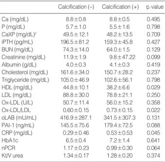
Ca, calcium; P, phosphate; CaXP, Calcium phosphate product; iPTH, intact parathyroid hormone; BUN, blood urea nitrogen; HDL, high den- sity lipoprotein cholesterol; LDL, low density lipoprotein cholesterol; Ox- LDL, oxidized LDL; oLAB, anti-oxidized LDL antibody; PAI-1, plasmino- gen activator inhibitor 1; CRP, C-reactive protein; nPCR, normalized pro- tein catabolic rate; Kt/V urea, fractional clearance of urea as a function of its distribution volume.
Calcification (-) Calcification (+) p value
Ca (mg/dL) 8.8±0.8 8.8±0.5 0.495
P (mg/dL) 5.7±1.0 5.5±1.6 0.798
CaXP (mg/dL)2 49.5±12.1 48.2±13.5 0.709 iPTH (pg/mL) 196.5±81.2 159.3±45.8 0.427
BUN (mg/dL) 74.3±14.0 64.0±1.5 0.129
Creatinine (mg/dL) 11.9±1.9 9.8±47.22 0.099
Albumin (g/dL) 4.0±0.3 4.1±0.3 0.419
Cholesterol (mg/dL) 161.6±34.0 150.7±28.2 0.237 Triglyceride (mg/dL) 105.0±46.9 102.6±56.1 0.798
HDL (mg/dL) 44.8±10.1 38.2±6.6 0.029
LDL (mg/dL) 88.8±30.0 78.8±21.1 0.250
Ox-LDL (U/L) 50.7±11.4 56.0±15.2 0.358
Ox-LDL/LDL 0.60±0.15 0.73±0.15 0.022
oLAB (mU/mL) 416.9±287.1 341.5±307.3 0.131 PAI-1 (ng/mL) 145.5±75.6 179.4±72.5 0.088
CRP (mg/dL) 0.29±0.46 0.53±0.53 0.045
HbA1c 6.5±0.4 7.2±1.4 0.641
nPCR 1.17±0.23 0.99±0.30 0.064
Kt/V urea 1.34±0.17 1.28±0.20 0.374
Table 2. Comparison of laboratory results between patients with vascular calcification and without vascular calcification
Fig. 2. Correlation between CRP, PAI-1 and HDL in hemodialysis patients.
CRP, C-reactive protein; PAI-1, plasminogen activator inhibitor 1; HDL, high density lipoprotein cholesterol.
PAI-1 (ng/mL)
350
300
250
200
150
100
50
0
20 30 40 50 60 70 80
HDL (mg/dL) r=-0.375, p=0.009
CRP (mg/dL)
2.5
2.0
1.5
1.0
0.5
0.0
20 30 40 50 60 70 80
HDL (mg/dL) r=-0.336, p=0.023
calcium channel blockers, calcium acetate phosphate binder (94.4% vs. 93.3%), statin and vitamin D was not significant- ly different among groups with VC and without VC (Table 1).
Patients with VC had much more arteriovenous graft as a vascular access compared to patients without VC. Patients with VC showed significantly higher oxidized LDL to LDL ratio and CRP levels, lower high density lipoprotein (HDL) cholesterol than patients without VC. Calcium, phosphate, iPTH, BUN, creatinine, serum albumin, total cholesterol, triglyceride, LDL, ox-LDL, anti-oxidized LDL antibody (oLAB), PAI-1, HbA1c, nPCR and Kt/V urea were not sig- nificantly different among groups with VC and without VC (Table 2).
Correlation between ox-LDL/LDL, HDL, CRP, and PAI-1
A negative association was found between ox-LDL and oLAB (r=-0.426, p=0.003). Negative associations were found between HDL and PAI-1(r=-0.375, p=0.009), CRP (r=-0.336, p=0.023) (Fig. 2). A positive association was found between PAI-1 and ox-LDL to LDL ratio (r=0.305, p=0.016).
Logistic regression analysis according to vascular calcification
Significant predictors detecting the presence of VC were diabetes mellitus, history of cardiovascular disease, HD dura- tion, pulse pressure, HDL and ox-LDL to LDL ratio in uni- variate analysis. History of cardiovascular disease was the only independent factor associated with the presence of VC (odds ratio: 55.71 and 95% confidence interval: 2.75-1130.46, p=0.009) by multivariate analysis including age and gender (Table 3).
DISCUSSION
VC in dialysis patients has been related to diabetes, CVD and
increased stiffness of artery (3-5, 8, 17, 18). In our study, his- tory of diabetes, CVD and high pulse pressure were related with the presence of medial artery calcification on the plain radiography of feet, but history of CVD was the only inde- pendent factor associated with the presence of VC. Thus we can consider evaluation of CVD if there is angiography like VC of dorsalis pedis artery on the plain radiography of feet in HD patients. A recent report mentioned that coexistence of coronary artery calcification (CAC) and peripheral artery calcification was common particularly in chronic kidney disease patients with diabetes, although VC detected by plain radiograph of foot was not an adequate marker for identify- ing patients with CAC (19). They checked a standardized plain radiograph of the left foot to determine if there was any calcification of the dorsalis pedis artery, not medial artery calcification.
Ox-LDL is known as a marker of atherosclerosis in HD patients (20). High levels of ox-LDL are associated with cal- cification of the aortic valve in patients with aortic valve stenosis (21). However, ox-LDL was not involved in coronary artery calcification in HD patients (22). Ox-LDL was not significantly different, but ox-LDL to LDL ratio was signifi- cantly different among groups with VC and without VC in our study. Ox-LDL to LDL ratio, an accurate estimation of in vivo LDL oxidation (23), indicates an environment of oxidative stress in HD patients with relatively low LDL lev- els. Thus, we suggest that the formation of medial artery calcification on the plain radiograph of feet may be affected by oxidative stress. In other aspect, if there are medial artery calcifications of feet, they reflect high oxidative stress condi- tion.
The higher titer of anti-oxidized LDL antibody in females and young persons had a negative association with atheroscle- rosis (24) and anti-oxidized LDL antibody was independently inversely associated with arterial wall thickness in HD patients (25). Ox-LDL had a reverse relationship with anti-oxidized LDL antibody in our HD patients. This result supports the anti-atherogenic role of anti-oxidized LDL antibody. But there is no evidence that anti-oxidized LDL antibody is related with VC in our study; it requires further study.
HDL was significantly associated with calcification in extra- coronary arteries evaluated by electron beam computed tomog- raphy (26). Furthermore, low HDL levels were associated with rapid progression of coronary artery calcification determined by electron-beam computed tomography in HD patients (27).
HD patients with VC exhibited significantly lower HDL lev- els than HD patients without VC in this study. Thus, low HDL which is common in dialysis patients may play a role in the formation of VC on the plain radiograph of feet.
HDL has anti-oxidant and anti-inflammatory properties.
The anti-oxidant effect of HDL was shown by inhibition of LDL oxidation in healthy young men (28). In our study there was no association between HDL and ox-LDL but a negative association was found between HDL and CRP as well as HDL
OR, odds ratio; CI, confidence interval; DM, diabetes mellitus; CVD, his- tory of cardiovascular disease; HD, hemodialysis; HDL, high density li- poprotein cholesterol; Ox-LDL, oxidized LDL; LDL, low density lipopro- tein cholesterol.
Variables OR (95% CI) p value
Age 1.03 (0.94-1.14) 0.513
Gender 0.91 (0.07-11.65) 0.940
DM 2.39 (0.07-80.69) 0.628
CVD 55.71(2.75-1,130.46) 0.009
HD duration (months) 1.01 (0.97-1.04) 0.688
Pulse pressure 1.03 (0.95-1.12) 0.445
HDL (mg/dL) 0.96 (0.80-1.16) 0.657
Ox-LDL/LDL 2,261.57 (0.62-8,312,558.7) 0.065 Table 3. Determinants of vascular calcification on the plain radio- graphy of feet by multivariate analysis including age and gender
and PAI-1, which are important markers of inflammation and atherosclerosis. PAI-1 levels are elevated in dialysis patients with developing atheromatous cardiovascular disease associ- ated with oxidative stress and inflammation (29). A recent report showed that PAI-1 was related with ox-LDL and inflam- mation in HD patients (30). PAI-1 had a positive relation with ox-LDL to LDL ratio in our study. This result supports that PAI-1 may be linked to oxidative stress. CRP levels were ele- vated in our HD patients with VC compared to HD patients without VC. Krasniak A et al. (22) reported that CRP was related with coronary artery calcification in HD patients, al- though CRP was not an independent factor. Finally, these observations suggest that ox-LDL/LDL, HDL, PAI-1, and CRP may be indirectly cross- linked in HD patients who have conditions of oxidative stress and inflammation.
This cross-sectional study has some limitations. The dura- tion of dialysis correlates with calcification in the peripheral arteries (8). But our study showed that HD duration was short- er in patients with VC than in patients without VC. This result is affected by diabetic HD patients, who had shorter HD duration and frequent VC in our study. And cardiovas- cular calcification was more prevalent in those receiving cal- cium containing phosphate binders (31). But our cross-sec- tional study showed that calcium, phosphate, calcium and phosphate product, iPTH, phosphate binder and vitamin D medication was not different among groups with VC and without VC. Thus prospective study will be needed to eval- uate the effect of hyperphosphatemia and phosphate binder in HD patients without vascular calcification.
In conclusion, Ox-LDL/LDL, HDL, CRP, and PAI-1 were closely related with one another in HD patients. History of CVD is the most important factor associated with the pres- ence of VC in the feet and low HDL and relatively high oxi- dized LDL/LDL ratio may affect VC formation on the plain radiography in the feet of HD patients.
REFERENCES
1. Foley RN, Parfrey PS, Sarnak MJ. Clinical epidemiology of cardio- vascular disease in chronic renal disease. Am J Kidney Dis 1998;
32 (5 Suppl 3): S112-S9.
2. Keith DS, Nichols GA, Gullion CM, Brown JB, Smith DH. Longi- tudinal follow-up and outcomes among a population with chronic kidney disease in a large managed care organization. Arch Intern Med 2004; 164: 659-63.
3. Blacher J, Guerin AP, Pannier B, Marchais SJ, Safar ME, London GM. Impact of aortic stiffness on survival in end-stage renal disease.
Circulation 1999; 99: 2434-9.
4. Blacher J, Guerin AP, Pannier B, Marchais SJ, London GM. Arteri- al calcifications, arterial stiffness, and cardiovascular risk in end-stage renal disease. Hypertension 2001; 38: 938-42.
5. Guerin AP, London GM, Marchais SJ, Metivier F. Arterial stiffen- ing and vascular calcifications in end-stage renal disease. Nephrol
Dial Transplant 2000; 15: 1014-21.
6. Adragao T, Pires A, Lucas C, Birne R, Magalhaes L, Goncalves M, Negrao AP. A simple vascular calcification score predicts cardiovas- cular risk in haemodialysis patients. Nephrol Dial Transplant 2004;
19: 1480-8.
7. Braun J, Oldendorf M, Moshage W, Heidler R, Zeitler E, Luft FC.
Electron beam computed tomography in the evaluation of cardiac calcification in chronic dialysis patients. Am J Kidney Dis 1996; 27:
394-401.
8. London GM, Guerin AP, Marchais SJ, Metivier F, Pannier B, Adda H. Arterial media calcification in end-stage renal disease: impact on all-cause and cardiovascular mortality. Nephrol Dial Transplant 2003;
18: 1731-40.
9. Coresh J, Longenecker JC, Miller ER, Young HJ, Klag MJ. Epidemi- ology of cardiovascular risk factors in chronic renal disease. J Am Soc Nephrol 1998; 9 (12 Suppl): S24-30.
10. Fleischmann EH, Bower JD, Salahudeen AK. Risk factor paradox in hemodialysis: better nutrition as a partial explanation. ASAIO J 2001; 47: 74-81.
11. Kalantar-Zadeh K, Block G, Humphreys MH, Kopple JD. Reverse epidemiology of cardiovascular risk factors in maintenance dialysis patients. Kidney Int 2003; 63: 793-808.
12. Stenvinkel P, Pecoits-Filho R, Lindholm B. Coronary artery disease in end-stage renal disease: no longer a simple plumbing problem. J Am Soc Nephrol 2003; 14: 1927-39.
13. Libby P. Inflammation in atherosclerosis. Nature 2002; 420: 868-74.
14. Hansson GK. Inflammation, atherosclerosis, and coronary artery dis- ease. N Engl J Med 2005; 352: 1685-95.
15. Ridker PM, Rifai N, Rose L, Buring JE, Cook NR. Comparison of C-reactive protein and low-density lipoprotein cholesterol levels in the prediction of first cardiovascular events. N Engl J Med 2002; 347:
1557-65.
16. Vaughan DE. PAI-1 and atherothrombosis. J Thromb Haemost 2005;
3: 1879-83.
17. Honkanen E, Kauppila L, Wikstrom B, Rensma PL, Krzesinski JM, Aasarod K, Verbeke F, Jensen PB, Mattelaer P, Volck B. CORD study group. Abdominal aortic calcification in dialysis patients: results of the CORD study. Nephrol Dial Transplant 2008; 23: 4009-15.
18. Toussaint ND, Kerr PG. Vascular calcification and arterial stiffness in chronic kidney disease: implications and management. Nephrol- ogy (Carlton) 2007; 12: 500-9.
19. Porter CJ, Stavroulopoulos A, Roe SD, Pointon K, Cassidy MJ. Detec- tion of coronary and peripheral artery calcification in patients with chronic kidney disease stages 3 and 4, with and without diabetes. Nephrol Dial Transplant 2007; 22: 3208-13.
20. Takenaka T, Takahashi K, Kobayashi T, Oshima E, Iwasaki S, Suzu- ki H. Oxidized low density lipoprotein (Ox-LDL) as a marker of at- herosclerosis in hemodialysis (HD) patients. Clin Nephrol 2002; 58:
33-7.
21. Cote C, Pibarot P, Despres JP, Mohty D, Cartier A, Arsenault BJ, Couture C, Mathieu P. Association between circulating oxidized low- density lipoprotein and fibrocalcific remodeling of the aortic valve in aortic stenosis. Heart 2008; 94: 1175-80.
22. Krasniak A, Drozdz M, Pasowicz M, Chmiel G, Michalek M, Szu-
′
′
. .
′
milak D, Podolec P, Klimeczek P, Konieczynska M, Wicher-Muni- ak E, Tracz W, Khoa TN, Souberbielle JC, Drueke TB, Sulowicz W. Factors involved in vascular calcification and atherosclerosis in maintenance haemodialysis patients. Nephrol Dial Transplant 2007;
22: 515-21.
23. Galland F, Duvillard L, Petit JM, Lagrost L, Vaillant G, Brun JM, Gambert P, Verges B. Effect of insulin treatment on plasma oxidized LDL/LDL-cholesterol ratio in type 2 diabetic patients. Diabetes Metab 2006; 32: 625-31.
24. Tinahones FJ, Gomez-Zumaquero JM, Garrido-Sanchez L, Garcia- Fuentes E, Rojo-Martinez G, Esteva I, Ruiz de Adana MS, Cardona F, Soriguer F. Influence of age and sex on levels of anti-oxidized LDL antibodies and anti-LDL immune complexes in the general popula- tion. J Lipid Re 2005; 46: 452-7.
25. Shoji T, Kimoto E, Shinohara K, Emoto M, Ishimura E, Miki T, Tsu- jimoto Y, Tabata T, Nishizawa Y. The association of antibodies against oxidized low-density lipoprotein with atherosclerosis in hemodialy- sis patients. Kidney Int Suppl 2003; 84: S128-30.
26. Allison MA, Pavlinac P, Wright CM. The differential associations between HDL, non-HDL and total cholesterols and atherosclerotic calcium deposits in multiple vascular beds. Atherosclerosis 2007;
194: e87-94.
27. Tamashiro M, Iseki K, Sunagawa O, Inoue T, Higa S, Afuso H, Fukiya-
ma K. Significant association between the progression of coronary artery calcification and dyslipidemia in patients on chronic hemodial- ysis. Am J Kidney Dis 2001; 38: 64-9.
28. Toikka JO, Ahotupa M, Viikari JS, Niinikoski H, Taskinen M, Irjala K, Hartiala JJ, Raitakari OT. Constantly low HDL-cholesterol con- centration relates to endothelial dysfunction and increased in vivo LDL-oxidation in healthy young men. Atherosclerosis 1999; 147:
133-8.
29. Segarra A, Chacon P, Martinez-Eyarre C, Argelaguer X, Vila J, Ruiz P, Fort J, Bartolome J, Camps J, Moliner E, Pelegri A, Marco F, Olmos A, Piera L. Circulating levels of plasminogen activator inhibitor type- 1, tissue plasminogen activator, and thrombomodulin in hemodialy- sis patients: biochemical correlations and role as independent pre- dictors of coronary artery stenosis. J Am Soc Nephrol 2001; 12: 1255- 63.
30. Calo LA, Naso A, Carraro G, Wratten ML, Pagnin E, Bertipaglia L, Rebeschini M, Davis PA, Piccoli A, Cascone C. Effect of haemodi- afiltration with online regeneration of ultrafiltrate on oxidative stress in dialysis patients. Nephrol Dial Transplant 2007; 22: 1413-9.
31. Asmus H-G, Braun J, Krause R, Brunkhorst R, Holzer H, Schulz W, Neumayer HH, Raggi P, Bommer J. Two year comparison of seve- lamer and calcium carbonate effects on cardiovascular calcification and bone density. Nephrol Dial Transplant 2005; 20: 1653-61.
′ ′
′
′
′ ′
′
′