INTRODUCTION
Liver cirrhosis is a major public health problem in Korea, where it is the fourth most common cause of death (1), and is probably linked to the high prevalence of hepatitis B virus (HBV) infection (2) and to the culture that encourages high alcohol consumption. According to the reports from western countries in the 1980s (3-5), the prognosis of cirrhosis was generally poor; the patients with compensated cirrhosis decom- pensated at a rate of 5-10% per year, and the 5-yr survival rate after decompensation was roughly 20%. Over the last 20 yr, noticeable progress has been made in the management of fatal complications, such as variceal bleeding and hepatocellular carcinoma (6, 7), and this might have considerably altered the survival of cirrhosis patients. Updated prognostic infor- mation is necessary for appropriate decision-making in the management of cirrhosis patients.
This study analyzed the prognosis of patients with cirrhosis who were managed during the last 10 yr. The aims of this study were 1) to evaluate survival rates and prognostic factors, 2) to investigate the pattern of development of major compli- cations and their impact on survival, and 3) to assess the ben- efits of two active measures designed to counter fatal compli- cations: surveillance for hepatocellular carcinoma (HCC) and endoscopic prophylaxis against first variceal hemorrhage.
MATERIALS AND METHODS Selection of Patients
The study examined a retrospective cohort of 823 adult patients with liver cirrhosis. This figure represents all the cirrhosis patients who were followed up in our department between September 1991 and July 1999, except those who had HCC at first presentation (n=623) or had certain types of cirrhosis, including primary biliary cirrhosis (n=5), Budd- Chiari syndrome (n=4), hemochromatosis (n=1), Wilson’s disease (n=1), and cardiac cirrhosis (n=1). In 675 patients, liver cirrhosis was newly diagnosed during this period, and the entry point was defined as the date of diagnosis of cirrho- sis. For the remaining 148 patients who had been managed for previously diagnosed cirrhosis, the entry point was defined as the first follow-up date after September 1, 1991. Ninety- seven patients (11.7%) dropped out during the study period, but all of their survival data were available from the National Death Index. For the entire cohort, the mean follow-up dura- tion was 48 (1-94) months.
Patient Assessments
The diagnosis of liver cirrhosis was made during admission.
Young Sun Kim, Soon Ho Um, Ho Sang Ryu, Jung Bok Lee*, Jae Won Lee*, Dong Kyu Park, Yong Sik Kim, Yoon Tae Jin, Hoon Jai Chun, Hong Sik Lee, Sang Woo Lee, Jai Hyun Choi, Chang Duck Kim, Jin Hai Hyun
Institution of Digestive Disease and Nutrition, Department of Internal Medicine, College of Medicine and Department of Statistics*, Korea University, Seoul, Korea
Received : 6 May 2003 Accepted : 8 August 2003
Address for correspondence Soon Ho Um, M.D.
Department of Internal Medicine, College of Medicine, Korea University, 126-1 Anam-dong 5 ga, Sungbuk-gu, Seoul 136-075, Korea
Tel : +82.2-920-5565, Fax : +82.2-953-1943 E-mail : umsh@korea.ac.kr
833
The Prognosis of Liver Cirrhosis in Recent Years in Korea
The survival of a recent series of 823 cirrhosis patients who were followed up for a mean of 48 months was analyzed. Cirrhosis was ascribed to alcohol (26%), hep- atitis virus B (58%), hepatitis virus C (11%) or both (2%), or was cryptogenic (3%).
Features of decompensation were observed in 51% of the patients at entry, and newly developed in 44% of compensated patients within 5 yr. The 5-yr survival after decompensation was 25%. The leading causes of death were liver failure (53%), hepatocellular carcinoma (HCC, 23%), and variceal bleeding (10%). Early detection of HCC significantly improved the survival of cirrhosis patients. Biannual ultrasonography increased the detection rate of small HCC. Mortality of variceal hemorrhage was much lower in patients with Child-Pugh scores from 5 to 8 than in those with scores above 8 (5% vs. 52%). Endoscopic prophylaxis significantly decreased the incidence of first variceal hemorrhage, but the effect was insufficient to improve the rate of survival. Mortality of first spontaneous bacterial peritonitis was 18%. These data suggest that the mortality of major complications of liver cirrhosis has considerably decreased during the last two decades, while there was no remarkable improvement in long-term survival. More efficient management of etiologic factors would be required.
Key Words : Liver Cirrhosis; Liver Cirrhosis, Alcoholic; Prognosis; Survival Rate; Survival Analysis;
Carcinoma, Hepatocellular; Esophageal and Gastric Varices; Hemorrhage
At admission, all the patients gave a full history, and under- went a complete physical and laboratory examinations includ- ing routine blood biochemistry, prothrombin time, blood cell count, and viral markers. Furthermore, upper gastrointestinal endoscopy, abdominal ultrasonography, and if needed, com- puterized tomography (CT) were performed. The diagnosis was biopsy-proven in 450 patients by using the histological criteria, proposed by Scheuer (8). In the remaining 373 pa- tients, the recognition of ascites and an irregular liver surface on imaging studies, in addition to esophageal varices on en- doscopy, was considered sufficient for the diagnosis.
HBV infection was recognized by identifying hepatitis B surface antigen (HBsAg) in the serum, and was further eval- uated by checking hepatitis B e antigen (HBeAg), anti-HBe antibody, and HBV DNA in serum. Hepatitis C virus (HCV) infection was recognized by detecting anti-HCV antibody in serum, and by confirming HCV RNA in serum with the poly- merase chain reaction. The etiology of liver cirrhosis was clas- sified as follows: HBV- or HCV-related cirrhosis was diagnosed in patients seropositive for HBsAg or anti-HCV, respectively.
Alcoholic cirrhosis was diagnosed in patients with a habit of alcohol abuse who were seronegative for HBsAg and anti- HCV. Alcohol abuse was defined as the consumption of more than 80 g of alcohol per day for more than 5 yr. Cryptogenic cirrhosis was diagnosed in nonalcoholic patients who were seronegative for viral markers.
Decompensation was defined as the presence of ascites, jaun- dice, encephalopathy, or variceal bleeding. Jaundice was de- fined as serum bilirubin levels >2 mg/dL. Ascites was detected by ultrasonography and confirmed by paracentesis. Sponta- neous bacterial peritonitis (SBP) was diagnosed according to Runyon (9). Encephalopathy was recognized using standard criteria (10). The prothrombin time was expressed as the activi- ty percentage. The degree of overall hepatic decompensation was assessed using the Child-Turcotte-Pugh score (11). Eso- phageal varices were described using Japanese guidelines with some modifications (12). In this study, varices were divided into two size categories: small (F1) or large (F2 or F3). Simi- larly, the red color (RC) sign was also categorized into two levels: positive or negative. When death occurred within 6 weeks of gastrointestinal bleeding, it was regarded as a bleed- ing-related death (13). HCC was diagnosed by ultrasound- or CT-guided biopsy, or by the combination of typical angio- graphic findings with an alpha-fetoprotein level over 400 ng/mL. Small HCC was defined as a single tumor <5 cm, or 2-3 tumors <3 cm without invasion of major veins larger than sub-segmental branches. These criteria are the same one ap- plied to define the early stage HCC in BCLC staging scheme (14). The tumors exceeding these limits were regarded as ad- vanced HCC. Cholelithiasis was detected by ultrasonography.
Follow-up was usually conducted in the outpatient clinic at 3-month intervals, but was more frequent for those with decompensated cirrhosis. On each occasion, the patients under- went physical and laboratory examinations to assess possible
alterations in hepatic function and the development of com- plications. For early detection of HCC, the serum alpha-feto- protein level and ultrasonography were checked in every 3 and 6 months, respectively, as a rule.
Treatments
Ascites, SBP, and encephalopathy were treated following textbook guidelines. If a subject had a sign of gastrointestinal bleeding, he or she was hospitalized immediately for emergen- cy endoscopy and adequate hemostatic treatment. For variceal bleeding, the patients underwent an emergency endoscopic band ligation or sclerotherapy, or supportive management using balloon tamponade or somatostatin or vasopressin fol- lowed by elective endoscopic treatment. Endoscopic treatment aimed to eradicate the varices, and was repeated, if needed.
Once HCC was diagnosed, hepatic resection was recommend- ed for the patients with operable tumors and compensated cirrhosis. For inoperable HCC, non-surgical treatments were considered, including transarterial oily chemo-embolization, local ablative therapies involving percutaneous ethanol injec- tion or radiofrequency wave ablation, or combined modalities.
Local ablation was generally used for small HCC.
Data Analysis
The cumulative survival rates and cumulative incidences of major complications were computed using the Kaplan-Meier method (15). The prognostic role of various clinical parame- ters for death and the development of HCC or variceal hem- orrhage was assessed using univariate and multivariate ana- lyses. In the univariate analysis, Kaplan-Meier curves were compared using the log rank test for each variable. Multivari- ate analyses were conducted using Cox’s regression model (16) with the variables for which p<0.1 in the univariate analyses.
The variables yielding continuous values were categorized into two levels in the univariate analysis using their median value, while they were introduced intact as continuous values in the multivariate analysis. Chi-square or Fisher’s exact tests were used to compare ratios, and ANOVA or Student’s t-test to compare averages. All the analyses were performed using the program SAS. The significance level was set at 0.05, and two-tailed tests were used.
RESULTS Initial Characteristics of the Patients
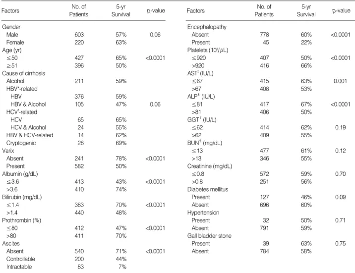
The initial characteristics of the patients at entry are sum- marized in Table 1. In the entire series, 603 (73%) of 823 patients were men. The mean age was 50 yr (range: 16-82).
Cirrhosis was compensated in 401 (49%) patients and decom- pensated in 422 (51%). Fifty patients in the compensated
group at entry had a previous history of decompensation. In the decompensated group at entry, ascites was found in 281 (67%), jaundice in 207 (49%), variceal bleeding in 119 (28%), and encephalopathy in 45 (11%). Esophageal varices were found in 582 (71%). The main comorbid conditions were diabetes mellitus in 127 (15%), cholelithiasis in 39 (5%), and hypertension in 32 (4%).
Cirrhosis was related to HBV in 481 (58%) patients, HCV
in 89 (11%), and HBV and HCV co-infection in 14 (2%);
alcohol abuse was recognized in 346 (42%) patients, including 211 (26%) with alcoholic cirrhosis; cirrhosis was cryptogenic in 28 (3%). The patients with alcoholic cirrhosis were predom- inantly male, while most of the patients with cryptogenic cir- rhosis were female as shown in Table 2. At the time of diag- nosis of cirrhosis, the mean age of patients was significantly different (p<0.001) depending on the etiologies of cirrhosis;
lowest in HBV-related cirrhosis and highest in HCV-related cirrhosis. Also, there were significant differences (p<0.001) in the incidence of decompensation or esophageal varices; high- est in the patients with alcoholic cirrhosis and lowest in those with HCV-related cirrhosis (Table 2).
Causes of Deaths
A total of 362 patients died during the study period, and the causes of death were identified from 310. Liver failure accounted for 165 deaths (53%), progression of HCC for 73 (23%), variceal bleeding for 28 (10%), infection for 13 (5%),
Factors No. of
Patients
5-yr
Survival p-value Factors No. of
Patients
5-yr
Survival p-value Gender
Male 603 57% 0.06
Female 220 63%
Age (yr)
≤50 427 65% <0.0001
≥51 396 50%
Cause of cirrhosis
Alcohol 211 59%
HBV*-related
HBV 376 59%
HBV & Alcohol 105 47% 0.06
HCV�-related
HCV 65 65%
HCV & Alcohol 24 55%
HBV & HCV-related 14 62%
Cryptogenic 28 69%
Varix
Absent 241 78% <0.0001
Present 582 50%
Albumin (g/dL)
≤3.6 413 43% <0.0001
>3.6 410 74%
Bilirubin (mg/dL)
≤1.4 383 70% <0.0001
>1.4 440 48%
Prothrombin (%)
≤80 412 47% <0.0001
>80 411 70%
Ascites
Absent 540 71% <0.0001
Controllable 200 44%
Intractable 83 7%
Encephalopathy
Absent 778 60% <0.0001
Present 45 22%
Platelets (103/ L)
≤920 407 50% <0.0001
>920 416 66%
AST�(IU/L)
≤67 415 63% 0.001
>67 408 53%
ALP�(IU/L)
≤81 417 67% <0.0001
>81 406 50%
GGT‖(IU/L)
≤62 414 62% 0.19
>62 409 55%
BUN¶(mg/dL)
≤13 477 61% 0.12
>13 346 55%
Creatinine (mg/dL)
≤0.8 572 59% 0.70
>0.8 251 56%
Diabetes mellitus
Present 127 46% 0.09
Absent 696 60%
Hypertension
Present 32 50% 0.71
Absent 791 59%
Gall bladder stone
Present 39 63% 0.75
Absent 784 58%
Table 1.Initial characteristics of patients in the series and the results of the univariate analysis of survival
*hepatitis virus B, �hepatitis virus C, �aspartate aminotransferase, �alkaline phosphatase, ‖gamma glutamyl transpeptidase, ¶blood urea nitrogen.
*Coinfection with HBV and HCV.
Alcoholic HBV HCV B&C* Cryptogenic
Number of patients 211 481 89 14 28
Age (Mean±SD) 52±10 46±10 60±9 51±10 57±11 Male patients (%) 200 (95) 331 (69) 53 (60) 12 (86) 7 (25) Decompensated 161 (76) 250 (52) 34 (38) 9 (64) 18 (64)
cirrhosis (%)
Esophageal 182 (86) 311 (65) 53 (60) 8 (57) 19 (68) varices (%)
Table 2.Patient characteristics at diagnosis of cirrhosis accord- ing to etiology
other digestive bleeding for 6 (2%), other malignancy for 4 (1%), and other causes for 21 (7%). Variceal bleeding was a terminal event in 12 of the patients who died of advanced HCC. Among the patients who died of liver failure, 55 (33%) and 29 (18%) were combined with SBP or hepatorenal syn- drome, respectively. In patients with alcoholic cirrhosis, va- riceal bleeding was a more frequent cause of death than HCC (18% vs. 8%).
Survival and Prognostic Indicators at Entry
The cumulative survival rates in the whole series were 94, 76, 58, and 46% at 1, 3, 5, and 7 yr after inclusion, respective- ly. Survival was much better in patients with compensated cirrhosis than in the decompensated group, with respective 5-yr survival rates of 74% and 43%. The Child-Turcotte-Pugh
scoring system allowed a more detailed prediction of survival depending on the severity of decompensation (Fig. 1). In this series, the 1-yr survival rate was 96% for patients with a Child- Turcotte-Pugh score of 6 to 8, and 40% for those with scores above 11.
In the univariate analysis of the entire series (Table 1), 13 of 18 variables at entry were linked to survival with p<0.1.
Ten of them had independent (p<0.05) relationships with survival in the multivariate analysis (Table 3). The rate of sur- vival was reduced for patients with male gender, older age, varix, ascites, prolonged prothrombin time (PT), lower platelet count, low serum albumin levels, and high levels of serum bilirubin or alkaline phosphatase (ALP). The patients with alcoholic cirrhosis had significantly better survival rate than those with HBV-related cirrhosis. In the multivariate analy- sis for subgroups, varix and ALP had a prognostic significance specifically for compensated cirrhosis, while bilirubin and PT were significant for decompensated cirrhosis.
The Status of Etiologic Factors during Follow-up and Their Impact on Survival
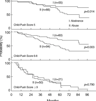
Sixty-one (28%) of 211 patients with alcoholic cirrhosis and 75 (56%) of 135 alcohol abusers with viral cirrhosis per- manently stopped drinking during the study period. They had a significantly better survival than those who continued to abuse alcohol (Table 4). In the subgroup analysis based on the Child-Turcotte-Pugh score, however, the benefit of absti- nence was recognized only for patients with a fairly good hep-
Survival Probability (%)
0 12 24 36 48 60 72 84 96
1. 367 363 351 311 261 194 135 82 2. 322 310 275 214 158 107 66 31
3. 109 92 68 44 25 11 7 3
4. 25 10 6 4 1 1 0
Months 100
90 80 70 60 50 40 30 20 10 0
Child-Push Score Score 5 (1) Score 6-8 (2) Score 9-11 (3) Score ≥12 (4)
3 4 p<0.0001
2 1
Fig. 1.Survival probability (Kaplan-Meier plot) in four subgroups based on the Child-Turcotte-Pugh scores. The numbers at the bot- tom of the figure are the numbers of patients at risk.
Variables Hazard ratio
(95% confidence interval) p value Gender (female/male) 0.687 (0.527-0.896) 0.0056
Age (yr) 1.037 (1.026-1.049) <0.0001
Varices (present/ absent) 1.552 (1.147-2.102) 0.0045 Albumin (g/dL) 0.581 (0.461-0.731) <0.0001 Bilirubin (mg/dL) 1.166 (1.076-1.264) 0.0002
PT (%) 0.990 (0.983-0.998) 0.0116
Ascites (intractable/ absent) 3.443 (2.517-4.710) <0.0001 Platelet count (103/ L) 0.962 (0.937-0.988) 0.0045
ALP (IU/L) 1.003 (1.001-1.004) 0.0006
Etiology of cirrhosis 1.608 (1.214-2.130) 0.0009 (HBV-related/alcoholic)
Table 3.Prognostic factors for survival at entry in the multivariate analysis for the entire series using the Cox regression model
Probability (%)
0 12 24 36 48 60 72 84 96
Months 100
50
0 100
50
0 100
50
0
Child-Push Score 5
I: Abstinence II: Abuse
Child-Push Score 6-8
Child-Push Score ≥9
Fig. 2.Survival probability (Kaplan-Meier plot) in alcoholic patients who continued to abuse or stopped abusing alcohol. The survival benefit of abstinence was significant only in patients with Child- Turcotte-Pugh scores of 5 to 8 at entry.
p=0.014 I (n=55)
II (n=66)
II (n=94)
II (n=50)
I (n=60)
I (n=21)
p=0.003
p=0.790
atic reserve, but not in those with advanced decompensation features, as shown in Fig. 2.
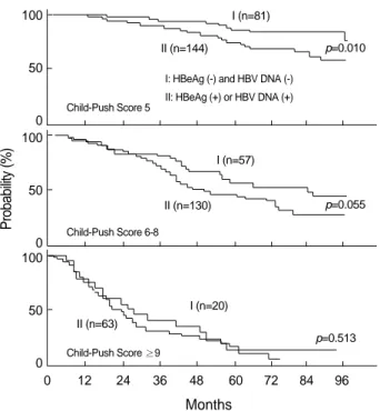
Of the 495 patients with HBsAg, 143 (29%) were seroneg- ative for both HBeAg and HBV DNA at entry, but 12 (8%) of them reverted to seropositive state thereafter. By contrast, 27 (8%) of 352 patients who had been seropositive for HBeAg or HBV DNA at entry became seronegative for both markers during follow-up. Overall, viral replication was suppressed in a total of 158 (32%) patients during the study period, and they showed a significantly improved survival rate compared to those with active viral replication (Table 4). However, this benefit was also significant only for the patients with compen- sated cirrhosis at entry (Fig. 3).
Development of Decompensation
Of the 351 patients who had never experienced decompen- sation at entry, 140 became decompensated during follow-up, with cumulative incidences of 13% and 44% at 1 and 5 yr after inclusion, respectively. The first sign of decompensation was ascites in 110 (79%), jaundice in 81 (58%), encephalopa- thy in 20 (14%), and variceal bleeding in 19 (14%). The 5- yr survival rate from entry was 97% in patients who did not develop decompensation, but 41% in those who decompen- sated. The survival rates after the initial episode of decompen- sation were 68% at 1 yr and 25% at 5 yr. Of note, the 5-yr survival after the appearance of ascites was 16%.
Development of Hepatocellular Carcinoma
One hundred and eighteen patients developed HCC during follow-up, with a cumulative incidence of 3% at 1 yr and 19%
at 5 yr after inclusion. HBV- or HCV-related cirrhosis was more frequently complicated by HCC than alcoholic cirrhosis, with the 5-yr cumulative incidence in each group being 24%, 28% and 5%, respectively. In addition, male gender, older age, esophageal varices, and high serum alkaline phosphatase level were independently associated (p<0.05) with a higher risk of HCC (Table 5).
At the initial diagnosis of HCC, 69 (59%) patients had small HCC, while 80 (68%) patients were in a decompensated state. Hepatic resection was performed in 7 (6%) patients, local ablation combined with chemo-embolization in 36 (31%), chemo-embolization alone in 37 (31%), and conservative management in 35 (30%); 3 patients dropped out after the diagnosis. Active treatment of HCC was attempted in 80%
of the cases with small HCC and in only 55% of advanced
Variables Hazard ratio
(95% confidence interval) p value Gender (female/male) 0.444 (0.278-0.712) 0.0007
Age (yr) 1.061 (1.040-1.082) <0.0001
Etiology of cirrhosis
(HBV-related/alcoholic) 11.598 (5.322-25.274) <0.0001 (HCV-related/alcoholic) 7.248 (3.004-17.486) <0.0001 Varices (present/absent) 1.823 (1.164-2.855) 0.0087
ALP (IU/L) 1.005 (1.002-1.008) 0.0018
Table 5.Risk factors for HCC using the Cox regression model
Probability (%)
0 12 24 36 48 60 72 84 96
Months 100
50
0 100
50
0 100
50
0
Child-Push Score 5
I: HBeAg (-) and HBV DNA (-) II: HBeAg (+) or HBV DNA (+)
Child-Push Score 6-8
Child-Push Score ≥9
Fig. 3.Survival probability (Kaplan-Meier plot) in patients with HBV-related cirrhosis according to the HBeAg status and HBV DNA in the serum during follow-up. A significant difference in sur- vival was observed between groups I and II only for the patients with a Child-Turcotte-Pugh score of 5 at entry.
p=0.010 I (n=81)
II (n=144)
II (n=130)
II (n=63)
I (n=57)
I (n=20)
p=0.055
p=0.513
Events No. of
patients
Survival
3-yr 5-yr p value
Alcohol abuse
Stop 136 82% 71% <0.0001
Continue 210 69% 48%
Clearance of HBeAg and HBV DNA
Absent 337 71% 52% 0.0002
Present 158 83% 66%
Hepatocellular carcinoma
Absent 705 78% 67% <0.0001
Small* 69 77% 37%
Advanced 49 57% 22%
Upper GI bleeding
Absent 687 79% 64% <0.0001
Present 136 64% 40%
SBP
Absent 674 82% 68% <0.0001
Present 149 50% 26%
Table 4.Univariate analysis of survival in relation to important clin- ical events during follow-up
*Hepatocellular carcinoma consisting of a single nodule <5 cm or 2-3 nodules <3 cm without invasion of major veins.
cases. The life expectancy following entry was longer in pa- tients in whom HCC was detected at the stage of small HCC than in those with HCC detected at advanced stage, although both groups had shorter survival times than those who did not develop HCC (Table 4, Fig. 4).
Development of Upper Gastrointestinal Bleeding
During follow-up, 163 upper gastrointestinal (GI) bleeds developed in 136 patients. The origins of the bleeding were identified in 157 cases by endoscopy: esophago-gastric varices in 138 (87.9%), gastric ulcer in 13 (8.3%), hemorrhagic gas- tropathy in 2 (1.3%), duodenal ulcer in 2 (1.3%), esophageal ulcer in 1 (0.6%), and esophagitis in 1 (0.6%).
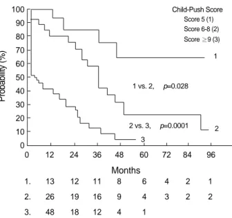
The patients who developed upper GI hemorrhages during follow-up had a lower survival rate than those without bleed- ing (Table 4). The overall mortality of variceal bleeding was 29% (40/138), and was almost the same in initial (25/87) and recurrent (15/51) episodes. By contrast, the mortality from non-variceal upper GI bleeding was 11% (2/19). The cumu- lative survival rate after the first variceal bleed was 71% at 6 weeks, 58% at 1 yr, and 20% at 5 yr, but there were signifi- cant differences (p<0.0001) depending on the severity of hep- atic decompensation at the time of bleeding (Fig. 5). For exam- ple, the 6-week survival rate was 95% (37/39) in patients with Child-Turcotte-Pugh scores from 5 to 8, but 52% (25/48) in those with scores above 8. The cumulative incidence of recur- rent variceal bleeding was 11% at 6 weeks, 25% at 1 yr, and 54% at 5 yr.
Development of Spontaneous Bacterial Peritonitis (SBP)
At least one episode of SBP occurred in 149 (30%) of the 502 patients with ascites during the investigation. The cumu- lative incidence of the first episode of SBP was 27% at 1 yr and 44% at 5 yr after developing ascites in 194 patients who experienced ascites for the first time during follow-up. At first presentation of SBP, most patients had advanced liver dysfunc- tion, and their mean (±SD) Child-Turcotte-Pugh score was 10.5±2.3. As expected, the patients who developed SBP during follow-up had reduced survival from entry (Table 4).
Twenty-seven (18%) of 146 patients died shortly after their first episode of SBP, while still in hospital. Of the 119 patients discharged alive, 58 (50%) developed recurrent SBP with a cumulative incidence of 44% at 1 yr and 68% at 5 yr. The survival rate after first SBP was 46% at 1 yr and 10% at 5 yr.
The Effectiveness of Surveillance Programs for Hepatocellular Carcinoma
We evaluated the effectiveness of our surveillance program for HCC, which involves regular ultrasonography and mea- surement of serum alpha-fetoprotein levels. Of 118 patients with HCC in this series, the most recent ultrasound examina- tion just before the diagnosis of HCC was 6 months or less in 35 patients (group I), 7 to 12 months in 46 patients (group II), and more than 1 yr in 37 patients (group III).
The frequency of small HCC in groups I or II was higher than in group III (69% or 63 vs. 43%, p<0.05). However, life expectancy after entry was similar in all three groups, as
Probability (%)
0 12 24 36 48 60 72 84 96
1. 705 42 551 440 325 222 151 82
2. 69 63 58 46 32 20 16 10
3. 49 45 38 28 20 9 5 4
Months 100
90 80 70 60 50 40 30 20 10 0
Without HCC (1) Small HCC (2) Advanced HCC (3)
3 2 vs. 3, p=0.008 1 vs. 2, p=0.0004
2 1
Fig. 4.Survival probability (Kaplan-Meier plot) of patients with or without hepatocellular carcinoma (HCC) during follow-up. The pa- tients who developed HCC were divided into two subgroups accord- ing to the tumor status when diagnosed: small or advanced HCC.
The numbers at the bottom of the figure are the numbers of patients at risk.
Child-Push Score Score 5 (1) Score 6-8 (2) Score ≥9 (3)
Probability (%)
0 12 24 36 48 60 72 84 96
1. 13 12 11 8 6 4 2 1
2. 26 19 16 9 4 3 2 2
3. 48 18 12 4 1
Months 100
90 80 70 60 50 40 30 20 10
0 3
2 vs. 3, p=0.0001 1 vs. 2, p=0.028
2 1
Fig. 5.Survival probability (Kaplan-Meier plot) after developing a first variceal hemorrhage according to the Child-Turcotte-Pugh score at the time of bleeding. The numbers at the bottom of the figure are the numbers of patients at risk.
indicated by the 5-yr survival rates, which were 30%, 33%, and 29% in groups I, II, and III, respectively. Although the prognostic factors at baseline were adjusted in the multivari- ate analysis, no significant benefit in survival was recognized.
The Benefit of Endoscopic Prophylaxis against First Variceal Bleed
We assessed the effect of endoscopic prophylaxis against first variceal bleeds for the patients who had large esophageal varices (Japanese classification F2 or F3) without coincident HCC or severe decompensated cirrhosis (Child-Turcotte-Pugh scores over 12). The effect of beta-blockers was not evaluated because there were too few cases and the schedule of medica- tions was not consistent.
Throughout the study period, 441 patients were found to have large esophageal varices (Japanese classification F2 or F3).
One hundred and eighty-seven of them, however, had previous or ongoing variceal bleedings at the time of diagnosis; 9 had coincident HCC; 8 presented the features of severe decompen- sation. Among the remaining 237 patients who met the above inclusion criteria, 75 underwent endoscopic prophylaxis (band- ligation in 50, sclerotherapy in 9, and a combination of the two methods in 16), 29 were given oral propranolol, and 133 were given no prophylaxis. At baseline, the endoscopy pro- phylaxis group had a higher prevalence of red color sign (67%
vs. 44%, p<0.01), and had a slightly lower mean (±SD) value of Child-Turcotte-Pugh score (6.5±1.7 vs. 7.1±1.8, p<0.05) compared with the no-prophylaxis group.
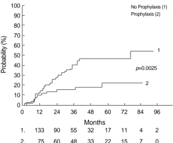
The cumulative incidence of first variceal bleed after the diagnosis of large varices was significantly lower in the pro- phylaxis group than in the no-prophylaxis group, with 3-yr
incidences of 18% and 42%, respectively (Fig. 6). This pre- ventive effect still remained significant in multivariate ana- lysis, which adjusted several potential risk factors of variceal hemorrhage at base line that had shown p<0.1 in univariate analysis, such as red color sign, serum bilirubin level, gender, aspartate aminotransferase, and gammaglutamyl transpepti- dase (Table 6).
However, there was no significant difference in the cumu- lative survival rates between the prophylaxis and no-prophy- laxis groups, with 3-yr survival rates of 68% and 58%, respec- tively. Furthermore, no survival benefit was observed in the univariate analysis considering the Child-Turcotte-Pugh scores at baseline or in the multivariate analysis adjusting the pro- gnostic factors at baseline.
DISCUSSION
In the present study, the main causes of liver cirrhosis were HBV, HCV, and alcohol, which accounted for the origin of ci- rrhosis in 60%, 13%, and 26% of the whole patients, respecti- vely. The predominant role of HBV in viral cirrhosis reflects the high prevalence of chronic HBV infection in this country (2).
Depending on the etiologies of cirrhosis some clinical fea- tures were distinctive. The patients with alcoholic cirrhosis were predominantly male (95%), presumably this being relat- ed with the cultural background in alcohol ingestion, and most of them (75%) showed decompensation features at first presentation, suggesting chronic alcoholics usually keep drink- ing without insight until decompensated. HBV-related cirrho- sis was diagnosed in mid-forties on average, relatively earlier than in other types of cirrhosis. This presumably may be asso- ciated with the vertical transmission of HBV, the main route of HBV infection in this country, which elicits chronic hep- atitis from earlier ages. In contrast, HCV-related cirrhosis was detected at the mean age of 60, and was compensated in 60%
of cases. Compared with alcoholic cirrhosis, viral cirrhosis was complicated by HCC approximately 5-fold more in frequency.
As a result, HCC was responsible for about one third of total deaths in viral cirrhosis, whereas only 8% of all deaths in alco- holic cirrhosis. Instead, variceal bleeding was a more dominant cause of death in alcoholic cirrhosis.
As expected from the causes of death in the cirrhosis patients,
No Prophylaxis (1) Prophylaxis (2)
Probability (%)
0 12 24 36 48 60 72 84 96
1. 133 90 55 32 17 11 4 2
2. 75 60 48 33 22 15 7 0
Months 100
90 80 70 60 50 40 30 20 10 0
1
Fig. 6.Cumulative probability (Kaplan-Meier plot) of developing a first variceal hemorrhage in two subgroups of patients with large esophageal varices: those who did and did not receive endoscopic prophylaxis. The numbers at the bottom of the figure are the num- bers of patients at risk.
p=0.0025 2
Variables Hazard ratio
(95% confidence interval) p value Bilirubin (mg/dL) 1.371 (1.090-1.725) 0.007 Red color sign 4.683 (2.516-8.718) 0.000
(present/absent)
Endoscopic prophylaxis 0.286 (0.151-0.542) 0.000 (present/absent)
Table 6.Risk factors for first variceal hemorrhage in patients with large esophageal varices using the Cox regression model (n=
208)
the clinical variables representing the severity of hepatic failure or portal hypertension, including the five variables used in the Child-Turcotte-Pugh system, esophageal varices, and throm- bocytopenia, are reported as the key prognostic indicators of liver cirrhosis (4, 5, 11, 17, 18), and their importance was reconfirmed in the present study. Also, the variables such as age, gender, and serum alkaline phosphatase level had prog- nostic significance, as previously reported (4, 5, 17, 18). Of interest, these three factors had a significant correlation with the risk of developing HCC as well.
It is well known that efficient management of etiologic factors is closely correlated with survival in cirrhosis patients (3, 4, 17-20). In this study, we were able to confirm that the prognosis was far better in the patients who stopped drinking during follow-up than in those who continued to drink, and the patients with inactive HBV replication had a higher sur- vival rate than those with consistent viral replication. Further- more, we demonstrated that these benefits were seen only in patients with compensated or mildly decompensated cirrhosis in the subgroup analysis. This emphasizes that the control of etiologic factors must be attempted at an earliest stage of cir- rhosis, if possible, to improve the rate of survival substantially.
In addition, the better prognosis of alcoholic cirrhosis in our study compared with HBV-related cirrhosis might be explain- ed by the differing intractability of etiologic factors, i.e., 28%
of the patients with alcoholic cirrhosis succeeded in abstain- ing from alcohol after entry, while only 8% of the patients with HBV-related cirrhosis showed HBeAg seroconversion during follow-up. Indeed, we could not find any significant difference in survival between alcoholic and HBV-related cir- rhosis when the patients who stopped drinking were excluded.
In this study, 44% of the patients with compensated cirrho- sis developed decompensation signs within 5 yr after entry, and the 5-yr survival rate after decompensation was 25%.
These data are similar to the figures reported in the 1980s (3-5); therefore, it is difficult to say that there has been a sub- stantial improvement in the long-term survival of cirrhosis patients during the last 20 yr. Although the 5-yr survival rate in patients who had been decompensated at entry appears higher in the present series as compared with previous results (40% vs. 16-21%), it can be noted that our series included a relatively lower percentage of patients with ascites and no patients with HCC at entry.
However, the short-term prognosis of some major compli- cations appears to have remarkably improved. For example, in this study, which took vigorous endoscopic measures to prevent variceal rebleeding, the rebleeding rate after a first variceal hemorrhage was 11% at 6 weeks and 25% at 1 yr.
These values are much lower than the figures of 20-50% at 6 weeks and 47-84% at 1 yr in untreated cases in earlier stud- ies (6). Correspondingly, the 6-week mortality after variceal hemorrhage was decreased to 29% (22% when terminal HCC patients were excluded) in our study, and to 5% in patients with Child-Turcotte-Pugh scores below 9, as compared to
33-71% in prior studies (6, 21). Similarly, the hospital mortal- ity of SBP in this study was reduced to 18% compared with 40-50% in previous reports (22-24).
After considering tumor stage and functional hepatic reserve, a mere 6% of the patients with HCC underwent partial hep- atectomy in this study, and another 62% were treated non-sur- gically. It is now evident that the development of non-surgi- cal modalities has enlarged the range of therapeutic indications for HCC patients, especially for cases that are inoperable due to impaired liver function or multiple lesions. Particularly, patients with small HCC had more opportunities for treatment than those with advanced HCC (80% vs. 55%). In concert with these findings, it is noteworthy that the patients with small HCC had a significantly longer life expectancy from entry, as well as after the diagnosis of HCC, than those with advanced HCC. This indicates that early detection of HCC can lead to a genuine survival benefit in cirrhosis patients, and provides good grounds for regular surveillance for the early detection of HCC. As far as we know, this study will be the first to demonstrate that early detection of HCC can improve the survival of cirrhosis patients beyond the lead-time bias (25). Unfortunately, however, biannual ultrasonography in this study failed to achieve a significant improvement in sur- vival, although it increased the detection rate of small HCC in the screened group by more than 20%. This is most like- ly because about 30% of cases of small HCC were still over- looked on ultrasound examination, probably due to limitations of sonography and the inexpertness of the examiners. There- fore, in order to improve the detection rate of small HCC in cirrhosis patients, it is necessary to introduce complementary imaging modalities with higher resolution and to develop highly sensitive tumor markers.
In this study, endoscopic prophylaxis for a first variceal bleed significantly reduced the bleeding rate, in spite of the higher incidence of the RC sign in the prophylaxis group at baseline. However, we did not find a significant improvement in survival, although there were fewer signs of decompensa- tion in the prophylaxis group at baseline. This concurs with previous studies (26-29). Of course, the possibility of type II error still exists in our study, since the prophylaxis group had a higher survival rate than the non-prophylaxis group by 10%
at 3 yr, although this was not significant. Nonetheless, the expected survival benefit of endoscopic prophylaxis does not seem very high, even if it has a statistical significance. This low potential benefit might be ascribed to the reduced bleed- ing-related mortality that has resulted from the progress in the management of variceal hemorrhage, and the development of means to prevent rebleeding, especially in patients with an otherwise good prognosis. Therefore, for endoscopic pro- phylaxis against a first variceal hemorrhage, a more discreet approach might be required that considers cost-effectiveness, and a stricter selection of target patients is desirable, since the 3-yr bleeding rate was only 42% in patients with large varices in this study.
In summary, this study provided a wide range of updated prognostic information that will be helpful for the appropriate management of cirrhosis patients. We found that 1) effective control of causative agents increased life expectancy in patients with compensated or mildly decompensated cirrhosis; 2) early detection of HCC led to a genuine survival benefit; 3) the short-term prognosis of some major complications, such as variceal hemorrhage and SBP, has considerably improved over the last two decades, while there was no remarkable improve- ment in the long-term survival of cirrhosis patients; and 4) biannual ultrasonography increased the detection rate of small HCC and endoscopic prophylaxis decreased the incidence of first variceal hemorrhage without improving survival. These results suggest that more efficient management of etiologic factors and complications is necessary to improve the prognosis of compensated or mildly decompensated cirrhosis. In severely decompensated cirrhosis, liver transplantation might be the only therapeutic option that alters the outcome.
REFERENCES
1. Korean National Statistical Office. Annual Report on the Causes of Death Statistics. Seoul 1999: 25-7 (in Korean).
2. Lee DH, Kim JH, Nam JJ, Kim HR, Shin HR. Epidemiological findings of hepatitis B infection based on 1998 National Health and Nutrition Survey in Korea. J Korean Med Sci 2002; 17: 457-62.
3. Saunders JB, Walters JRF, Davies AP, Paton A. A 20-year prospec- tive study of cirrhosis. Br Med J 1981; 282: 263-6.
4. D’Amico G, Morabito A, Pagliaro L, Marubini E, the liver study group of ‘‘V. Cervello’’ Hospital. Survival and prognostic indicators in compensated and decompensated cirrhosis. Dig Dis Sci 1986;
31: 468-75.
5. Gines P, Quintero E, Arroyo V, Teres J, Bruguera M, Rimola A, Caballeria J, Rodes J, Rozman C. Compensated cirrhosis: natural history and prognostic factors. Hepatology 1987; 7: 122-8.
6. D’Amico G, Pagliaro L, Bosch J. The treatment of portal hyperten- sion: a meta-analytic review. Hepatology 1995; 22: 332-54.
7. Farinati F, Gianni S, Marin G, Fagiuoli S, Rinaldi M, Naccarato R.
Does the choice of treatment influence survival of patients with small hepatocellular carcinoma in compensated cirrhosis? Eur J Gastroenterol Hepatol 2001; 13: 1217-24.
8. Scheuer PJ. Liver biopsy in the diagnosis of cirrhosis. Gut 1970;
11: 275-8.
9. Runyon BA. Spontaneous bacterial peritonitis: An explosion of information. Hepatology 1988; 8: 171-5.
10. Sherlock S, Dooley J. Disease of the liver and biliary system, 11th ed. Oxford: Blackwell Science, 2002: 93-109.
11. Pugh RNH, Murray-Lyon IM, Dawson JL, Pietroni MC, Williams R. Transection of the oesophagus for bleeding oesophageal varices.
Br J Surg 1973; 60: 646-9.
12. Beppu K, Inokuchi K, Koyanagi N, Nakayama S, Sakata H, Kitano S, Kobayashi M. Prediction of variceal hemorrhage by esophageal
endoscopy. Gastrointest Endosc 1981; 27: 213-8.
13. de Franchis R. Updating consensus in portal hypertension: report of Baveno III consensus workshop on definitions, methodology and therapeutic strategies in portal hypertension. J Hepatol 2000; 33:
846-52.
14. Llovet JM, Bru C, Bruix J. Prognosis of hepatocellular carcinoma:
the BCLC staging classification. Semin Liver Dis 1999; 19: 329-38.
15. Kaplan GL, Meier P. Nonparametric estimation from incomplete observation. J Am Stat Assoc 1958; 53: 457-81.
16. Cox DR. Regression models and life tables. J R Stat Soc B 1972;
34: 187-220.
17. Christensen E. Prognostic models in chronic liver disease: validity, usefulness and future role. J Hepatol 1997; 26: 1414-24.
18. Christensen E, Schlichting P, Andersen PK, Fauerholdt L, Schou G, Pedersen BV, Juhl E, Poulsen H, Tygstrup, N Copenhagen study group for liver disease. Updating prognosis and therapeutic effect evalua- tion in cirrhosis with Cox’s multiple regression model for time-depen- dent variables. Scand J Gastroenterol 1986; 21: 163-74.
19. de Jongh FE, Janssen HL, de Man RA, Hop WC, Schalm SW, van Blankenstein M. Survival and prognostic indicators in hepatitis B surface antigen-positive cirrhosis of the liver. Gastroenterology 1992; 103: 1630-5.
20. Powell WJ Jr, Klatskin G. Duration of survival in patients with Laennec’s cirrhosis. Influence of alcohol withdrawal, and possible effects of recent changes in general management of the disease. Am J Med 1968; 44: 406-20.
21. Burroughs AK, D’heygere F, McIntyre N. Pitfalls in studies of pro- phylactic therapy for variceal bleeding in cirrhotics. Hepatology 1986; 6: 1407-13.
22. Hoefs JC, Canawati HN, Sapico FL, Hopkins RR, Weiner J, Mont- gomerie JZ. Spontaneous bacterial peritonitis. Hepatology 1982; 2:
399-407.
23. Runyon BA, Hoefs JC. Culture-negative neutrocytic ascites: a variant of spontaneous bacterial peritonitis. Hepatology 1984; 4: 1209-11.
24. Tito L, Rimola A, Gines P, Llach J, Arryo V, Rodes J. Recurrence of spontaneous bacterial peritonitis in cirrhosis: Frequency and predictive factors. Hepatology 1988; 8: 27-31.
25. Sherman M. Screening for hepatocellular carcinoma. Bailliere’s Clinical Gastroenterology 1999; 13: 623-35.
26. Piai G, Cipolletta L, Claar M, Marone G, Bianco MA, Forte G, Iodice G, Mattera D, Minieri M, Roccop P, et al. Prophylactic sclerotherapy of high risk esophageal varices: Results of a multicentric prospective controlled trial. Hepatology 1988; 8: 1495-500.
27. Kobe E, Zipprich B, Schentke KU, Nilius R. Prophylactic endo- scopic sclerotherapy of esophageal varices. A prospective random- ized trial. Endoscopy 1990; 22: 245-8.
28. Sarin SK, Guptan RK, Jain AK, Sundaram KR. A randomized con- trolled trial of endoscopic variceal band ligation for primary pro- phylaxis of variceal bleeding. Eur J Gastroenterol Hepatol 1996; 8:
337-42.
29. Lo GH, Lai KH, Cheng JS, Lin CK, Hsu PI, Chiang HT. Prophylac- tic banding ligation of high-risk esophageal varices in patients with cirrhosis: a prospective, randomized trial. J Hepatol 1999; 31: 451-6.
′
′ ′
′
′
′ ′