Kor. J. Fertil. Steril., Vol. 30, No. 2, 2003, 6
Expression of Id-1 Gene in Mouse Uterus
Hee Young Nah
1, Seok Ho Hong
1,2, Ji Yoon Lee
1, Hee Dong Chae
1, Byung Moon Kang
1, Chung Hoon Kim
11Department of Obstetrics and Gynecology, College of Medicine, Ulsan University, Asan Medical Center, Seoul, 2Department of Life Science, College of Natural Sciences,
Hanyang University, Seoul, Korea
생쥐 자궁에서의 Id-1 유전자의 발현
울산의대 서울아산병원 산부인과학교실1, 한양대학교 자연과학대학 생명과학과2
나희영
1・홍석호
1,2・이지윤
1・채희동
1・강병문
1・김정훈
1연구 목적: Microarray data에 의해 밝혀진 생쥐자궁에서의 Id 유전자의 hormonal effect와 implantation process 동안의 관계를 조사하고자 실험을 수행하였다.
연구재료 및 방법: 난소절제한 생쥐에 estrogen을 주사하고 6시간, 12시간이 지난 후 자궁을 적출하 여 두 가지 방법으로 sample을 준비하였다. 먼저 자궁전체의 RNA를 추출하여 실험하거나 laser capture microdissection (LCM) 방법으로 자궁내막상피세포, 자궁기질세포, 자궁근육층으로 분리하여 RNA를 추출 하고 semi-quantative RT-PCR을 수행하여 Id 유전자의 발현을 조사하였다. 임신 4.5일째 생쥐에 Chicago blue dye를 주사하여 착상부위와 비착상부위를 분리하고 RNA를 추출하여 Id 유전자의 발현을 semi- quantitative RT-PCR 방법으로 실험하였다.
결 과: Estrogen을 처리한 난소절제된 생쥐 자궁에서의 cDNA microarray 자료에서 Id-1 mRNA는 점진 적으로 두 배 이상 증가하였고 Id-2 mNRA는 반대로 시간이 지날수록 두 배 이상 감소하였다. Microarray 자료를 재확인하기 위해 semi-quantitative RT-PCR을 이용하여 실험하였고, 그 결과 Id-1 유전자는 estrogen 처리 6시간까지는 큰 변화가 없었으나 12시간에서는 4배 이상의 높은 발현을 보였으며, Id-2 mRNA의 발현은 estrogen 처리 6시간과 12시간 모두에서 대조군에 비해 4배 가량 감소하였다. 이 실 험군을 LCM을 이용하여 자궁내막상피세포, 자궁기질세포, 자궁근육층을 각각 분리하여 실험한 결과 estrogen 처리군에서 Id-1의 발현은 자궁내막상피세포에서만 높은 발현을 보였으며, estrogen 처리 6시간 과 12시간에는 큰 차이를 보이지 않았다. 그러나, Id-2 mRNA는 자궁내막상피세포에서 estrogen 처리 6, 12시간 모두에서 높은 발현을 보였고, 근육세포층에서는 estrogen 처리 6시간에서는 변화가 거의 없었 으나 12시간에는 현저하게 증가하였다. 단, 자궁기질세포에서는 대조군에 비해 estrogen 6, 12시간에서 Id-2 mRNA의 발현이 감소하였다. 임신한 생쥐 자궁의 착상부위에서는 Id-1 mNRA의 발현은 비착상부 위보다 월등하게 높은 증가를 보였다.
결 론: 난소절제 생쥐를 이용한 실험에서 Id-1, -3는 estrogen에 의해 발현이 증가하고, Id-2는 발현 이 감소하였다. LCM을 이용한 실험에서는 Id-2는 이와는 달리 부위별 발현양상은 다르게 나타났지만 이는 넓은 부위를 차지하는 자궁기질에서의 발현감소가 전체적인 Id-2의 발현양상으로 나타난 것으 연 락 저 자: 나희영, 우) 138-736 서울특별시 송파구 풍납동 388-1, 서울아산병원 산부인과 체외수정실
Tel: (02) 3010-5053, Fax: (02) 3010-5059, e-mail: [email protected]
주관책임자: 홍석호, 우) 138-736 서울특별시 송파구 풍납동 388-1, 서울아산병원 산부인과 체외수정실 Tel: (02) 3010-5053, Fax: (02) 3010-5059, e-mail: [email protected]
로 추측된다. 착상부위에서의 Id의 발현은 Id-1만이 유일하게 월등한 증가를 보였다.
위의 결과를 종합해 볼 때 생쥐 자궁에서 Id 유전자는 estrogen에 의해 조절되며 직, 간접적으로 착 상시기에 다양한 작용을 할 것으로 사료된다.
Key Words: Id-1, Estrogen, Uterus, Implantation
Steroid hormone actions in target tissues are normally mediated via binding to nuclear receptors that are ligand-inducible transcription factors.
1,8,15The uterus is composed of heterogenous cell types that undergo dynamic changes to support embryo development and implantation by harmony of ovar- ian steroid hormones. Implantation is a complex process involving various molecular "cross-talk"
between the blastocyst and the uterus. The sites of blastocyst attachment in uterus is visualized by in- creased stromal vascular permeability and undergo morphological and biochemical changes to receive the blastocyst under the control of ovarian steroid hormone.
7,19,20These changes suggest that cell pro- liferation and differentiation at this sites is asso- ciated with up- and down-regulation of a many of genes, which are poorly understood. We have recently performed cDNA microarray to screen estrogen-responsive genes in the mouse uterus and so shown that Id genes were differentially expres- sed in the estrogen-treated ovariectomized mouse uterus. Id, inhibitors of basic helix-loop-helix (bHLH), are positive and negative regulators of proliferation, differentiation, cell cycle, apoptosis and angiogenensis.
3,14Four members, Id1 through Id4, have been identified in mammals.
2,4,17,18In the present study, we examined the expression patterns of Id genes according to uterine specific cell type and the difference between implantation sites and interimplantation sites using laser capture microdissection (LCM) and RT-PCR.
MATERIALS AND METHODS
1. Animals and Uterus preparation
ICR mice were housed within temperature- and
light-controlled conditions under the supervision of a licenced veterinarian. Mice were maintained on a 12L:12D photoperiod and provided with food and water ad libitum. Female mice (6~7 weeks of age) were ovariectomized (OVX) and rested for 14 days before receiving estrogen treatment. They were injected with oil (0.1 ml) and 17β-estradiol (Sigma, St. Louis, MO; 300 ng/mouse) which was dissolved in sesame oil and injected (0.1 ml/mouse) subcutaneously. Mice (n=9) were killed and uter- ine horns were collected at 6 h or 12 h after in- jection.
To obtain pregnant mice, adult female mice (6~
8 weeks old) were placed with fertile males of the same strain, and the day that a vaginal plug was found was considered as day 1 of pregnancy. On the evening of day 4 (22:00~23:00) at the time of blastocyst attachment, implantation sites were vi- sualized by intravenous injection (tail vein) of Chi- cago Blue B solution (1% in saline) 10 min before killing the mice. Implantation segments containing implanting embryo were finely separated from non- implantation segments, and both segments were then snap-frozen in liquid nitrogen until utilized for total RNA extraction. All animal experiments were performed in accordance with the Guide of Ulsan University for Care and Use of Laboratory Animals.
2. cDNA microarray and Data analysis
Uterine tissues from nine female mice were
pooled, snap frozen and homogenized by mortar
in liquid nitrogen. For cDNA microarray analysis,
total RNA was extracted using TRIZOL reagent
(InVitrogen, Carlsbad, CA) and purified using
RNeasy total RNA isolation kit (Qiagen, Valencia,
CA) according to the manufacturers instructions.
Total RNA was quantified by spectrophotometer and its integrity was assessed by running on a de- naturing 0.8% agarose gel. Prior to use in cDNA microarray analysis, each RNA sample via OVX/
estrogen treatment/6 h/12 h protocol was validated by assaying for up-regulation (asparagine synthe- tase and lactoferrin) or down-regulation genes (glu- tathione S-transferase and secreted frizzled-related sequence protein 2) as markers of estrogen efficacy.
Profiling of estrogen-regulated gene expression was analyzed with a TwinChip Mouse-7.4 K (Digital Genomics, Seoul) consisting of 7616 mouse cDNA clones. Twenty µg RNA was reverse-transcribed with Cy3- or Cy5-conjugated dUTP (Amersham Pharmacia Biotech, Piscataway, NJ) respectively, using SuperScript II (Gibco BRL, Rockville, MD) and oligo(dT)
18primer (Ambion, Austin, TX) in a reaction volume of 20 µl according to the method suggested by the manufacturer. After the labeling reaction for 1 h at 42℃, unincorporated fluorescent nucleotide was cleaned up using Microcon YM-30 column (Millipore, Bedford, MA). The Cy3- and Cy5-labeled cDNA probes were mixed together and hybridized to a microarray slide. After over- night at 65℃, the slide was washed twice with 2×
SSC containing 0.1% SDS for 5 min at 42℃, once with 0.1× SSC containing 0.1% SDS for 10 min at room temperature, and finally with 0.1× SSC for 1 min at room temperature. Slide was dried by centrifugation at 650 rpm for 5 min. Hybridization images on the slide were scanned by Scanarray lite (Packard Bioscience, Boston, MA) and analy- zed by GenePix Pro3.0 software (Axon Instrument, Union City, CA). Three independent experiments were performed, and the ratio of Cy3 and Cy5 signal intensity was calculated for each spot. These ratio was log
2-transformed and normalized by sub- tracting the average of log
2(Cy3/Cy5) values for internal control genes using Excel (Office 2000, Microsoft Corp.). For each gene, the mean values
were then calculated and a difference of two-fold was applied to select up- or down-regulated genes by estrogen.
3. Laser capture microdissection
Uterine horns were embedded in Tissue-Tek (Sakura Finetek, Torrance, CA) and frozen in liquid nitrogen. Cryosections (thickness, 6 µm) were cut and mounted onto clean glass slides. After the sec- tions were conterstained with Mayers hematoxylin, each population of uterine cells (luminal epithelial, muscle and stromal cells) was isolated from these sections using P.A.L.M. Robot-Microbeam version 4.0 (P.A.L.M. Microlaser Technologies AG, Bern- ried, Germany). For each cell, an average of 150 laser shots were transferred onto 0.5 ml tube cap and stored at -70℃ until utilized for total RNA extraction.
4. Confirmation of microarray data with semiquantitative RT-PCR analysis
Total RNA was extracted from whole uterine
tissues or each population of uterine cells using
TRIZOL reagent and purified using RNeasy total
RNA isolation kit following the manufacturers
instructions. One µg of RNA was reverse-transcri-
bed at 42℃ for 60 min in 20 µl reaction mixture
consisting of oligo(dT)-adapter primer (Takara,
Shiga, Japan) and AMV reverse transcriptase XL
(Takara). The following PCR was performed in a
total volume of 40 µl with 2 µl of the RT reaction
mixture, 2 µl 25 mM MgCl
2, 4 µl 10× PCR buffer
(100 mM Tris-HCl, 500 mM KCl, pH 8.3), 4 µl
2.5 mM dNTPs, 10 pmole forward and reverse
primer and 1.25 U Taq polymerase (Takara). The
sequences of the primers used were described in
Table 1. An increasing number of cycles was tes-
ted to assess the best conditions of achieve linear
amplification. The thermal cycling parameters were
consisted of 22~26 cycles of denaturing (94℃,
30 sec); annealing (60℃, 30 sec); and extension
(72℃, 30 sec). The PCR products were separated by electrophoresis on a 1.2% TBE agarose-ethi- dium bromide gels and visualized under UV light.
The images were quantified by densitometric scan- ning followed by BioID image analysis software (Vilber-Lourmat, Mama La Vallee, Cedex, France) and gene expression was normalized against the density of the corresponding ribosomal protein L- 7 (rpL7) PCR product as internal control.
RESULTS
1. Changes of Id genes by estrogen in ovar- iectomized mouse uterus
We have performed cDNA microarray to screen
estrogen-regulated genes in the mouse uterus (data not shown) and then unexpectedly, Id genes were showed differential expression pattern (Table 2).
In particular, the expression level of Id-1 mRNA was increased in proportion as estrogen-treated time in the estrogen-induced uterus and Id-2 decre- ased. Although there is no significance, the level of Id-3 mRNA was decreased in the estrogen- induced uterus. However, the expression of Id-4 gene made a no difference between the groups. To confirm microarray data, the expression patterns of Id-1 and -2 using semi-quantitative RT-PCR were shown in Figure 1. All groups were mixed with five mice to each other. The RT-PCR data was dis- played just Id-1 and -2 which were detected signi-
Table 1. Primer sequences for RT-PCRGene Forward (F) and Reverse (R) primer sequences Product size (bp) F GATCATGAAGGTCGCCAGTG
Id-1
R TCCATCTGGTCCTCAGTGC 703
F GTGACCAAGATGGAAATCCT Id-2
R TTTATTTAGCCACAGAGTAC 523
F GAAGGCGCTGAGCCCGGTGC Id-3
R GTTCGGGAGGTGCCAGGACG 383
F GCGATATGAACGACTGCTAC Id-4
R TCACCCTGCTTGTTCACGGC 616
F CTCCTCCAAGCCACAAGGCC HB-EGF
R CTAGCCACGCCCAACTTCAC 389
F TCAATGGAGTAAGCCCAAAG rpL7
R CAAGAGACCGAGCAATCAAG 246
Table 2. Change of Id genes by estrogen in ovariectomized mouse uterus Fold change
Gene name
6 H 12 H Genbank No.
Inhibitor of DNA binding 1 (Id-1) 1.23 2.01 U43884
Inhibitor of DNA binding 2 (Id-2) -1.26 -2.26 AF077860 Inhibitor of DNA binding 3 (Id-3) -1.48 -1.69 NM008321
Inhibitor of DNA binding 4 (Id-4) 1.17 1.07 AF077859
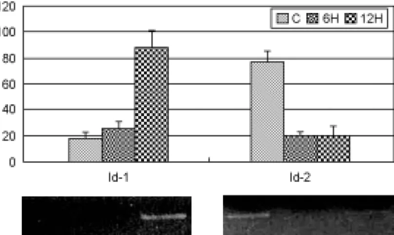
ficantly difference. After injection of estrogen, the level of Id-1 mRNA were few difference at 6 h, increased more than four-fold at 12 h. In contrast, the expression of Id-2 mRNA was remarkably de- creased than control both 6 h and 12 h after injec- tion of estrogen. In comparison with Id-1 and Id-2, Id-2 mRNA was rapidly responsed to estrogen, but Id-1 was relatively showed slow response to estrogen.
2. Differential Expression of Id genes in the uterine specific cell types
Id genes were displayed differential expression
according to cell type in the estrogen-treated OVX mouse uterus (Figure 2). Surprisingly, Id-1 gene was showed the specific expression in luminal epithelial cell. However, the expression of Id-2 mRNA in the uterine specific cell types was sho- wed different pattern in contrast with the whole uterus data.
Although the level of Id-2 mRNA in the stro- mal cells was similar to the whole uterus data, expression of Id-2 in the luminal epithelial cells and muscle cells were the very reverse to the data.
In the mouse uterus, stromal cells exist broadly compared with epithelial cells. Therefore, these results suggest that low expression of Id-2 mRNA
Figure 1. Semiquantitative RT-PCR analysis confirming Id-1 and -2 expression level in cDNA microarray result.Values of each band were normalized to rpL7 for the same sample. Data were mean±SEM from three replicate experiments.
Id-1 Id-2
rpL7 LE S M LE S M LE S M
C E6H E12H
Figure 2. Expression patterns of Id-1 and -2 mRNA in uterine specific cell types obtained with LCM. C, oil- treated group; E6H, post-estrogen 6 h; E12H, post- estrogen 12 h; LE, luminal epithelial cells; S, stromal cells; M, muscle cells.
Id-1
HB-EGF
rpL7 I IT
Figure 3. RT-PCR analysis of Id-1 mRNA obtained from implantation (I) and interimplantation (IT) sites of pregnancy day 4 night. The expression level of HB-EFG mRNA was examined as marker gene.
in the broad stromal cell may be compensates for highly expression in the epithelial cell.
3. Expression patterns of Id-1 in the Implan- tation and the Interimplantation Sites
We compared the mRNA expression pattern of implantation and interimplantation sites on day 4.5 pregnant (vaginal plug = day 1) in mouse using RT-PCR. Total RNA was isolated in individual uterine tissues obtained from sixteen female mice on day 4.5 pregnant and submitted to RT-PCR amplification (Figure 3). The expression level of Id-2 mRNA (0.89±1.79) was shown to make a slightly difference between the two. Id-4 mRNA was almost never detected. However, the expres- sion levels of Id-1 and -3 mRNA (1.96±0.75 and 1.83±0.77) were shown to upregulate at the sites of implantation in all mice and similar to that of heparin-binding EGF-like growth factor (HB-EGF, 2.05±0.62). The amplification of the rpL7 mRNA was served as an internal control in the RT-PCR.
DISCUSSION
In the mouse, the main distinguished signs of blastocyst attachment sites are an angiogenesis and apoptosis. With regard to the angiogenesis, recent work has shown that the expression of Id-1 and 3 is required for the proper angiogenesis in the neur- oectoderm during embryo development and part- icularly the partial reduction of Id-1 dosage also results in an angiogenic defect in adult mice which blocks the vascularization of tumour xenografts.
13More interestingly, the thickening of the extracell- ular matrix surrounding endothelial cells in Id kno- ckout mice was observed and thus Id expression may regulate the expression of integrin and metal- loproteinase MMP2 which are required for tumour angiogenesis. VEGF system is important for uter- ine vascular permeability and angiogenesis during implantation. Others have also shown the expres-
sion of VEGF and its receptors in the uterus as a whole during pregnancy and in response to steroid hormones.
10Estrogen rapidly stimulates uterine vascular permeability and VEGF expression, and because vascular permeability is considered a pre- requisite for angiogenesis. It is widely believed that estrogen is potent stimulator of uterine angiogene- sis during normal reproductive process in vivo.
10,11However, The role for estrogen in uterus angioge- nesis are still unclear.
Id-1 mediates the effects of sex steroid hormo- nes on T47D cells, particularly on hormonal con- trol of cell proliferation. Estrogen stimulates Id-1 expression and activates cell proliferation. Proge- sterone represses Id-1 expression and inhibits cell growth.
5Loss of Id function leads to a decrease in VEGF expression.
13Mobilization of CFPs is regu- lated by VEGF/VEGFR2 signaling pathway and also proteins of Id-1 and -3 is controlled the regu- lation of mobilization of CEPs. Perhaps the most unsuspected activity of Id proteins has been their essential role in angiogenesis, both in the forebrain during development and during the growth and metastasis of tumors in adults.
2In the mammary glands, Id-1 mRNA is highly expressed in the de- veloping gland and also during involution, a phase of extensive remodeling characterized by degrada- tion of ECM and epithelial cell death by apoptosis.
12,16
Some of the activities of Id-2, such as induc- tion of apoptosis, have been shown to reside in the N-terminal domain and are independent of HLH- mediated dimerization.
9Implantation process is similar to tumor that (which) angiogenesis and apo- ptosis act in the capacity of key feature.
In this present study, we hypothesized that Id
genes under the estrogenic control might be im-
portant in early implantation process, in specially
related angiogenesis. The expression patterns of Id
family were analyzed in uterine tissues obtained
from sixteen pregnant mice considering the varia-
tion in each mouse by semiquantitative RT-PCR.
These previous data provide the possibility that the steroid-regulated and site-restricted expression of Id mRNAs is contributed to the differential ex- pression between the implantation and the interim- plantation sites in each mouse. Id-1 and -3 was showed the differential expression up-regulated in the implantation sites compare to the interimplan- taiton sites and the expression levels of them was similar to that of HB-EGF.
The expression level of HB-EGF mRNA was examined as control because HB-EGF gene was demonstrated to induced in the mouse uterine luminal epithelium temporally by the blastocyst soley at the site of its attachment on day 4 of pre- gnancy.
6In addition, estrogenic effect of Id genes was revealed it using estrogen-treated OVX mouse model. After estrogen injection, Id-1 was shown to gradually up-regulation and Id-2 presented opposite results.
These results strongly suggest that endometrial angiogenesis during implantation period may be directly or indirectly regulated by Id expression under the control of ovarian steroid hormone.
The observations presented in the current study implicate that Id genes may have a distinct func- tion in uterine physiological events, such as im- plantation process and estrous cycle, contrary to the hypothesis that Id genes have redundant func- tions and may play a critical role for blastocyst attachment or angiogenesis at implantation process.
However, we do not know the molecular mecha- nisms which regulate the expression of Id genes between the uterus and the blastocyst and how steroid hormone might regulate the expression of Id genes is not clear.
Therefore, further work is needed to localize Id mRNA and proteins in uterus, to define factors which control expression of Id proteins and to identify target genes whose expression is regulated by Id at the site of blastocyst attachment in im-
plantation process.
Acknowledgements
We thank Prof. Kang Hee-Gyoo for contributing to LCM experiment and critical suggestions in Department of Biomedical Laboratory Science, Seoul Health College.
REFERENCES
1. Beato M. Gene regulation by steroid hormones. Cell 1989; 56: 335-44.
2. Benezra R, Davis RL, Lockshon D, Turner DL, Weintraub H. The protein Id: A negative regulator of helix-loop-helix DNA binding proteins. Cell 1990; 61: 49-59.
3. Benezra R, Rafii S, Lyden D. The Id proteins and angiogenesis. Oncogene 2001; 20: 8334-41.
4. Christy BA, Sanders LK, Lau LF, Copeland NG, Jenkins NA, Nathans D. An Id-related helix-loop- helix protein encoded by a growth factor-inducible gene. Proc Natl Acad Sci 1991; 88: 1815-9.
5. Coppe J-P, Smith AP, Desprez P-Y. Id proteins in epithelial cells. Exp Cell Res 2003; 285: 131-45.
6. Das SK, Wang J, Paria BC, Damm D, Abraham JA, Klagsbrun M, et al. Heparin-binding EGF-like gro- wth factor gene is induced in the mouse uterus temporally by the blastocyst soley at the site of its apposition: a posible ligand for interaction with blastocyst EGF-receptor in implantation. Develo- pment 1994; 120: 1071-83.
7. Das SK, Das N, Wang J, Lim H, Schryver B, Plo- wman GD, et al. Expression of betacellulin and epiregulin genes in the mouse uterus temporally by the blastocyst soley at the site of its apposition is coincident with the window of implantation. Dev Biol 1997; 190: 178-90.
8. Evans RM. The steroid and thyroid hormone rece- ptor superfamily. Science 1988; 240: 889-95.
9. Florio M, Hernandez MC, Yang H, Shu HK, Cleve- land JL, Israel MA. Id2 promotes apoptosis by a
novel mechanism independent of dimerization to basic helix-loop-helix factors. Mol Cell Biol 1997;
18: 5435-44.
10. Hyder SM, Stancel GM. Regulation of angiogenic growth factors in the female reproductive tract by estrogen and progesterone. Mol Endocrinol 1999;
13: 806-11.
11. Hyder SM, Nawaz Z, Chiappetta C, Stancel GM.
Identification of functional estrogen response ele- ments in the gene coding for the potent angiogenic factor vascular endothelial growth factor. Cancer Res 2000; 60: 3183-90.
12. Lund LR, Romer J, Thomasset N, Solberg H, Pyke C, Bissell MJ, et al. Two distinct phases of apo- ptosis in mammary gland involution: proteinase- independent and -dependent pathways. Development 1996; 122: 181-93.
13. Lyden D, Young AZ, Zagzag D, Yan W, Gerald W, OReilly R, et al. Id1 and Id3 are required for neuro- genesis, angiogenesis and vascularization of tumour xenografts. Nature 1999; 401: 670-77.
14. Norton JD. Id helix-loop-helix proteins in cell gro- wth, differentiation and tumoriogenesis. J Cell Sci 2000; 113: 3897-905.
15. O'Malley BW. The steroid receptor superfamily:
more excitement predicted for the future. Mol Endo- crinol 1990; 4: 363-9.
16. Parrinello S, Lin CQ, Murata K, Itahana Y, Singh J, Krtolica A, et al. Id-1, ITF-2, and Id-2 comprise a network of helix-loop-helix proteins that regulate mammary epithelial cell proliferation, differentiation, and apoptosis. J Biol Chem 2001; 19: 39213-9.
17. Riechmann V, Van Cruchten I, Sablizky F. The ex- pression pattern of Id4, a novel dominant negative helix-loop-helix protein, is distinct from Id1, Id2 and Id3. Nucleic Acids Res 1994; 22: 749-55.
18. Sun XH, Copeland NG, Jenkins NA, Baltimore D.
Id proteins Id1 and Id2 selectively inhibit DNA binding by one class of helix-loop-helix proteins.
Mol Cell Biol 1991; 11: 5603-11.
19. Suzuki N, Nadano D, Paria BC, Kupriyanov S, Sugihara K, Fukuda MN. Trophinin expression in the mouse uterus coincides with implantation and is hormonally regulated but not induced by implanting blastocyst. Endocrinology 2000; 141: 4247-54.
20. Zhang Z, Funk C, Roy D, Glasser S, Mulholland J.
Heparin-binding epidermal growth factor-like growth factor is differentially regulated by progesterone and estradiol in rat uterine epithelial and stromal cells.
Endocrinology 1994; 134: 1089-94.