大 韓 不 妊 學 會 誌 : 第 32 卷 第 3 號 2005 Kor. J. Fertil. Steril., Vol. 32, No. 3, 2005, 9
쥐의 초기 난포 발달에 관여하는 Cell Size Growth 및 CCN Family 유전자에 관한 연구
포천중문의과대학교 생명과학전문대학원
1, 차병원 여성의학연구소
2김경화
1·박창은
1,2·윤세진
2·이경아
1,2Characterization of Genes Related to the Cell Size Growth and CCN Family According to the Early Folliculogenesis in the Mouse Kyeoung-Hwa Kim
1, Chang-Eun Park
1,2, Se-Jin Yoon
2, Kyung-Ah Lee
1,2Graduate School of Life Science and Biotechnology, Pochon CHA University College of Medicine
1Infertility Medical Center, CHA General Hospital
2Objectives: Previously, we sought to compile a list of genes expressed during early folliculogenesis by using cDNA microarray to investigate follicular gene expression and changes during primordial- primary follicle transition and development of secondary follicles (Yoon et al., 2005). Among those genes, a group of genes related to the cell size growth was characterized during the ovarian development in the present study.
Methods: We determined ovarian expression pattern of six genes related to the cell size growth (cyr61, emp1, fhl1, socs2, wig1 and wisp1) and extended into CCN family (connective tissue growth factor/cysteine-rich 61/nephroblastoma-overexpressed), ctgf, nov, wisp2, wisp3, including cyr61 and wisp1 genes. Expression of mRNA and protein according to the ovarian developmental stage was evaluated by in situ hybridization, and/or semiquantitative reverse transcriptase polymerase chain reaction (RT-PCR), and immunohistochemistry, respectively.
Results: Among 6 genes related to the cell size growth, cyr61 and wisp1 mRNA was detected only in oocytes in the postnatal day5 mouse ovaries. cyr61 mRNA expression was limited to the nucleolus of oocytes, while wisp1 was expressed in the cytoplasm and nucleolus of oocytes, except nucleus. cyr61 mRNA expression, however, was found in granulosa cells from secondary follicles. The rest 4 genes in the cell size growth group were detected in oocytes, granulosa and theca cells. Cyr61 and Wisp1 proteins were expressed in the oocyte cytoplasm from primordial follicle stage. Especially, Cyr61 protein was detected in pre-granulosa cells, Wisp1 protein was not. By using RT-PCR, we evaluated and decided that Cyr61 protein is produced by their own mRNA in pre-granulosa cells that was not detected by in situ hybridization. cyr61 and wisp1 genes are happen to be the CCN family members. The other members of CCN family were also studied, but their expression was detected in oocytes, granulose and theca cells.
Conclusions: We firstly characterized the ovarian expression of genes related to the cell size growth
주관책임자: 이경아, 우) 135-081 서울시 강남구 역삼1동 606-13, 포천중문의과대학교 차병원 여성의학연구소 Tel: (02) 3468-3440, Fax: (02) 563-2028, e-mail: [email protected]
*이 논문은 2004년 정부(교육인적자원부)의 재원으로 한국학술진흥재단의 지원을 받아 수행된 연구임 (KRF-2004-041-E00189).
and CCN family according to the early folliculogenesis. Cyr61 protein expression in the pre-granulosa cells is profound in meaning. Further functional analysis for cyr61 in early folliculogenesis is under investigation.
Key Words: Early folliculogenesis, Cell size growth, cyr61, wisp1, CCN family
포유류의 난소에 존재하는 제한된 수의 원시난포 들은 태어날 때 성장이 정지되어 있는 상태로 존재 하며 , 이때 난자는 감수분열 전기의 diplotene 시기 에 멈춰있는 상태다. 난포의 성장 과정은 난자와 난자 주변을 둘러싸고 있는 과립 세포 (granulosa cells)와 협막 세포 (theca cells)의 상호 협동 과정에 의해 이루어진다. 원시난포가 1차 난포로 성장을 재 개하게 되면 난자를 둘러싸고 있는 몇 개의 pre- granulosa cell이 입방형으로 변화하게 되고, 휴면 상 태의 난자 또한 성장을 시작하게 된다.
1여성의 가 임기간 동안 몇 백 만개의 난포는 두 번의 선택 과 정에 의해 400~500개의 난포만이 배란되고 나머지 는 atresia라는 일종의 세포사멸 과정에 의해 소멸 되게 된다.
2난포 발달 과정은 다양한 요소에 의해 조절이 되며 이 과정에서 처음으로 대두 되는 문제 는 원시난포의 휴면 상태 유지와 성장 개시의 조절 이 어떠한 방법으로 이루어지는가 하는 것이다.
현재까지 후기 난포 발달에 관한 연구는 많이 진 행되어 있는 반면 초기 난포 발달에 관한 연구는 그 의미가 매우 중요한 과정임에도 불구하고 상대 적으로 미흡한 상태이다.
3,4본 연구실에서는 이전 의 실험에서 원시난포와 1차 난포 그리고 2차 난포 를 분리하고 cDNA microarray를 시행하여 초기 난 포 발달 과정에 따라 다르게 발현하는 유전자의 목 록을 얻었다.
5본 연구는 이렇게 얻어진 cell size growth에 관련된 유전자들과 이 유전자들 중 cyr61 과 wisp1이 포함되어 있는 CCN family 유전자들이 원시난포 성장과 발달에 어떻게 연관되어 있는지 알아보기 위해 수행하였다.
연구 대상 및 방법
1. 실험 동물 및 total RNA 분리
생후 1일, 5일, 2주령, 3주령, 그리고 4주령 된 ICR 생쥐의 암컷으로부터 얻은 난소에 Trizol (In- vitrogen, Carlsbad, CA) 용액을 넣고 균질화 시킨 후
상온에서 5분간 두었다가 15분 후 4℃에서 12,000 g 로 20분간 원심분리한 뒤, RNA를 포함하는 무색 의 상층액을 새로운 튜브로 옮겼다. 여기에 동일한 양의 isopropanol과 15 µg/µl의 glycoblue (Ambion, Austin, TX) 1 µl를 첨가하여 상온에서 10분간 두었 다가 4℃에서 12,000 g로 10분간 원심분리하여 RNA 를 침전시켰다. 상층액을 따라 버린 후 75% ethanol 을 넣고 4℃에서 10분간 8,000 g로 원심분리하여, RNA 침전물을 공기 중에서 건조 시킨 후 DEPC 처 리된 증류수로 용해시켜 사용 전까지 -70℃에 보관 하였다.
2. In situ hybridization
생쥐 난소 조직을 4% paraformaldehyde에서 하루 동안 고정시킨 후 파라핀 블록을 제작하였으며, 파 라핀 침투 조직은 5 µm 두께의 연속 절편을 슬라이 드 (Fisher Scientific, Pittsburgh, PA)에 부착하여 사용 전까지 4℃에서 보관하였다. RNA probe는 in vitro transcription kit (Promega, Madison, WI)을 이용하여 제작하였다 . 이때 1 µg/µl DNA template (1 µl), 5X Trans buffer (4 µl), RNAsin (2 µl), T7 또는 SP6 RNA polymerase (2 µl), DIG RNA labeling mix (Boehringer Mannheim, Indianapolis, IN; 2 µl), 100 mM DTT (2 µl) 및 DEPC-H
2O를 전체 20 µl가 되도록 첨가하여 37℃에서 6시간 이상 합성한 후, RNase-free DNase I (Invitrogen, Carlsbad, CA)을 처리하였다. 합성된 RNA probe의 생성 효율을 확인하기 위하여 반응물 1 µl를 1% agarose gel에 전기 영동하여 확인하였다.
Probe 반응물을 G-50 Columns (Amersham Pharmacia
Biotech Ltd., Piscataway, NJ)으로 정제한 후 농도가
100 µg/ml이 되도록 Hybe buffer (50% formamid, 5X
SSC, 1 µg/ml Torula yeast RNA, 100 µg/ml heparin, 1X
Denhardt's solution, 0.1% Tween-20, 0.1% CHAPS, 0.5
mM EDTA)로 희석하였다. 파라핀 절편 슬라이드를
Xylene에 처리하여 파라핀을 제거하고, 알코올 처
리 과정과 D-PBS 용액에 세척한 후, 4% paraformal-
dehyde 용액으로 실온에서 10분간 재고정 하였다.
그리고 0.1 M triethanolamine (TEA) 용액으로 5분간 반응시키고, 0.25% acetic acid가 포함된 0.1 M TEA 용액으로 실온에서 10분간 처리하였다. 알코올 처리 과정을 거쳐 탈수시킨 후 Hybe buffer에 RNA probe 를 희석하여 조직 위에 처리하였다. RNA probe를 처리한 후 조직이 마르지 않도록 파라 필름을 덮어 서 65℃ humid chamber에서 하루 동안 반응시켰다.
이후 65℃로 예열된 2X SSC-50% formamide 용액 으로 30분간 처리한 후 blocking reagent (20% sheep serum, 2% BMB; Boehringer Mannheim Blocking bu- ffer)가 들어있는 MAB (100 mM maleic acid in 150 mM NaCl, pH 7.5) 용액으로 실온에서 1시간 반응 시켰다 . 여분의 용액을 제거하고 anti-DIG alkaline phosphatase-conjugated Fab antibody fragments (anti- DIG-AP, Roche, Indianapolis, IN, 1:1000)를 blocking reagent가 포함된 MAB 용액으로 희석하여 실온에 서 1시간 반응시켰다. MAB 용액으로 10분씩 4회 수세하고 BCIP-NBT (Sigma-Aldrich Co., St. Louis, MO)로 실온에서 1시간 발색시켰다. 발색 후 PBS로 수세하고 , Nuclear Fast Red로 대조 염색 후 수용성 봉입제 (DAKO, Carpinteria, CA)로 봉입하였다.
3. Immunohistochemistry
ICR 생쥐의 생후 5일과 2주령 난소 조직을 10%
neutrally buffered formalin에서 하루 동안 고정시킨 후 파라핀 블록을 제작하였으며, 파라핀 침투 조직 은 5 µm 두께의 연속 절편을 슬라이드 (Fisher Sci- entific, Pittsburgh, PA)에 부착하여 사용 전까지 4℃
에서 보관하였다. 파라핀 절편 슬라이드를 Xylene 에 처리하여 파라핀을 제거하고, 알코올 처리 과정 을 거친 후, 3% H
2O
2가 포함된 methanol 용액으로 실온에서 20분간 방치하여 내인성 peroxidase 활성 을 억제시킨다. Blocking buffer (DAKO, Carpenteria, CA)로 실온에서 15분간 처리한 후 Cyr61 항체 (H- 78, sc-13100, Santa Cruz Biotechnology, Inc., Santa cruz, CA)와 Wisp1 항체 (H-57, sc-25441, Santa Cruz Biotechnology, Inc., Santa cruz, CA)를 dilution buffer 로 희석하여 실온에서 1시간 동안 처리하였다. PBS 로 슬라이드를 세척하고, biotin이 부착된 2차 항체 를 실온에서 20분간 처리한 후 streptavidin-biotin-
peroxidase 혹은 alkaline phosphatase complex로 실온 에서 20분간 반응시켰다. Peroxidase 활성은 AEC+
(DAKO, Carpenteria, CA)로 alkaline phosphatase 활 성은 Nuclear Fast Red (DAKO, Carpinteria, CA)를 이용하여 발색하였다. 발색 후 멸균된 3차 증류수 로 세척하고 hematoxylin으로 대조 염색 후 수용성 봉입제로 봉입하였다. 음성 대조군 슬라이드는 1차 항체를 처리하지 않은 dilution buffer로 반응시켰다.
4. 원시난포의 난자와 pre-granulosa cells이 포함된 somatic cell의 분리
생후 1일된 ICR 생쥐의 난소 10개에서 물리적 · 효소적인 방법으로 난자와 pre-granulosa cell이 포함 된 somatic cell을 분리하였다.
6분리된 단일 세포를 하루 동안 배양하면 somatic cell은 배양 접시에 붙게 되는 반면 난자는 붙지 않아 분리가 용이하게 된다.
이렇게 원시난포에서 분리한 난자와 pre-granulosa
cell이 포함된 somatic cell을 250 µl lysis/binding bu-
ffer (100 mM Tris-HCl pH 7.5, 500 mM LiCl, 10 mM
EDTA, 1% LiDS, 5 mM DTT)에 용해하고 5분 후
Dynabeads mRNA Direct Kit를 이용해 mRNA를 추
출하였다 .
7전 처리된 20 µl의 dynabeads oligo (dT
25)
와 함께 실온에서 5분간 합성한 후, Dynal MPC-S
(magnetic particle concentrator)에 의해 bead를 포획
하고 2번의 세척 과정을 거친다. poly(A)+ RNAs는
10 µl Tris-HCl (10 mM Tris-HCl, pH 7.5)를 첨가하고
65℃에서 2분간 방치한 후 정제된 mRNA를 얻은
즉시 semiquantitative RT-PCR을 수행하였다. 전체
반응 용액이 20 µl가 되도록 DNase I (Invitrogen,
Carlsbad, CA) (1 U/µl)이 처리된 total RNA에 oligo
(dT)
20(1 µl)를 첨가하여 70℃에서 10분간 수행하고,
M-MLV reverse transcriptase (Promega, Madison, WI)
200 U/µl (1 µl), 5X RT buffer (4 µl) 10 mM dNTP (1
µl), DEPC-treated water (2 µl)를 첨가하여 42℃에서
1시간, 94℃에서 2분간 반응하여 역전사 반응을 수
행하였다 . PCR은 cDNA 1 µl에 각 유전자의 25 pmol
forward/reverse primer (1 µl), 25 mM MgCl
2(2 µl),
10X buffer (2 µl), 10 mM dNTP (0.4 µl), 5 U/µl Taq
DNA polymerase (Promega, Madison, WI) (0.2 µl), 그
리고 멸균된 3차 증류수로 전체 반응 용액을 20 µl
으로 맞추어 수행하였다. PCR 조건은 94℃에서 5분,
94℃에서 40초, 각 annealing 온도에서 40초, 72℃
에서 40초, 72℃에서 2분간 수행하였다. PCR 수행 후 최종산물 확인은 1.5% agarose gel에서 전기 영 동하여 각 유전자의 발현을 분석하였다.
결 과
1. Cell size growth 관련 유전자
cDNA microarray 방법으로 얻어진 결과 중에서 cell size growth에 관련된 유전자는 9개 였으며 이 들 중 3개의 유전자 (igfbp2, igfbp5, riken cDNA 2010308M01)를 제외한 6개의 유전자 (cyr61, emp1, fhl1, socs2, wig1, wisp1)를 대상으로 연구를 수행하 였다 . Heat map 결과, cyr61, fhl1, emp1, wig1, wisp1 은 원시난포에서 2차 난포로 성장할 때까지 계속적 으로 증가하는 반면 socs2의 경우에는 원시난포에 서 1차 난포로 성장하는 시기에는 증가하나 1차 난 포에서 2차 난포로 발달하는 시기에는 감소하는 것 으로 나타났다 (Figure 1).
emp1 fhl1
wig1
socs2 wisp1
cyr61
A B C
D E F
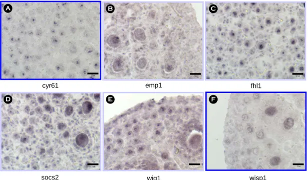
Figure 2. The expression pattern of genes related to the cell size growth in the postnatal day5 mouse ovaries by in situ hybridization. Sections were counterstained with nuclear fast red. (A) cyr61, (B) emp1, (C) fhl1, (D) socs2, (E) wig1, and (F) wisp1. Scale bars indicate 25 µm.
Figure 1. Heat map of genes related to the cell size
growth obtained from the previous study (Yoon et al.,
2005). Red indicates increase in expression, while green
indicates decrease in expression. Numbers on the colu-
mns indicating primordial (1), primary (2), and secon-
dary (3) follicles.
Cyr61 Wisp1 B
A
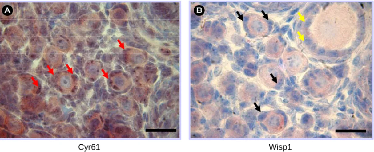
Figure 3. The expression pattern of Cyr61 (A) and Wisp1 (B) protein in the postnatal day5 mouse ovaries by immunohistochemistry. Sections were counterstained with hematoxylin. Cyr61 protein was expressed in pre-granulosa cells (red arrow). Wisp1 protein was not detected in pre-granulosa cells of the primordial follicle (black arrow), but detected in granulosa cells from primary follicle stage (yellow arrow). Scale bars indicate 25 µm.
cyr61 wisp1
1-day-old mouse ovary
Dissociation Overnight culture incubated at 37℃
separation of oocytes and pre-granulosa cells
cyr61
wisp1 fig α
g3pdh Oocyte Somatic
cells
A B
C D
Figure 4. The expression pattern of cyr61 and wisp1 mRNA. cyr61 (A) and wisp1 (B) mRNA was detected only in
oocytes in the postnatal day5 mouse ovaries by in situ hybridization. (C) The experimental scheme to obtain dissociated
oocytes and somatic cells including pre-granulosa cells from neonatal ovaries. (D) Relative abundance of cyr61 and
wisp1 mRNA in oocytes and somatic cells including pre-granulosa cells from neonatal ovaries by RT-PCR. Expression
of the fig α, oocyte specific gene, was measured to confirm the complete separation of oocyte from somatic cell. The
expression of g3pdh was used as an internal standard.
2. Cell size growth 관련 유전자의 in situ hy- bridization 발현 양상
생후 5일령 ICR 생쥐 난소에서 6개 유전자에 대 한 in situ hybridization 결과, cyr61과 wisp1 두 유전 자는 난자에서만 발현하는 것을 관찰하였다. 세부 적으로 살펴보면, cyr61은 난자의 인과 과립 세포에 서 발현하였고 wisp1은 난자의 핵을 제외한 인과 세포질에서 발현하고 있는 것으로 나타났다. 그러나 cyr61의 경우에는 14일령 생쥐 난소에 존재하는 2 차 난포의 과립 세포의 인에서만 발현하는 것을 관 찰하였다 (data not shown). 그러나 나머지 네 개의 유전자 (emp1, fhl1, socs2, wig1)는 난자와 과립 세포 에서 모두 발현하는 것으로 나타났고 (Figure 2) 더 발달된 난포에서는 협막 세포에서도 발현하는 것을 관찰하였다 .
3. Cyr61과 Wisp1 단백질 발현
위의 in situ hybridization 연구 결과에 따라, Cyr61 과 Wisp1 단백질의 발현 양상을 알아 보기 위해서 immunohistochemistry를 수행하였다. Cyr61의 경우, 원시난포 내 난자의 세포질에서 높게 발현하였고, 특히 pre-granulosa cell에서 강하게 단백질이 발현하 는 것을 확인할 수 있었다 (Figure 3A). Wisp1의 단 백질은 원시난포의 난자 세포질에서는 발현을 확인 하였지만 Cyr61과는 다르게 pre-granulosa cell에서 발현을 관찰할 수 없었으며 1차 난포의 과립 세포 에서부터 발현이 나타나기 시작하였다 (Figure 3B).
4. Semiquantitative RT-PCR
Pre-granulosa cell에서 발현하는 Cyr61 단백질의 근원을 알아 보기 위해 생후 1일자 생쥐 난소에서
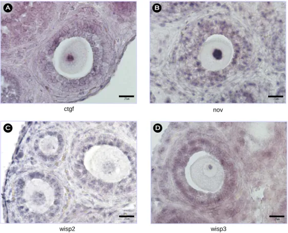
ctgf nov
wisp3 wisp2