D I A B E T E S & M E T A B O L I S M J O U R N A L
This is an Open Access article distributed under the terms of the Creative Commons Attribution Non-Commercial License (http://creativecommons.org/licenses/by-nc/4.0/) which permits unrestricted non-commercial use, distribution, and reproduction in any medium, provided the original work is properly cited.
A Journey to Understand Glucose Homeostasis:
Starting from Rat Glucose Transporter Type 2 Promoter Cloning to Hyperglycemia
Yong Ho Ahn
Department of Biochemistry and Molecular Biology, Integrated Genomic Research Center for Metabolic Regulation, Yonsei University College of Medicine, Seoul, Korea
My professional journey to understand the glucose homeostasis began in the 1990s, starting from cloning of the promoter region of glucose transporter type 2 (GLUT2) gene that led us to establish research foundation of my group. When I was a graduate stu- dent, I simply thought that hyperglycemia, a typical clinical manifestation of type 2 diabetes mellitus (T2DM), could be caused by a defect in the glucose transport system in the body. Thus, if a molecular mechanism controlling glucose transport system could be understood, treatment of T2DM could be possible. In the early 70s, hyperglycemia was thought to develop primarily due to a defect in the muscle and adipose tissue; thus, muscle/adipose tissue type glucose transporter (GLUT4) became a major research interest in the diabetology. However, glucose utilization occurs not only in muscle/adipose tissue but also in liver and brain. Thus, I was interested in the hepatic glucose transport system, where glucose storage and release are the most actively occurring.
Keywords: Adipogenesis; Diabetes mellitus, type 2; Glucokinase; Gluconeogenesis; Glucose transporter type 2; Glycolysis; Tran- scription factors
Corresponding author: Ahn Yong Ho https://orcid.org/0000-0002-4133-0757 Department of Biochemistry and Molecular Biology, Integrated Genomic Research Center for Metabolic Regulation, Yonsei University College of Medicine, 50 Yonsei-ro, Seodaemun-gu, Seoul 03722, Korea
E-mail: [email protected]
INTRODUCTION
In β-cells of pancreas and liver, glucose transport machinery consists of glucose transporter type 2 (GLUT2) and glucoki- nase (Gck), which is also called a glucose sensor [1]. But in the 1980s, the mechanism of the regulation of gene expression of hepatic glucose transport system in the insulin resistant, dia- betic states was largely unknown. Thus, we started to clone up- stream regions (promoters) of GLUT2, Gck, and glucose-6- phosphatase (G6Pase) and searched for potential trans-acting factors regulating the expression of these genes. In this review, I will introduce my research path which has mainly focused on the characterization of the transcriptional network of genes in- volved in the glucose homeostasis.
STARTING FROM RAT GLUT2 PROMOTER
In the 1990s, cloning of genes with upstream regulatory ele- ments was the mainstream biochemistry field. Because it was the previous era of the Genome Project we had to manually se- quence a lot of short gene fragments using Sanger’s dideoxy method to clone the whole rat GLUT2 gene, which was labori- ous and time consuming. Dr. Gil-Soo Han, my first research associate, started the project, but left my Lab in 1993 before the work was completed. So, we had to determine sequence of exon 1 with 5´-flanking region. Repeated screenings of lambda phage clones finally revealed the exon 1 and upstream se- quences, and furthermore, using rapid amplification of 5´
complementary DNA ends (5´-RACE) and primer extension, https://doi.org/10.4093/dmj.2018.0116
pISSN 2233-6079 · eISSN 2233-6087
we were able to identify complete structure of rat GLUT2 gene, which has four split exon 1 (1a to 1d). This discovery let us to obtain the rat GLUT2 promoter region, which conveys many potential transcription regulatory elements [2]. Next, we tried to find the mechanism of regulation of GLUT2 gene expression in liver. GLUT2 is a major glucose transporter in liver and pan- creatic β-cells, and we initially focused to liver because nuclear extract from this organ is easier to prepare to study DNA-pro- tein interaction, including DNase I footprinting and electro- phoretic mobility shift assay (EMSA). The DNase I footprint- ing assay was a difficult and laborious experiment and took a long time, but finally we demonstrated CCAAT/enhancer binding protein (C/EBP) elements in the GLUT2 promoter.
Furthermore, it was also identified that the regulation by C/
EBPs were gradually attenuated in the primary cultured hepa- tocytes, providing an evidence why primary hepatocytes on culture dishes lose their ability to express GLUT2 [3]. These earlier studies became foundation stone of my research.
EXPANSION TO HUMAN GLUT2 PROMOTER
GLUT2 gene is mainly expressed in liver and pancreatic β- cells; however, the transcription factors and cis-elements regu- lating the tissue-specific expression of the GLUT2 were largely unknown. Transfection studies using pancreatic β-cells (HIT- T15, MIN6), hepatoma cell (HepG2), and fibroblast cells (NI- H3T3, HeLa) revealed the differences in the promoter activi- ties and site C (87 to 132) responsible for tissue-specific ex- pression of the human GLUT2 gene [4]. We identified that both hepatocyte nuclear factor 1 (HNF1) and HNF3 function as transcriptional activators in GLUT2 gene expression. The GLUT2 mRNA level was well correlated with the status of HNF1 and HNF3 mRNA levels regardless of the origins or types of cells. Furthermore, we showed that the promoter ac- tivity of the mutG construct containing the 103A→G muta- tion, which was found in a Korean non-insulin-dependent dia- betes mellitus (NIDDM) patients, was in part compensated by the binding of nuclear factor Y (NF-Y) instead of HNF1 or HNF3 [5].
NUCLEAR RECEPTOR AND GLUCOSE SENSOR REGULATION IN LIVER AND β-CELLS
In the late 1990s, peroxisome proliferator-activated receptor γ
(PPARγ) was identified as a target for thiazolidinedione (TZD), the first insulin sensitizing agent [6]. Anti-diabetic ac- tion of TZD was thought to be mediated by PPARγ in adipose tissues until we identified that TZD restores the GLUT2 ex- pression in pancreatic β-cells of type 2 diabetes mellitus (T2DM) model mice. While searching for transcription factors regulating GLUT2 gene, we noticed the presence of PPARγ re- sponse element (PPRE) in 5´ untranslated region of the GLUT2 gene. Although PPARγ in β-cells is not abundant, TZD treat- ment directly up-regulated PPARγ-dependent GLUT2 expres- sion and restored glucose sensing ability of β-cell in diabetic condition [7]. Subsequently, we identified PPRE in β-cell spe- cific glucokinase (βGck) promoter and showed its functional relevance in β-cells [8].
After that, we studied liver glucokinase (L-Gck) promoter, because we wanted to stay focused in the hepatic glucose sen- sor in connection with glucose homeostasis. In liver, we were able to identify and characterize the role of PPARγ in the regu- lation of L-Gck [9,10]. Next, we demonstrated the interrela- tionship among PPARγ, liver X-receptor α (LXRα), sterol reg- ulatory element binding protein-1c (SREBP-1c), and small heterodimer partner (SHP) in the regulation of hepatic gluco- kinase expression [11].
SREBP-1c AND GLUCOSE METABOLISM
The regulation of hepatic glucose metabolism is important in glucose homeostasis and L-Gck plays a central role in this pro- cess. L-Gck expression is known to be regulated by insulin and SREBP-1c was suggested to be a transcription factor that medi- ates the action of insulin on Gck transcription in liver. Howev- er, the mechanism how SREBP-1c induce hepatic Gck expres- sion in response to insulin is not well characterized. Using DNA-protein interactions techniques, we identified sterol reg- ulatory elements (SREs) in rat L-Gck promoter and present ev- idences that insulin increases the binding of SREBP-1c on L- Gck promoter resulting in the increase of the L-Gck transcrip- tion [12].
In 2004, we performed an experiment whether SREBP-1c could be a potential trans-acting factor regulating GLUT2 in liver. At that time, GLUT2 gene expression was not known to be regulated by insulin. And thus, SREBP-1c could not be a transcriptional coactivator of GLUT2 gene expression. Howev- er, we observed that both GLUT2 and SREBP-1c gene expres- sion were increased in hyperglycemic states. Moreover, we
demonstrated that SREBP-1c binding region (SRE) is present in the mouse GLUT2 promoter. With these data, we concluded that glucose-stimulated activation of GLUT2 gene expression might be induced by SREBP-1c in primary cultured hepato- cytes. Adenoviral expression of the dominant negative form of SREBP-1c suppressed glucose-stimulated GLUT2 mRNA level in primary cultured hepatocytes [13].
We also showed regulatory mechanism of GLUT4 gene ex- pression in adipocytes. With increased GLUT4 expression, mRNA levels of SREBP-1c in adipose tissue were proportion- ally increased by refeeding. Insulin treatment increased mRNA levels of GLUT4 in adipose tissue. Because SREBP-1c gene ex- pression is regulated by insulin, it is thought that GLUT4 might be regulated by SREBP-1c. On this basis, we examined presence of a potential site binding SBREP-1c in the GLUT4 promoter and found highly conserved SRE. Through this study, we published a paper entitled, “Insulin-mediated activa- tion of human GLUT4 promoter is directly induced by SREBP-
1c in adipocytes” [14].
Subsequent to this study, we hypothesized that transcription factor E3 (TFE3) could be able to up-regulate Gck because TFE3 was shown to up-regulate genes of insulin signaling pathway [15]. Using adenoviral transduction of TFE3, we demonstrated the presence of TFE3 binding site (E-box) in the Gck and TFE3 up-regulates Gck with amelioration of hypergly- cemia in the db/db and ob/ob mice [16]. Fig. 1 shows a sum- mary of cis-elements and trans-acting factors acting on the glucose sensor genes in liver explored by my team.
ROLE OF PPARα AND PROLACTIN
REGULATORY ELEMENT-BINDING ON THE GLUCONEOGENESIS
When glucose level is low, blood glucose level is maintained by hepatic gluconeogenesis. Because increase in a hepatic glucose production occurs in the post-absorptive state of T2DM, it is
Fig. 1. Schematic summary of the regulation of liver type glucose transporter (glucose transporter type 2 [GLUT2]) and glucoki- nase (Gck) promoter. During the long journey to understand glucose homeostasis, main interest of my group was how glucose sensors, GLUT2 and glucokinase, are regulated by transcription factors. Tissue-specific study, and disease-specific alteration as well, revealed that many transcriptional factors are interconnected in the regulation of glucose sensor in liver. C/EBP, CCAAT/
enhancer binding protein; PPARγ, peroxisome proliferator-activated receptor γ; RXRα, retinoid X receptor α; PPRE, PPARγ re- sponse element; SREBP-1c, sterol regulatory element binding protein-1c; SRE, sterol regulatory element; HNF1α, hepatocyte nu- clear factor 1 α; HNF, hepatocyte nuclear factor; FOXA2, forkhead box A2; LXRα, liver X-receptor α; SHP, small heterodimer partner; L-GcK, liver glucokinase; TFE3, transcription factor E3; LXRE, liver X-receptor response element.
required to unveil the mechanisms of how hyperglycemia de- velops. To explore a pathogenesis, we are interested in a role of PPARα and prolactin regulatory element-binding (PREB).
First, we have performed experiments based on the previous observation that PPARα-null mice shows severe hypoglycemia following 24-hour fasting, characterized by a 50% drop in blood glucose level, suggesting a potential role of PPARα in glucose metabolism [17]. Using PPARα knockout (KO) mice as an animal model, we were able to demonstrate presence of PPRE and indicated that PPARα might be responsible for glu- cose production through the regulation of hepatic G6pase ex- pression [18]. Most recently, we observed that PREB protein is down-regulated in the liver of db/db, ob/ob, and high-fat diet- induced obese mice. This observation led us to explore a possi- bility that PREB could be a transacting factor down-regulating gluconeogenic. Indeed, we found a cis-element, prolactin core- binding element in the promoters of gluconeogenic genes, which is responsible for down-regulation of these genes, that is, decreased PREB in the liver of insulin resistant animal can- not repress the expression of gluconeogenic genes (G6Pase and phosphoenolpyruvate carboxykinase [Pck]), resulting in the hyperglycemia, a typical manifestation of the diabetic model mice or high fat induced obesity mice [19].
ROLE OF PROTEIN INTERACTIONS AND GLUCOSE HOMEOSTASIS
Glucose homeostasis is of prime importance in maintaining whole body metabolism. In the insulin resistant states, a bal- ance in the intermediary metabolism may be disturbed. Dur- ing prolonged starvation, hepatic gluconeogenesis is a major pathway that maintains normal blood glucose levels. In the postprandial state, temporarily increased glucose needs to be disposed by liver to prevent the cells from glucotoxicity. In this process, Gck is known to play the most important role [20].
Although we have been stayed focus on the transcriptional aspects of metabolic genes in liver, we have also searched for the possibility that post-translational modification of metabol- ic enzymes/protein could also be a novel regulatory mecha- nism. Using current proteome technology, we were able to demonstrate that glucokinase regulatory protein (GKRP) in liver is acetylated by acetyltransferase p300. Acetylated GKRP decreases ubiquitination of the protein itself with an increase in its affinity to Gck, resulting in increased nuclear retention of Gck and decreased glycolytic flux [21].
To understand this, we have done couple of studies to eluci- date mechanisms of how hepatic gluconeogenesis occurs.
From the beginning, we showed that insulin-mediated repres- sion of G6pase and Pck gene expression is restored by resvera- trol, which might be associated with sirtuin 1 (Sirt1) mediated forkhead box O1 (FOXO1) deacetylation [22]. And then, we found a role of thioredoxin-interacting protein (TXNIP) in the hyperglycemia. The increased TXNIP in the hyperglycemic mice model, impairs glucose and insulin tolerance in mice by up-regulating G6pase through interaction with SHP. Binding of TXNIP to SHP resulted in the degradation of SHP through ubiquitination, allowing FOXO1 to up-regulate gluconeogenic gene expression [23].
ROLE OF CREB3L4 ON THE ADIPOGENESIS
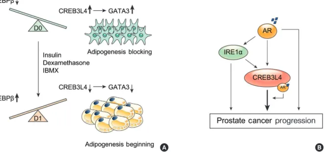
Another field of our research was transcriptional connections in adipogenesis. We were interested in obesity problem, which became a worldwide epidemic. To this end, we wanted to ex- plore signaling networks governed by transcription factors in- volved in the adipogenesis, ultimately to understand the patho- physiology of obesity and T2DM. Using cyclic AMP-respon- sive element-binding protein 3 (CREB3)-like 4 (CREB3L4) KO mice, we demonstrated that CREB3L4 acts as a negative regu- lator of adipogenesis [24]. In this paper we demonstrated that CREB3L4 binds C/EBPβ, promoting its ubiquitination, result- ing in the inhibition of adipogenesis and obesity in mice (Fig.
2A). Thus, if a measure to trigger CREB3L4 expression is taken, it could be one of novel way to treat obesity or T2DM. In con- nection with this work, we published a paper entitled, “The ef- fects of low fat diet (LFD) or aging on the metabolic profiles of Creb3l4 KO mice,” which showed significant weight gain and adiposity with impaired glucose tolerance and decreased insu- lin sensitivity in Creb3l4 KO mice [25]. Furthermore, we dem- onstrated that CREB3L4 is required for proliferation of pros- tate cancer cells and CREB3L4 is a crucial activator of andro- gen receptor (AR) function. CREB3L4 directly interacts with, and facilitates, AR recruitment to the androgen responsive ele- ments (AREs) of AR target genes. In addition, indirectly, through inositol requiring enzyme 1α signaling, a distinct AR- endoplasmic reticulum stress-CREB3L4 regulatory axis also plays a role in prostate cancer proliferation (Fig. 2B) [26]. Table 1 shows a summary of major works done by my team for the last 25 years.
Fig. 2. Role of cyclic AMP-responsive element-binding protein 3 (CREB3)-like 4 (CREB3L4) on the adipogenesis. (A) In preadi- pocytes, CREB3L4 acts as a negative regulator of adipogenesis by both regulating the stability of CCAAT/enhancer binding pro- tein β (C/EBPβ) protein and increasing GATA binding protein 3 (GATA3) expression. (B) Proposed mechanism of action of CREB3L4 in prostate cancer progression. Adapted from Kim et al. [24,26]. IBMX, 3-isobutyl-1-methylxanthine; AR, androgen receptor; IRE1α, inositol requiring enzyme 1α.
A B
Table 1. Summary of major research papers of my team
Year Journal Research subject Contributor(s)
1995 Arch Biochem Biophys L-GLUT2 promoter cloning Ahn et al. [2]
1998 Biochem J L-GLUT2 promoter, C/EBP Kim et al. [3]
2000 J Biol Chem L-GLUT2 liver specific expression, HNF1&3 Cha et al. [4]
2000 Diabetes L-GLUT2 promoter, PPRE Kim et al. [7]
2002 Diabetes Β-Glucokinase, PPARγ Kim et al. [8]
2004 Diabetes L-GcK promoter, PPARγ Kim et al. [10]
2004 J Biol Chem L-GcK promoter, SREBP-1c Kim et al. [12]
2005 Diabetes L-GLUT2 promoter, SREBP-1c Im et al. [13]
2006 Biochem J GLUT4 promoter, SREBP-1c adipocytes Im et al. [14]
2009 J Biol Chem L-GcK promoter, LXR, SREBP-1c, PPARγ Kim et al. [11]
2011 J Biol Chem G6Pase promoter, PPARα Im et al. [18]
2013 Diabetologia L-GcK gene, TFE3 Kim et al. [16]
2013 Diabetologia Liver gluconeogenic gene, Txnip Jo et al. [23]
2014 Cell Death Dis Adipogenesis and CREB3L4 Kim et al. [24]
2015 Sci Rep L-GcK and GKRP acetylation Park et al. [21]
2017 Sci Rep Prostate cancer and CREB3L4 Kim et al. [26]
2018 BBA Mol Basis Dis Liver PREB and glucose homeostasis Park et al. [19]
L-GLUT2, liver-glucose transporter type 2; C/EBP, CCAAT/enhancer binding protein; HNF, hepatocyte nuclear factor; PPRE, PPARγ response element; PPAR, peroxisome proliferator-activated receptor; L-GcK, liver glucokinase; SREBP-1c, sterol regulatory element binding protein-1c;
LXR, liver X-receptor; G6Pase, glucose-6-phosphatase; TFE3, transcription factor E3; CREB3L4, cyclic AMP-responsive element-binding pro- tein 3 (CREB3)-like 4; GKRP, glucokinase regulatory protein; PREB, prolactin regulatory element-binding.
SUMMARY
My team has been exploring the roles of transcription factors regulating the genes involved in glucose metabolism for the last 30 years. The main field of study can be summarized as (1) identification of promoters of genes of glucose homeostasis, (2) exploration of interaction between promoters and transcrip- tion factors leading to tissue-specific gene expression, (3) modification and interactions between transcription factors in metabolic diseases such as diabetes or obesity, and (4) role of a transcription factor in the adipocyte differentiation and devel- opment of obesity. While studying transcriptional regulation, my team members were interconnected as if we were tran- scription factors coordinating with each other. As an analogy, I might act as a basal transcription factor and my team-mates, wherever they are, played co-activator role. Without their de- votion in science, the works of my group could not be made possible.
CONFLICTS OF INTEREST
No potential conflict of interest relevant to this article was re- ported.
ACKNOWLEDGMENTS
I would like to thank all the people who worked with me at the same time or at different times. Also, I would like to apologize that I could not mention all the researches in this article, in which every single study carried out was meaningful. The manuscript was mainly prepared by Drs, Mi Young Kim, Seung Soon Im, Ha Il Kim, Ji Young Cha, and Jae Woo Kim.
We thank Dong-Su Jang for the schematic representation of the model of CREB3L4 action.
REFERENCES
1. Schuit FC, Huypens P, Heimberg H, Pipeleers DG. Glucose sensing in pancreatic beta-cells: a model for the study of other glucose-regulated cells in gut, pancreas, and hypothalamus.
Diabetes 2001;50:1-11.
2. Ahn YH, Kim JW, Han GS, Lee BG, Kim YS. Cloning and characterization of rat pancreatic beta-cell/liver type glucose transporter gene: a unique exon/intron organization. Arch Biochem Biophys 1995;323:387-96.
3. Kim JW, Ahn YH. CCAAT/enhancer binding protein regulates the promoter activity of the rat GLUT2 glucose transporter gene in liver cells. Biochem J 1998;336(Pt 1):83-90.
4. Cha JY, Kim H, Kim KS, Hur MW, Ahn Y. Identification of transacting factors responsible for the tissue-specific expres- sion of human glucose transporter type 2 isoform gene. Coop- erative role of hepatocyte nuclear factors 1alpha and 3beta. J Biol Chem 2000;275:18358-65.
5. Cha JY, Kim HS, Kim HI, Im SS, Kim SY, Kim JW, Yeh BI, Ahn YH. Analysis of polymorphism of the GLUT2 promoter in NI- DDM patients and its functional consequence to the promoter activity. Ann Clin Lab Sci 2002;32:114-22.
6. Nolan JJ, Ludvik B, Beerdsen P, Joyce M, Olefsky J. Improve- ment in glucose tolerance and insulin resistance in obese sub- jects treated with troglitazone. N Engl J Med 1994;331:1188- 93.
7. Kim HI, Kim JW, Kim SH, Cha JY, Kim KS, Ahn YH. Identifi- cation and functional characterization of the peroxisomal pro- liferator response element in rat GLUT2 promoter. Diabetes 2000;49:1517-24.
8. Kim HI, Cha JY, Kim SY, Kim JW, Roh KJ, Seong JK, Lee NT, Choi KY, Kim KS, Ahn YH. Peroxisomal proliferator-activated receptor-gamma upregulates glucokinase gene expression in beta-cells. Diabetes 2002;51:676-85.
9. Kim HI, Ahn YH. Role of peroxisome proliferator-activated receptor-gamma in the glucose-sensing apparatus of liver and beta-cells. Diabetes 2004;53 Suppl 1:S60-5.
10. Kim SY, Kim HI, Park SK, Im SS, Li T, Cheon HG, Ahn YH.
Liver glucokinase can be activated by peroxisome proliferator- activated receptor-gamma. Diabetes 2004;53 Suppl 1:S66-70.
11. Kim TH, Kim H, Park JM, Im SS, Bae JS, Kim MY, Yoon HG, Cha JY, Kim KS, Ahn YH. Interrelationship between liver X re- ceptor alpha, sterol regulatory element-binding protein-1c, peroxisome proliferator-activated receptor gamma, and small heterodimer partner in the transcriptional regulation of gluco- kinase gene expression in liver. J Biol Chem 2009;284:15071- 83.
12. Kim SY, Kim HI, Kim TH, Im SS, Park SK, Lee IK, Kim KS, Ahn YH. SREBP-1c mediates the insulin-dependent hepatic glucokinase expression. J Biol Chem 2004;279:30823-9.
13. Im SS, Kang SY, Kim SY, Kim HI, Kim JW, Kim KS, Ahn YH.
Glucose-stimulated upregulation of GLUT2 gene is mediated by sterol response element-binding protein-1c in the hepato- cytes. Diabetes 2005;54:1684-91.
14. Im SS, Kwon SK, Kang SY, Kim TH, Kim HI, Hur MW, Kim
KS, Ahn YH. Regulation of GLUT4 gene expression by SREBP- 1c in adipocytes. Biochem J 2006;399:131-9.
15. Nakagawa Y, Shimano H, Yoshikawa T, Ide T, Tamura M, Fu- rusawa M, Yamamoto T, Inoue N, Matsuzaka T, Takahashi A, Hasty AH, Suzuki H, Sone H, Toyoshima H, Yahagi N, Yamada N. TFE3 transcriptionally activates hepatic IRS-2, participates in insulin signaling and ameliorates diabetes. Nat Med 2006;12:
107-13.
16. Kim MY, Jo SH, Park JM, Kim TH, Im SS, Ahn YH. Adenovi- rus-mediated overexpression of Tcfe3 ameliorates hyperglycae- mia in a mouse model of diabetes by upregulating glucokinase in the liver. Diabetologia 2013;56:635-43.
17. Kersten S, Seydoux J, Peters JM, Gonzalez FJ, Desvergne B, Wahli W. Peroxisome proliferator-activated receptor alpha me- diates the adaptive response to fasting. J Clin Invest 1999;103:
1489-98.
18. Im SS, Kim MY, Kwon SK, Kim TH, Bae JS, Kim H, Kim KS, Oh GT, Ahn YH. Peroxisome proliferator-activated receptor {alpha} is responsible for the up-regulation of hepatic glucose- 6-phosphatase gene expression in fasting and db/db mice. J Biol Chem 2011;286:1157-64.
19. Park JM, Kim MY, Kim TH, Min DK, Yang GE, Ahn YH. Pro- lactin regulatory element-binding (PREB) protein regulates hepatic glucose homeostasis. Biochim Biophys Acta 2018;1864
(6 Pt A):2097-107.
20. Niswender KD, Shiota M, Postic C, Cherrington AD, Magnu- son MA. Effects of increased glucokinase gene copy number on glucose homeostasis and hepatic glucose metabolism. J Biol Chem 1997;272:22570-5.
21. Park JM, Kim TH, Jo SH, Kim MY, Ahn YH. Acetylation of glucokinase regulatory protein decreases glucose metabolism by suppressing glucokinase activity. Sci Rep 2015;5:17395.
22. Park JM, Kim TH, Bae JS, Kim MY, Kim KS, Ahn YH. Role of resveratrol in FOXO1-mediated gluconeogenic gene expres- sion in the liver. Biochem Biophys Res Commun 2010;403:329- 34.
23. Jo SH, Kim MY, Park JM, Kim TH, Ahn YH. Txnip contributes to impaired glucose tolerance by upregulating the expression of genes involved in hepatic gluconeogenesis in mice. Diabeto- logia 2013;56:2723-32.
24. Kim TH, Jo SH, Choi H, Park JM, Kim MY, Nojima H, Kim JW, Ahn YH. Identification of Creb3l4 as an essential negative regulator of adipogenesis. Cell Death Dis 2014;5:e1527.
25. Kim TH, Park JM, Jo SH, Kim MY, Nojima H, Ahn YH. Effects of low-fat diet and aging on metabolic profiles of Creb3l4 knockout mice. Nutr Diabetes 2015;5:e179.
26. Kim TH, Park JM, Kim MY, Ahn YH. The role of CREB3L4 in the proliferation of prostate cancer cells. Sci Rep 2017;7:45300.
![Fig. 1. Schematic summary of the regulation of liver type glucose transporter (glucose transporter type 2 [GLUT2]) and glucoki- glucoki-nase (Gck) promoter](https://thumb-ap.123doks.com/thumbv2/123dokinfo/5221483.123590/3.892.101.777.556.928/schematic-regulation-transporter-glucose-transporter-glucoki-glucoki-promoter.webp)