서 론
1)중격-시신경 형성장애(septo-optic dysplasia)는 시신경 형성부전과투명중격(septum pellucidum)의 결손을 동반한 선천 질환으로1956년 de Morsier[1]에 의하여 최초로 명명 되었다. 이 질환의 원인은 중추 신경계 형성기의 발달 이상 으로 추정되는데 선천적으로 시력장애, 안구 진탕, 경련, 뇌 하수체기능저하증 등의 임상 증상과 시신경이나 시교차부 형성부전, 투명중격 결손 등의 병리소견을 나타내는 질환이
접수일자: 2007년 6월 2일 통과일자: 2007년 7월 27일
책임저자: 정태식, 경상대학교 의과대학 내과학교실
다[2,3]. 인구 100,000명 당 2명 정도의 유병률을 갖는 비 교적 드문 질환으로 5세 이전의 영유아기에 주로 발견된 다[4~7]. 상당수에서 뇌 실질 이동장애의 하나로 드문 선천 성 뇌 기형인 분열뇌증(schizencephaly)과 동반된다고 하며 이런 경우에는 신경학적 증상과 안과적 이상이 더 많은 경 향이 있다[4,8,9].
대부분의 환자들은 시력 저하나 사시, 안구 진탕 그리고 간질발작 등의 이상소견 발견 후 방사선 영상 검사로 영유 아기에 발견되었다[2,5~7]. 국내 보고 역시 안과적 증상을 위주로 영유아기의 환자들이 보고되었으나[10~12], 본 환자 와 같이 출산 경험이 있는 성인에서 중추성 요붕증과 분열 뇌증이 함께 보고된 예가 국내에는 없어 문헌고찰과 함께 보고하는 바이다.
31세 여자 환자에게서 발견된 중격-시신경 형성장애와 분열뇌증을 동반한 중추성 요붕증 1예
경상대학교 의과대학 내과학교실1, 건강과학연구원2
김수경
1․정태식
1,2․함종렬
1,2․이상민
1․문성원
1․이경주
1․정순일
1,2A Case of Central Diabetes Insipitus Combined with Septo-Optic Dysplasia and Schizencephaly in 31-year-old Woman
Soo Kyoung Kim1, Tae Sik Jung1,2, Jong Ryeal Hahm1,2, Sang Min Lee1, Sung Won Moon1, Kyeong Ju Lee1, Soon Il Chung1,2
Department of Internal Medicine1, Institute of Health Sciences, Gyeongsang National University School of Medicine2
ABSTRACT
A 31-year-old woman was referred to our hospital for evaluation and management of poorly controlled epilepsy. The patient had been taking anti-epileptic drugs for six years. An MRI imaging study showed septo-optic dysplasia (SOD) and schizencephaly. SOD is a syndrome characterized by agenesis of the septum pellucidum or corpus callosum, optic nerve dysplasia and congenital hypothalamic-pituitary insufficiency. The patient was referred to the endocrine clinic for exclusion of any pituitary hormonal deficiencies. In a systemic review, the patient complained of polydipsia and polyuria for 20 years. In laboratory tests, measurements showed a serum osmolarity of 281 mOsm/kg, a serum sodium concentration of 144.7 mmol/L, a spot urine osmolarity of 183 mOsm/kg and a spot urine sodium concentration of 32 mmol/L. The patient underwent a water deprivation test, and was diagnosed with central diabetes insipidus.
We report a case of central diabetes insipitus combined with SOD, schizencephaly and epilepsy. (J Kor Endocrine Soc 22:339~343, 2007)
ꠏꠏꠏꠏꠏꠏꠏꠏꠏꠏꠏꠏꠏꠏꠏꠏꠏꠏꠏꠏꠏꠏꠏꠏꠏꠏꠏꠏꠏꠏꠏꠏꠏꠏꠏꠏꠏꠏꠏꠏꠏꠏꠏꠏꠏꠏꠏꠏꠏꠏꠏꠏꠏꠏꠏꠏꠏꠏꠏꠏꠏꠏꠏꠏꠏꠏꠏꠏꠏꠏꠏꠏꠏꠏꠏꠏꠏꠏꠏꠏꠏꠏꠏꠏꠏꠏꠏꠏꠏꠏꠏꠏꠏꠏꠏꠏꠏꠏꠏ Key Words: Diabetes Insipidus, Epilepsy, Septo-optic dysplasia
증 례
환 자: 31세 여자
주 소: 잘 조절되지 않는 경련
현병력: 내원 6년 전부터 간간히 발생한 경련으로 인근병 원에서 치료 받아 왔으나 최근 경련의 빈도가 늘고 조절이 잘 되지 않아 본원 신경과를 방문하였다. 뇌 자기공명영상 (MRI) 촬영과 안과 검진에서 중격-시신경 형성장애 진단받 고 호르몬 이상을 배제하기 위해서 내분비-대사 내과에 의 뢰되었다. 문진에서 환자는 하루 4L이상의 다음을 호소하였 는데, 다음은 다뇨와 야뇨의 증상과 함께 약 10세 경부터 시 작되었다. 환자는 규칙적인 생리를 하고 있었고, 8년 전 남 편과 결혼하여 현재 6세 된 딸을 키우고 있으며, 분만 후 모 유 수유를 하였다고 한다.
과거력: 본인의 주산기 시절의 특별한 병력은 없었고, 6 년 전부터 간질약을 복용 중이었다.
가족력: 간질이나 요붕증을 의심할 만한 병력은 없었다.
사회력: 고등학교를 졸업 후 현재 전업 가정주부로 지내 오고 있다.
진찰 소견: 내원 당시의 생체 활력 징후는 체온 36.6℃,
맥박 80회/분, 호흡 16회/분, 혈압 120/80 mmHg이었고, 의 식은 명료하였다. 키 159 cm이고 체중 50 kg이었다. 신경학 적 이상이나 특이한 안과적 이상 소견은 없었다. 호흡음, 심 음, 장음은 정상적으로 청진되었고 복부의 팽만, 간 및 비장 비대 등은 관찰되지 않았다.
검사 소견: 말초혈액검사에서 백혈구 4,830/mm3, 혈색소 11.6 g/dL, 혈소판 22,400/mm3 이었다. 생화학 검사에서 혈액요소질소 4.9 mg/dL, 크레아티닌 0.6 mg/dL, 나트륨 144.7 mmol/L, 칼륨 4.8 mmol/L, 염소 109.6 mmol/L, 혈 액 삼투압 농도 281 mOsm/kg, 소변 검사에서 비중 1.004, 삼투압 농도 183 mOsm/kg, 나트륨은 32 mmol/L, 칼륨 15.3 mmol/L, 염소 33.2 mmol/L, 크레아티닌 45.5 mg/dL 이였다. 성장호르몬 0.62 ng/mL, 인슐린유사성장인자-I 342.90 ng/mL (정상범위: 79~384), 프로락틴 10.19 ng/mL, 코르티솔 12.03 μg/dL, 부신피질자극호르몬 18.66 pg/mL, 난 포자극호르몬 4.65 mIU/mL, 황체형성호르몬 6.75 mIU/mL, 갑상선자극호르몬 0.77mIU/L 그리고 유리 T4 1.23 ng/dL 이었다. 뇌 MRI 촬영에서는 양측 측두엽에 막힌 형태의 분 열뇌증과 함께 투명중격의 완전 결손이 관찰되었다(Fig. 1).
또한 뇌파검사에서 부분 간질에 합당한 우반구 측두엽의 부 A
B
C
Fig. 1. Brain MRI showed schizencephaly (A) and absence of septum pellucidum (B and C). Arrows in picture A indicate closed lip type of schizocephalic clefts of lateral parietal regions in horizontal view, and arrows in picture B and C indicate absence of septum pellucidum in coronal view (B) and in sagittal view (C).
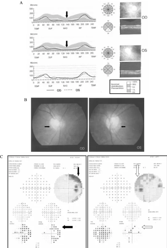
분발작 소견이 관찰 되었다. 안과에서 시행한 광간섭성 단층 촬영(optical coherence tomogaphy) 검사 및 시야검사에서 시신경의 경한 위축 소견을 확인하였다(Fig. 2).
임상경과 및 치료: 환자는 수분제한 검사를 하였는데, 검 사 시작 4시간 후에 체중의 5% 에 해당하는 체중 감소 소견 관찰되었고 소변 삼투압이 131 mOsm/kg에서 197 mOsm/kg A
B
C
Fig. 2. Ophthalmologic examinations of the patient. (A) Optical coherence tomography showed partial optic nerve atrophy. Arrow indicates abnormal thin thickness of red-free retinal nerve fiber layer at nasal area. (B) Disc photography showed peri-papillary degeneration (arrows). C/D (Cup/Disc) ratio is 0.5 and it suggests optic nerve atrophy. (C) Visual field analysis showed right nasal side arcuate scotoma (black arrows) and left superior arcuate scotoma (white arrows) and this finding was compatible with optic nerve atrophy.
으로 농축이 관찰되지 않아 검사를 중지하고 바소프레신 5 단위를 피하주사 하였다. 바소프레신 주사 후로 요 삼투압이 197 mOsm/kg에서 358 mOsm/kg로 50% 이상 증가하였으 며, 바소프레신 투여 직전 혈청 바소프레신 수치는 1.00 pg/mL (0~7.60) 미만이었다(Table 1).
환자는 중추성 요붕증, 투명중격 결손과 시신경 위축이 동반된 중격-시신경 형성장애, 분열뇌증 그리고 부분 간질로 진단되었다. 환자는 desmopressin acetate 0.2 mg 하루 2회 복용 후 다음, 다뇨 그리고 야뇨 증상은 없어 졌으며 현재까 지 복용 중이다. 간질 역시 하루 phenobarbital 45 mg, valproic acid 900 mg 및 lamotrigin 100 mg을 복용 후 더 이상의 경련 발작은 없어졌으며 외래에서 경과 관찰 중이다.
고 찰
중격-시신경 형성장애는 구개열, 합지증, 귀의 형성장애, 양안 격리증, 시신경 위축, 왜소 고환, 무후각증 등의 임상 양상과 함께 뇌하수체 기능부전으로인한 요붕증, 성장호르 몬 결핍, 단신, 드물게 갑상선자극호르몬 결핍을 보이기도 한다[3].
1941년 7개월된 영아에서 시신경 형성부전과 투명중격의 결손이 동반된 예를 Reeves 등[4]이 처음 보고하였고, 1956 년 de Morsier[1]에 의해 중격-시신경 형성장애라는 하나의 증후군으로 명명되었다. 중격-시신경 형성장애의 원인으로 임신 중 음주, quinidine, phenytoin 등의 약물 복용과도 관 련이 있다는 보고가 있고[13,14], 당뇨병 등의 산전병력을 가진 엄마에서 태어난 아이에서 그 빈도가 높다는 보고가 있다[15]. 최근에는 드물게 전뇌 배부분의 발달에 관여하는 HESX1 유전자 변이와의 연관성이 보고되고 있으나[3,16]
이 환자의 경우는 이런 원인과의 연관성을 찾기는 어려웠고 유전자검사도 시행하지 않았다.
시신경 형성부전에 의한 시력장애의 정도는 거의 정상적 인 시력에서부터 실명에 이르기까지 다양하며 시신경 크기 와는 연관이 없다[2,5,9]. 국내에는 안과 이상을 보여 영유아
기에 진단된 경우가 대부분이고[10~12], 성인이 되어서 발 견된 경우도 어릴 때부터 안과 증상이나 신경 이상이 존재 하였다[17,18]. 환자는 일반적인 중격-시신경 형성장애 환자 보다 진단이 늦어졌는데 그 이유로 안과 증상이 없었고, 간 질의 발작 역시 25세로 늦게 시작되었으며, 유아기 때부터 중추성 요붕증이 있었다고 추정되나 31세까지 적절한 진단 을 받지 못했던 것으로 사료된다. 중격-시신경 형성장애 환 자에서 분열뇌증이 5~50%에서 같이 동반된다고 하며, 분열 뇌증의 약 1/3에서 시신경 발육부전이 동반된다고 한다[9].
중격-시신경 형성장애에서 호르몬 이상이 약 2/3 에서 관찰 되는데 이 중 성장호르몬 결핍이 가장 흔하고, 그 외 다른 뇌하수체 전엽 호르몬의 결핍이나 중추성 요붕증이 있을 수 있다[6,14,15]. 이 환자의 경우 기초 호르몬 검사에서 이상 이 없었고 정상적인 성장, 규칙적인 생리, 임신, 출산 그리고 모유 수유 등으로 미루어 복합뇌하수체 자극검사를 하지는 않았지만 뇌하수체 전엽의 호르몬 이상은 없을 것이라 추정 된다. 하지만 6년 전부터 간질이 발생하였으므로 최근 뇌하 수체 기능에 대한 평가와 특히 상대적으로 빈도가 높은 성 장호르몬 분비에 대해서 자극 검사를 할 수 있었으면 더 좋 았을 것이나 잘 조절되지 않는 간질로 인하여 저혈당 유발 검사를 할 수 없었다는 아쉬운 점이 있다.
대부분의 중격-시신경 형성장애 환자들이 안과 증상이나 신경증상으로 영유아기에 발견되는 것과는 달리 본 증례는 성인이 되어서 출산 후 간질의 원인을 진단하는 과정에서 중추성 요붕증, 분열뇌증과 함께 우연히 발견되었다. 이번 증례와 같이 성인이 되어서 발견된 중격-시신경 형성장애는 아주 드물게 보고되고 있다[19]. 20세에 발견된 여자 환자는 어릴 때부터 다음과 다뇨의 요붕증 증상이 있었고, 142 cm (3% 이하)로 저신장이 있었으나 간질 등의 신경학적 증상은 없었다. 29세에 발견된 여자 환자는 요붕증은 없었고, 136 cm (3% 이하)로 저신장이 있었으며, 부분 발작이 4년 전부 터 있었다. 본 증례의 환자는 요붕증이 소아기 때부터 있었 던 것으로 추정되며, 병력에서 간질의 진단이 임신 보다 빨 라서 임신이 요붕증이나 간질을 유발하거나 악화시킨 것은 Table 1. Serial flow chart of water deprivation test
Time Body weight
(kg)
Serum osmolarity (mosm/kg)
Urine osmolarity (mosm/kg)
Hourly urine (mL)
Vasopressin (pg/mL)
8 AM 48.30 290 131 300 < 1.00
9 AM 47.50 300 158 250
10 AM 47.10 302 178 300 < 1.00
11 AM 46.50 304 189 250
12 AM 46.05 309 197 300
Vasopressin 5 unit subcutaneous injection
1 PM 45.75 310 358 40 76.61
2 PM 45.75 309 435 100 29.79
아닌 것으로 판단된다. 세 환자 모두 늦게 발견된 이유는 안 과 증상이나 신경증상이 유아기에 뚜렷하지 않아서 성인이 되어서야 발견되어 진단이 늦어졌다. 드물기는 하지만 중추 성 요붕증이 간질과 같은 신경학적 이상을 동반한 경우는 중격-시신경 형성장애와 같은 선천성 기형에 대해서 고려해 보아야 할 것이다.
요 약
저자 등은 간질 조절을 주소로 내원한 31세 여자 환자에 서 중격-시신경 형성장애, 분열뇌증과 중추성 요붕증을 진단 하였다. 중격-시신경 형성 장애와 분열뇌증은 선천적인 기형 이고, 간질은 성인이 되어서 발병하였다. 중격-시신경 형성 장애는 시신경의 형성 부전이 있었으나 안과이상 증상이 없 어서 진단이 늦어졌다. 중추성 요붕증은 수분제한 검사로 확 진하였고, 약 20년 전부터 증상이 있었으나 적절한 검사를 받지 못해 진단이 늦어졌다. 현재 요붕증은 desmopressin acetate 사용하면서 외래 경과 관찰 중이다.
참 고 문 헌
1. de Morsier G: Studies on malformation of cranio- encephalic sutures. III. Agenesis of the septum lucidum with malformation of the optic tract. Schweiz Arch Neurol Psychiatr 77:267-292, 1956
2. Skarf B, Hoyt CS: Optic nerve hypoplasia in children.
Association with anomalies of the endocrine and CNS.
Arch Ophthalmol 102:62-67, 1984
3. Melmed S, Jameson JL: Disorders of the anterior pituitary and hypothalamus. In: Kasper DL, Braunwald E, Fauci AS, Hauser SL, Longo DL, Jameson JL ed.
Harrison’s Principles of Internal Medicine. 16th ed.
pp2077, Seoul, McGraw-Hill press, 2005
4. St John Jr, Reeves DL: Congenital absence of the septum pellucidum: a review of the literature with case report. Am J Surg 94:974-980, 1957
5. Siatkowski RM, Sanchez JC, Andrade R, Alvarez A:
The clinical, neuroradiographic, and endocrinologic profile of patients with bilateral optic nerve hypoplasia.
Ophthalmology 104:493-496, 1997
6. Haddad NG, Eugster EA: Hypopituitarism and neuro- developmental abnormalities in relation to central nervous system structural defects in children with optic
nerve hypoplasia. J Pediatr Endocrinol Metab 18:853- 858, 2005
7. Willnow S, Kiess W, Butenandt O, Dorr HG, Enders A, Strasser-Vogel B, Egger J, Schwarz HP:
Endocine disorders in septo-optic dysplasia (De Morsier syndrome)-evaluation and follow up of 18 patients. Eur J Pediatr 155:179 -184, 1996
8. Barkovich AJ, Fram EK, Norman D: Septo-optic dysplasia: MR imaging. Radiology 171:189-192, 1989 9. Kuban KC, Teele RL, Wallman J: Septo-optic-
dysplasia-schizencephaly. Radiographic and clinical features. Pediatr Radiol 19:145-150, 1989
10. Kim WJ, Yu YS, Chang BL: Septo-optic dysplasia. J Korean Ophthalmol Soc 32:327-330, 1991
11. Lee WH, Park SH, Shin H: A case of septo-optic dysplasia. J Korean Ophthalmol Soc 32:332-338, 1991 12. Han JW, Lee YC: De Morsier’s syndrome expressed as congenital exotropia. J Korean Ophthalmol Soc 40:
869-874, 1999
13. Parson SH, Dhillon B, Findlater GS, Kaufman MH:
Optic nerve hypoplasia in the fetal alcohol syndrome:
a mouse model. J Anat 186:313-320, 1995
14. Hoyt CS, Billson FA: Maternal anticonvulsant and optic nerve hypoplasia. Br J Opthalmol 62:3-6, 1978 15. Donat JF: Septo-optic dysplasia in an infant of a
diabetic mother. Arch Neurol 38:590-591, 1981 16. McNay DE, Turton JP, Kelberman D, Woods KS,
Brauner R, Papadimitriou A, Keller E, Keller A, Haufs N, Krude H, Shalet SM, Dattani MT: HESX1 mutations are an uncommon cause of septooptic dysplasia and hypopituitarism. J Clin Endocrinol Metab 92:691-697, 2007
17. Kim JM, Na DR, Park SH, Lee KW, Lee SB, Myung HJ: Two case of septo-optic dysplasia. J Korean Neurol Assoc 4:255-259, 1986
18. On KK, An JW, Myung NH, Kim JD: A case of septo-optic dysplasia. J Korean Ophthalmol Soc 34:
366-370, 1993
19. Lam KS, Wang C, Ma JT, Leung SP, Yeung RT:
Hypothalamic defects in two adult patients with septo-optic dysplasia. Acta Endocrinol 112:305-309, 1986