6
(NMP22), Fluorescence in Situ Hybridization (FISH)의 효용성 비교
Comparison of the Efficacy of Urine Cytology, Nuclear Matrix Protein 22 (NMP22), and Fluorescence in Situ Hybridization (FISH) for the Diagnosis of Bladder Cancer
Won Tae Kim, Kyeongmee Park2, Nam Hoon Cho1, Won Sik Ham, Jin Sun Lee, Hee Jeong Ju, Yong Uk Kwon3, Young Deuk Choi From the Department of Urology, Urological Science Institute, 1Department of Pathology, Yonsei University College of Medicine, 2Department of Pathology, Inje University College of Medicine, Seoul, 3Department of Urology, College of Medicine, Kwandong University, Goyang, Korea
Purpose: We compared the efficacy of urine cytology, nuclear matrix protein 22 (NMP22), and fluorescence in situ hybridization (FISH) for the detection of bladder cancer.
Materials and Methods: Washing urine samples from 156 patients were evaluated for the detection of bladder cancer. Patients were divided into 3 groups. Group 1 was 106 patients with bladder cancer, group 2 was 30 patients with benign prostatic hyperplasia who underwent tra- nsurethral resection of the prostate without bladder cancer, and group 3 had gross hematuria without bladder cancer. The sensitivity and specificity of cytology, NMP22, and FISH were compared. NMP22 positivity was defined as ≥10U/ml. FISH was done with the UroVysionⓇ system and FISH positivity was defined as ≥2 abnormal urothelial cells with an abnormal signal from any out of 4 probes.
Results: The overall sensitivity of urine cytology, NMP22, and FISH was 60.4%, 75.5%, and 84.9%, respectively (p<0.001). The overall specificity of cytology, NMP22, and FISH was 96.7%, 83.3%, and 93.3%, respectively (p=0.168). In group 3, the false-positive rates of cytology, NMP22, and FISH were 20.0%, 55.0%, and 10.0%, respectively. In these patients with gross hematuria, the false-positive rate with NMP22 was significantly higher than with cytology or FISH (p=0.004). The sensitivity of cytology, NMP22, and FISH in low-grade bladder cancer patients was 25.9%, 51.9%, and 77.8%, respectively, and that in pTa-1 bladder cancer patients was 40.6%, 65.6%, and 78.1%, respectively. In low-grade or in pTa-1 patients, the sensitivity of the three diagnostic tools was significantly different (low grade; p<0.001, pTa-1; p<0.001).
Conclusions: FISH is more sensitive in low-grade bladder cancer than is urine cytology and can be used as a diagnostic tool for the detection of primary and recurrent bladder cancer. NMP22 was affected by gross he- maturia and thus has limitations for screening of bladder cancer. However, it can be used to follow-up bladder cancer. (Korean J Urol 2009;50:6-11) Key Words: Transitional cell carcinoma; In situ hybridization, fluorescence;
Nuclear matrix protein 22
대한비뇨기과학회지 제 50 권 제 1 호 2009
연세대학교 의과대학 비뇨기과학교실,
비뇨의과학연구소, 1병리학교실,
2인제대학교 의과대학
병리학교실, 3관동대학교
의과대학 비뇨기과학교실
김원태ㆍ박경미2ㆍ조남훈1
함원식ㆍ이진선ㆍ주희정
권용욱3ㆍ최영득
접수일자:2008년 6월 25일 채택일자:2008년 11월 5일
교신저자: 최영득
연세대학교 의과대학 비뇨기과학교실
서울시 서대문구 성산로 250 120-752
TEL: 02-2228-2317 FAX: 02-312-2538 E-mail: youngd74@yuhs.ac
서 론
방광암은 처음 진단 시 약 70-75%가 표재성방광암 (non- muscle invasive; non-MIBC, stage Ta, T1, or Tis)이며, 이 중 에서 70% 이상이 재발하고, 재발의 20-30%가 진행한다. 따 라서 표재성방광암은 조기 발견에 따른 적절한 처치 후 세 밀한 추적 관찰이 필요하다.1-5
요세포검사는 방광경의 보조적 진단법으로 일차성 방광 암의 진단이나 상피내암과 같은 잘 보이지 않는 병소의 진 단 및 방광암의 내시경 처치 후 방광암 재발을 발견하기 위하여 주로 사용되어 왔다. 그러나 요세포검사는 높은 특 이도를 보이는 반면, 저등급 분화도 암에서 민감도가 낮다.
이에 방광암 진단에 있어서 민감도가 높은 진단기법이 필 요한 경우가 많다.6,7
요세포검사의 낮은 민감도를 보완하기 위해, bladder tumor antigen (BTA), telomerase, urinary bladder cancer antigen (UBC), nuclear matrix protein 22 (NMP22)와 fluorescence in situ hy- bridization assay (FISH) 등 다양한 검사법이 개발, 시도되고 있다.
FISH는 소변 내 비정상적인 요상피세포를 발견하는 보조 적인 방법으로 요상피세포의 유전자 이상을 발견하여 직접 형광접합법을 통해 유전자 이상을 확인하는 방법으로 방광 암 진단에 있어 민감도와 특이도가 높은 검사법이다. 현재 는 3번, 7번, 17번, 그리고 9p21번 염색체 위치에 4가지 색깔 의 직접형광접합을 시행하는 multitarget FISH (M-FISH), UroVysionⓇ(Abbott/Vysis, Downers Grove, USA)이 개발되 어 사용되고 있다. FISH는 요세포검사를 보완하여 병행 시 행하면 방광암의 진단과 재발, 추적관찰에 효과적이라는 보고가 많다.6,8
한편 방광암의 종양 지표로 현재 많이 사용되고 있는 NMP22는 핵 내부 구조 골격의 부분으로 DNA 합성 및 복 제 조절, RNA 전사, 그리고 유사분열에 관여한다. 방광암 환자의 요중에는 NMP22 값이 상승하는 것으로 알려져 있 어 방광암의 진단 및 재발 등에 이용되고 있다.9
본 연구에서는 방광암의 진단에 있어 방광경검사와 병용 되는 기존의 요세포검사와 비교하여, NMP22 및 FISH의 사 용에 따른 각각의 유용성에 대해서 알아보고자 하였다.
대상 및 방법 1. 환자 선택 및 검체 채취
소변검체로 요세포검사 및 NMP22, FISH를 동시에 시행 한 156명의 환자를 대상으로 하였다. 방광암으로 경요도방
광종양절제술을 시행 받고 방광이행상피암으로 진단받은 106명을 대상군으로 하였고 (1군), 특이도를 분석하기 위한 대조군으로 과거 방광암의 병력이 없고, 현재 방광암이 없 는 전립선비대증 환자에서 경요도전립선절제술을 시행 받 는 30명의 환자를 2군으로 하였다. 방광암 대상군의 경우 신장결석, 신장염, 신장암 등의 신장질환의 기왕력이 있거 나, 방광수술 기왕력, 방사선치료, 항암치료, 육안적 혈뇨, 요로감염이 있는 경우는 본 연구에서 제외하였다. 육안적 혈뇨가 검사 결과에 미치는 영향을 알아보기 위해 육안적 혈뇨가 있고, 전산화단층촬영 및 방광경에서 방광암이 없 는 환자 20명을 3군으로 하여 위양성률을 분석하였다.
소변검체는 전산화단층촬영이나 초음파, 방광경에서 방 광암이 의심되어 경요도방광종양절제술을 시행 받는 환자 에서 경요도방광종양절제술을 시행하기 전 방광을 세척하 여 소변검체를 얻었고, 검체는 요세포검사, NMP22, FISH의 검사를 위해 3종류의 샘플로 나누어 각각의 검사실로 보냈 다. 이후 경요도방광종양절제술을 실시하여, 조직검사 결과 를 확인하였다. 방광암의 형태 및 분화도 분석은 1998년 World Health Organization/International Society of Urological Pathology (WHO/ISUP)의 분류를 따랐고,10 방광암의 병기는 American Joint Committee on Cancer (AJCC)의 TNM cla- ssification을 따랐다.11 대조군 (2군)의 소변검체는 경요도전 립선절제술을 시행하기 전에 방광세척을 통해 검체를 얻었 고, 위양성률 확인을 위한 3군 환자는 방광경 시에 방광세 척을 통해 검체를 얻었다.
2. 요세포검사, NMP22, FISH의 검사 방법 및 분석
요세포검사는 통상적인 방법으로 관찰하였으며, 암세포 가 의심이 될 경우부터 양성으로 간주하였다.
FISH를 위한 검체는 10분 동안 원심분리하고 침전물을 슬라이드에 도말 건조시켰다. 슬라이드를 73oC에서 2분 동 안 2x standard saline citrate (SSC) 용액에 담근 후, 10분 동안 37oC, protease 용액 (Vysis, Dowmers Grove, USA)에서 처리 시켰다. 이후 실온에서 PBS로 5분 동안 세척하고 5분 동안 1% 포름알데히드용액에서 고정한 후 다시 실온에서 PBS 용액으로 세척했다. Carnoy 고정액으로 고정하고, 70, 80, 100% acetic acid로 각각 5분간 탈수해 고정과 탈수를 3회 반복하였다. 이후 슬라이드를 73oC 2xSSC/70% 포름알데히 드용액에 5분간 담가 변성시키고 70%, 85%, 100% 에탄올 에 차례로 1분씩 탈수시켰다. 건조시킨 슬라이드에 5μl의 Vysis probe (Vysis, Downers Grove, USA)를 떨어뜨리고 하 룻밤을 hybridization시켜, NP-40 용액으로 헹굼, 건조시켰 다. DAPI용액 (4,6-diamidine, 2-phenylindole dihydrochloride) 으로 대조 염색을 하였다. 각각의 염색체는 Red (3번), Green
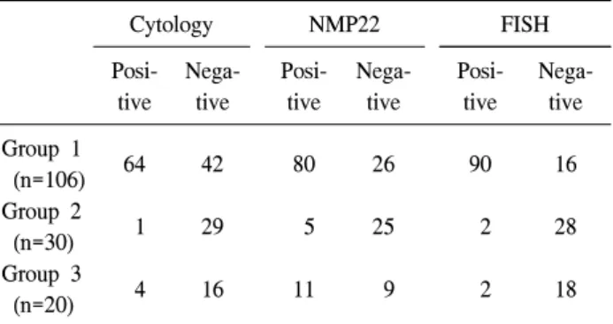
Table 1. Overall results of cytology, NMP22, and FISH of each group
Cytology NMP22 FISH
Posi- tive
Nega- tive
Posi- tive
Nega- tive
Posi- tive
Nega- tive Group 1
(n=106) 64 42 80 26 90 16
Group 2
(n=30) 1 29 5 25 2 28
Group 3
(n=20) 4 16 11 9 2 18
NMP: nuclear matrix protein, FISH: fluorescence in situ hy- bridization, Group 1: bladder tumor patients group, Group 2:
benign prostatic hyperplasia group, Group 3: hematuria or in- fection group
Table 2. Overall sensitivity, specificity, positive predictive value, and negative predictive value of urine cytology, NMP22, and FISH for the detection of bladder tumors
Cytology NMP22 FISH p-value
Sensitivity (%) 60.4 75.5 84.9 <0.001
Specificity (%) 96.7 83.3 93.3 0.168
PPV (%) 98.5 94.1 97.8 0.250
NPV (%) 40.8 49.0 63.6 0.059
NMP: nuclear matrix protein, FISH: fluorescence in situ hybridization, PPV: positive predictive value, NPV: negative predictive value
Table 3. False-positive rates of group 2 and group 3
Cytology NMP22 FISH p-value False positive rate of
group 2 (%) 3.3 16.7 6.6 0.168
False positive rate of
group 3 (%) 20.0 55.0 10.0 0.004
NMP: nuclear matrix protein, FISH: fluorescence in situ hybridization
(7번), Aqua (17번), Yellow (9p21번)로 염색되었다. 염색체 3, 7, 17에서 각각의 형광표식인자가 3개 이상으로 증가하거 나, 염색체 9p21에서 형광표식인자가 2개 이상 증가하거나 감소하는 경우를 비정상 세포 즉 방광암 세포로 판정, FISH 양성으로 하였다.
NMP22 검사는 NMP22Ⓡ test kit (Matritech Inc., Newton, USA)를 이용하여 세척을 통해 얻은 소변을 원심분리하여 상층액으로 EIA (enzyme immunoassay) 방법을 이용하여 요 중 NMP22 값을 구하였고, NMP22의 값이 10 이상일 경우 NMP22 양성으로 하였다.
3. 자료분석
요세포검사, NMP22, FISH의 민감도와 특이도의 분석을 위한 통계분석은 SPSS 12K를 이용하였고, chi-square test로 분석하였으며, p값이 0.05 미만인 경우를 통계적으로 의의 있는 것으로 판정하였다.
결 과
1. 환자분석
환자들의 평균 나이는 62.9±13.4세였고, 경요도방광종양 절제술 후 방광암의 분화도는 저등급 분화도가 54명, 고등 급 분화도가 52명이었다. 방광암의 병기는 pTa-1이 64명, pT2-4가 42명이었다.
2. 요세포검사, NMP22, FISH의 민감도 및 특이도 분석
방광암 환자 106명에서 요세포검사의 민감도는 60.4%
(64명), NMP22 75.5% (80명), FISH 84.9% (90명)로 통계적으 로 유의한 차이를 보였다 (p<0.001). 2군에서 각 검사의 특
이도는 요세포검사 96.7%, NMP22 83.3%, FISH 93.3%로 NMP22가 상대적으로 낮았지만, 통계적으로 유의한 차이는 없었다 (p=0.168) (Table 1, 2).
3. 요세포검사, NMP22, FISH의 위양성률 분석
2군에서의 위양성률은 요세포검사, NMP22, FISH가 각각 3.3%, 16.7%, 6.6%로 각 군 간에 통계적으로 유의한 차이가 없었다 (p=0.168). 3군에서 위양성률은 요세포검사, NMP22, FISH가 각각 20.0%, 55.0%, 10.0%로 각 군 간에 통계적으로 유의한 차이가 있었다 (p=0.004). 요세포검사, NMP22, FISH 결과에 육안적 혈뇨가 미치는 영향을 보기 위한 2군과 3군 을 비교한 결과 육안적 혈뇨가 있는 경우 특히 NMP22에서 위양성률이 상대적으로 높게 증가하였다 (Table 3).
4. 방광암의 분화도와 병기에 따른 요세포검사, NMP22, FISH의 민감도
저등급 분화도 54명의 환자에서 민감도는 요세포검사 25.9%, NMP22 51.9%, FISH 77.8%로 통계적으로 유의한 차 이가 있어 (p<0.001), 저등급 분화도 방광암의 경우 FISH의 민감도가 의의있게 높았다. 고등급 분화도 52명의 환자에 서 민감도는 요세포검사 96.2%, NMP22 100%, FISH 92.3%
로 고등급 분화도 방광암의 경우 검사 간에 통계적으로 유 의한 차이가 없었다 (p=0.125). 그러나 전체적으로 고등급
Table 6. Sensitivity of combination of cytology, NMP22, and FISH for the detection of bladder tumors according to grade and stage
Cyto+
NMP22 Cyto+
FISH
NMP22+
FISH
Cyto+
NMP22+
FISH
p-value
Grade Low
(n=54) (%) 18.5 22.2 48.1 18.5 0.001
High
(n=52) (%) 96.2 88.5 92.3 88.5 0.444
Stage pTa-1
(n=64) (%) 34.4 34.4 59.4 31.3 0.003
pT2-4
(n=42) (%) 90.5 85.7 85.7 85.7 0.890
Cyto: cytology, NMP: nuclear matrix protein, FISH: fluorescence in situ hybridization
Table 4. Sensitivity of cytology, NMP22, and FISH for the detection of bladder tumors according to grade and stage
Cytology NMP22 FISH p-value
Grade
Low (n=54) (%) 25.9 51.9 77.8 <0.001
High (n=52) (%) 96.2 100 92.3 0.125
Stage
pTa-1 (n=64) (%) 40.6 65.6 78.1 <0.001
pT2-4 (n=42) (%) 90.5 90.5 95.2 0.648
NMP: nuclear matrix protein, FISH: fluorescence in situ hybridization
Table 5. The overall sensitivity, specificity, positive predictive value, and negative predictive value of combination of cytology, NMP22, and FISH for the detection of bladder tumors
Cyto+
NMP22 Cyto+
FISH
NMP22+
FISH
Cyto+
NMP22+
FISH
p-value
Sensitivity (%) 56.6 54.7 69.8 52.8 0.051
Specificity (%) 96.7 96.7 100 100 0.565
PPV (%) 98.4 98.3 100 100 0.535
NPV (%) 38.7 37.7 48.4 37.5 0.522
Cyto: cytology, NMP: nuclear matrix protein, FISH: fluorescence in situ hybridization, PPV: positive predictive value, NPV:
negative predictive value
분화도를 보이는 경우 민감도는 증가하였다 (Table 4).
방광암의 병기에 따른 민감도의 분석 결과 64명의 pTa-1 의 경우 요세포검사가 40.6%, NMP22 65.6%, FISH 78.1%를 보여 각 검사 간에 통계적으로 유의한 차이가 있었다 (p<
0.001). 42명의 pT2-4 방광암 환자에서 민감도는 요세포검 사 90.5%, NMP22 90.5%, FISH 95.2%로 각 검사 간에 통계 적으로 유의한 차이가 없었다 (p=0.648). 병기 역시 전체적 으로 병기가 증가함에 따라 각 검사의 민감도가 증가하였다.
5. 두 가지 이상 검사를 병행했을 경우의 결과
방광암 진단에 있어 요세포검사, NMP22, FISH 중 두 가 지 검사를 병행한 경우에서 민감도는 요세포검사와 NMP22 를 병행 시에 56.6%, NMP22와 FISH를 병행 시에 54.7%, 요 세포검사와 FISH를 병행 시에 69.8%, 그리고 3가지 검사를 병행 시에 52.8%로 통계적으로 유의한 차이가 없었다 (p=0.051). 특이도 역시 96.7%, 96.7%, 100%, 100%로 통계적 으로 유의한 차이가 없었다 (p=0.565). 양성예측도 (positive predictive value; PPV)는 98.4%, 98.3%, 100%, 100%로 나타 났고 (p=0.535), 음성예측도 (negative predictive value; NPV) 는 38.7%, 37.7%, 48.4%, 37.5%로 각각 통계적으로 유의한
차이가 없었다 (p=0.522).
두 가지 이상의 검사를 병행할 때 분화도와 병기에 따른 민감도를 보면 요세포검사와 NMP22를 병행 시, 요세포검 사와 FISH를 병행 시, NMP22와 FISH를 병행 시, 그리고 3 가지 검사를 병행 시에 저등급 분화도 방광암의 경우 각각 18.5%, 22.2%, 48.1%, 18.5%로 NMP22와 FISH를 병행 시에 민감도가 통계적으로 유의하게 높았다 (p=0.001). 고등급 분 화도 방광암의 경우는 각각 96.2%, 88.5%, 92.3%, 88.5%로 각 검사 간 통계적으로 유의한 차이는 없었다 (p=0.444). 또 한 pTa-1인 경우 각각 34.4%, 34.4%, 59.4%, 31.3%로 역시 NMP22와 FISH를 병행 시에 민감도가 통계적으로 유의하 게 높았다 (p=0.003). pT2-4인 경우는 각각 90.5%, 85.7%, 85.7%, 85.7%로 높은 병기에서는 두 가지 이상의 검사를 병 행하는 경우 통계적으로 유의한 차이가 없었다 (p=0.890) (Table 5, 6).
고 찰
NMP22는 핵유사분열장치 (nuclear mitotic apparatus; NuMA) 의 구성 요소로서, 정상요로상피세포에 비해 방광암세포에 서 약 25배의 세포 내 농도가 증가한다.12 또한 이미 방광암 세포의 NMP22의 측정값이 방광암의 분화도와 연관이 있어 분화도가 나쁜 방광암에서 증가한다.13 그러나, NMP22의 경우 육안적 혈뇨 시에 위양성률이 높아, 다른 연구들에서 이런 환자들을 배제하면 특이도와 양성예측값이 증가한
다.14,15 본 연구에서도 육안적 혈뇨의 유무에 따라 위양성률
이 16.7%에서 55.0%로 증가를 보였다. 그러나, 요세포검사
나 FISH는 의미 있는 증가를 보이지 않아 상대적으로 육안 적 혈뇨에 영향을 적게 받음을 알 수 있었다.
1980년대부터 방광암을 포함한 이행세포암의 9번 장완과 단완의 염색체 변화에 대한 연구가 시작되었고, 최근 기술 의 발전으로 홑염색체 (monosomy) 9번에 의해 시작, 세염색 체 (trisomy) 7번에 의해 좀 더 침습성을 지니는 2개의 세포 유전경로를 파악하였다.16,17 다양한 세포 유전학적인 데이 터를 얻을 수 있게 되면서 다인자탐색법의 개발이 시작되 었고, 이것은 방광암 환자에서 진단, 추적 관찰의 보조적인 방법으로 사용되게 되었다. 또한 Meloni 등18은 최초로 방광 암 환자의 소변이나 세척뇨에서 나온 세포를 이용하여 FISH 와 방광암의 조직을 동시에 비교하였다.
FISH는 높은 민감도와 특이도를 보여 방광암 환자의 소 변이나 세척뇨를 통해 비침습적으로 방광암을 발견할 수
있다.1,6,19-21 최근에는 방광경 검사에서 음성을 보이는 방광
암 환자의 재발 위험을 예측하는 데 도움이 된다는 연구들 이 나오고 있다.1,22,23 본 연구에서도 저등급 분화도 방광암 환자에서 요세포검사의 낮은 민감도에 비해, FISH가 비교 적 높은 민감도를 보여 방광경 검사에서 육안적으로 관찰 하기 어려운 병변 등에서 FISH가 방광암의 조기진단에 유 용하리라 생각한다.
본 연구에서는 방광암 진단 시 요세포검사 및 NMP22, FISH를 동시에 시행하여 이들의 결과를 비교 분석하였다.
이미 국내에서도 방광암에서의 요세포검사 및 NMP22, FISH 등의 결과가 보고된 바 있으나 이는 NMP22 및 FISH에 대 한 각각 결과들만이 보고되었다. 국내에서 보고된 FISH의 유용성에 대한 연구는 Choi 등20이 FISH의 민감도와 특이도 를 각각 88.9%와 100%로 보고하였고, Kim 등21은 FISH의 민감도와 특이도를 각각 50%와 97.5%로 보고하였다. 본 연 구에서 FISH의 민감도와 특이도는 각각 84.9%와 93.3%를 보여, Choi 등20과 비슷한 결과를 보였고, Kim 등21에 비해 다소 높은 민감도를 보였다. 이러한 차이는 아마도 배뇨소 변과 방광경 시 세척소변에 대한 검체의 차이와 고등급 분 화도 및 고병기 환자의 분포 차이에 의한 것으로 여겨진다.
발표 연구에 약간의 차이는 있으나 방광암에서 특이도는 요세포검사, NMP22, FISH 모두 높아 통계적 차이는 없으나 민감도는 요세포검사 및 NMP22에 비해 FISH가 통계적으 로 의의있게 민감도가 높다. 따라서 FISH의 경우 높은 민감 도와 특이도로 방광암의 조기진단에 이용할 수 있을 것으 로 생각한다.
NMP22에 대한 국내 연구는 Kim 등24이 기준값을 7.70U/ml 로 할 때 80%의 민감도를 보고했고, 다른 연구들에서 기준 값을 10U/ml로 할 때 Kim 등25은 73.1%와 67.7%의 민감도와 특이도를, Park 등26은 96.7%와 65.5%의 민감도와 특이도를,
Kwon과 Hong27은 61.8%와 95.8%의 민감도와 특이도를 각 각 보고했다. 본 연구에서는 75.5%와 83.3%의 민감도와 특 이도를 보여 비슷한 값을 보였으나, 위양성률이 50%로 높 게 나타나 진단을 위한 screening에는 제한이 있을 것으로 생각한다. 본 연구에서 2개 이상의 검사를 병행하는 경우 양성의 진단기준을 두 가지 검사에서 모두 다 양성으로 나 오는 것으로 삼아 상대적으로 민감도와 특이도가 증가하지 못했다. 요세포검사의 민감도가 저등급 방광암에서 상대적 으로 낮아서 요세포검사와 다른 검사를 병행할 경우 결과 가 더 낮게 나왔으나, 저등급 방광암을 제외하면 통계적인 유의성은 없었다. 따라서 각각의 검사의 특성을 파악하여 NMP22의 경우 높은 위양성률로 방광암의 진단 screening에 적용하기 보다는 방광암 치료 후 재발 추적관찰에 이용하 는 것이 도움이 되리라 생각한다.
각 검사에 따른 경제적 관점을 고려할 경우, 현재 요세포 검사의 검사비는 6,930원 (보험가), NMP22의 검사비는 17,630 원 (보험가)이다. FISH의 경우는 아직까지 고가 (약 20만원) 의 경제적 문제로 연구용으로 주로 시행되고 있으며, 다른 검사에 비해 상대적으로 많은 시약과 장비 및 노력이 요구 되어 방광암 진단에 손쉽게 사용되기에는 현실적으로 어려 움이 있다. 많은 연구들로 좀 더 간편하고 저렴하게 시행할 수 있는 FISH 검사법이 등장한다면 추후에 방광경 검사를 대체할 수는 없지만, 방광경 검사의 횟수를 줄여 주고, 요세 포검사를 대체할 수 있는 screening법 및 방광암의 재발을 추적 관찰할 수 있는 검사법이 될 수 있을 것으로 생각한다.
결 론
저등급 방광암의 경우 요세포검사에 비해 FISH의 민감도 가 높아 FISH는 방광경과 병용하여 일차성 방광암의 진단 이나 방광암의 재발에 유용한 방법으로 생각한다. 그러나 FISH의 경우는 경제적 측면을 고려할 때 좀 더 많은 개발이 요구된다. NMP22의 경우는 육안적 혈뇨에 대한 위양성률 이 높아 방광암의 진단을 위한 screening에는 제한이 있으며 방광암의 재발 추적관찰에 유용하리라 생각한다.
REFERENCES
1. Bubendorf L, Grilli B, Sauter G, Mihatsch MJ, Gasser TC, Dalquen P. Multiprobe FISH for enhanced detection of bladder cancer in voided urine specimens and bladder washings. Am J Clin Pathol 2001;116:79-86
2. Dalbagni G, Herr HW. Current use and questions concerning intravesical bladder cancer group for superficial bladder cancer.
Urol Clin North Am 2000;27:137-46
3. Donat SM. Evaluation and follow-up strategies for superficial bladder cancer. Urol Clin North Am 2003;30:765-76 4. Lee R, Droller MJ. The natural history of bladder cancer.
Implications for therapy. Urol Clin North Am 2000;27:1-13 5. Soloway MS. Overview of treatment of superficial bladder
cancer. Urology 1985;26(4 Suppl):18-26
6. Halling KC, King W, Sokolova IA, Meyer RG, Burkhardt HM, Halling AC, et al. A comparison of cytology and fluorescence in situ hybridization for the detection of urothelial carcinoma.
J Urol 2000;164:1768-75
7. Grossman HB, Messing E, Soloway M, Tomera K, Katz G, Berger Y, et al. Detection of bladder cancer using a point-of-care proteomic assay. JAMA 2005;293:810-6
8. Lübbe L, Nowack R, May M, Ullmann K, Gunia S, Kaufmann O, et al. FISH--a new noninvasive method for the diagnosis of urinary bladder carcinomas. Clin Lab 2004;50:395-402 9. Menendez V, Filella X, Alcover JA, Molina R, Mallafre JM,
Ballesta AM, et al. Usefulness of urinary nuclear matrix protein 22 (NMP22) as a marker for transitional cell carcinoma of the bladder. Anticancer Res 2000;20:1169-72
10. Epstein JI, Amin MB, Reuter VR, Mostofi FK. The World Health Organization/International Society of Urological Pathology consensus classification of urothelial (transitional cell) neoplasms of the urinary bladder. Bladder Consensus Conference Com- mittee. Am J Surg Pathol 1998;22:1435-48
11. Greene F, Page L, Fleming L, Fritz AG, Balch CM, Haller DG. AJCC Cancer Staging Manual. 6th ed. New York: Springer Verlag; 2002
12. Keesee SK, Meneghini MD, Szaro RP, Wu YJ. Nuclear matrix proteins in human colon cancer. Proc Natl Acad Sci U S A 1994;91:1913-6
13. Di Carlo A, Terracciano D, Mariano A, Oliva A, D'Armiento M, Macchia V. Role of cytokeratins, nuclear matrix proteins, Lewis antigen and epidermal growth factor receptor in human bladder tumors. Int J Oncol 2003;23:757-62
14. Sharma S, Zippe CD, Pandrangi L, Nelson D, Agarwal A.
Exclusion criteria enhance the specificity and positive predictive value of NMP22 and BTA stat. J Urol 1999;162:53-7 15. Ponsky LE, Sharma S, Pandrangi L, Kedia S, Nelson D,
Agarwal A, et al. Screening and monitoring for bladder cancer:
refining the use of NMP22. J Urol 2001;166:75-8
16. Gibas Z, Prout GR Jr, Connolly JG, Pontes JE, Sandberg AA.
Nonrandom chromosomal changes in transitional cell carcinoma of the bladder. Cancer Res 1984;44:1257-64
17. Vanni R, Scarpa RM, Nieddu M, Usai E. Cytogenetic in- vestigation on 30 bladder carcinomas. Cancer Genet Cytogenet 1988;30:35-42
18. Meloni AM, Peier AM, Haddad FS, Powell IJ, Block AW, Huben RP, et al. A new approach in the diagnosis and follow-up of bladder cancer. FISH analysis of urine, bladder washings, and tumors. Cancer Genet Cytogenet 1993;71:105-18 19. Skacel M, Fahmy M, Brainard JA, Pettay JD, Biscotti CV,
Liou LS, et al. Multitarget fluorescence in situ hybridization assay detects transitional cell carcinoma in the majority of patients with bladder cancer and atypical or negative urine cytology. J Urol 2003;169:2101-5
20. Choi YD, Cho NH, Chang SY, Rha SY, Chung HC, Park K.
Comparison of the efficacy of urine cytology and fluorescence in situ hybridization (FISH) for the detection of bladder urothelial carcinoma. Korean J Urol 2004;45:410-5
21. Kim JY, Kim SH, Choi HY, Lee HM. Clinical utility of fluorescence in situ hybridization for voided urine for the diagnosis and surveillance of bladder cancer. Korean J Urol 2008;49:307-12
22. Sarosdy MF, Schellhammer P, Bokinsky G, Kahn P, Chao R, Yore L, et al. Clinical evaluation of a multi-target fluorescent in situ hybridization assay for detection of bladder cancer. J Urol 2002;168:1950-4
23. Pycha A, Lodde M, Comploj E, Negri G, Egarter-Vigl E, Vittadello F, et al. Intermediate-risk urothelial carcinoma: an unresolved problem? Urology 2004;63:472-5
24. Kim JS, Lee HM, Lee KH. The significance of urinary nuclear matrix protein (NMP) as a Marker for transitional cell carcinoma of the bladder. Korean J Urol 1997;38:259-62
25. Kim HY, Chang SG, Kim JI. The significance of urinary NMP22 in patients with bladder tumor as a diagnostic test.
Korean J Urol 1998;39:450-3
26. Park JO, Moon DG, Cheon J, Kim JJ, Yoon DK. Urinary NMP (nuclear matrix protein) 22 in screening and post-treatment follow-up of bladder cancer. Korean J Urol 1999;40:551-6 27. Kwon DH, Hong SJ. The clinical utility of BTA TRAK, BTA
stat, NMP22 and urine cytology in the diagnosis of bladder cancer: a comparative study. Korean J Urol 2003;44:721-6