두경부 편평세포암에서 F‐18 FDG PET/CT의 임상적 역할
Clinical Role of F-18 FDG PET/CT in Squamous Cell Carcinoma of Head and Neck
최윤영·장성준
한양대학교 의과대학 핵의학과, 국군대전병원 핵의학과
Yun-Young Choi, M.D., PhD.
Sung-June Jang, M.D.
Department of Nuclear Medicine, Hanyang University College of Medicine, Seoul, Korea
Department of Nuclear Medicine, Armed Forces Daejeon Hospital, Daejeon, Korea
책임저자 주소: 최윤영, 133-792, 서울시 성동구 행당동 17번지 한양대학교 의과대학 핵의학과
Tel: 02-2290-9265, Fax: 02-2281-0475 E-mail: [email protected]
투고일자: 2009년 6월 22일, 심사일자: 2009년 7월 29일, 게재확정일자: 2009년 8월 10일
Abstract
Diagnostic imaging plays an important role in accurate staging, restaging, and treatment monito- ring, and is essential in both planning adequate treatment and minimizing treatment-related toxicity and functional impairment in head and neck squa- mous cell carcinoma (HNSCC). MRI and CT remains the primary imaging modalities for the assessment of HNSCC, but F-18 FDG PET/CT had emerged as a vital adjunct when used in the appropriate clinical setting, such as: delineation of extent of regional lymph node involvement, detection of distant meta- stasis, identification of an unknown primary tumor, detection of an occasional synchronous primary tumor, monitoring of the treatment response, and long-term surveillance for recurrence and metas-
tases. In this manuscript, clinical application of F-18 FDG PET/CT on HNSCC in initial staging, radiothe- rapy planning, carcinoma of unknown primary of squamous cell origin, evaluation of response to radiation and/or chemoradiation therapy, and pre- diction of prognosis will be discussed and other promising PET radiotracers will be introduced.
Key Words: Head and neck squamous cell car- cinoma, F-18 FDG, PET/CT
서 론
주로 50~60대 남성에서 호발하였던 두경부암이 최근에 는 청소년과 여성의 흡연증가로 젊은 층에서 폐암과 함께 그 발병률이 증가하고 있다.1 본 종설에서는 두경부암의 90%이상을 차지하는 편평세포암에서 FDG PET, PET/CT 의 임상적 역할을 알아보고, FDG이외에 개발되고 있는 방 사성의약품을 소개하고자 한다.
본 론
1. 병기결정
두경부편평세포암 환자에서 원발 종양의 범위와 림프절 전이는 수술범위와 방사선치료의 표적 설정에 있어 매우 중요하다. 원발 종양의 진단에 있어 PET/CT가 MRI나 CT 와 비슷한 정도의 예민도를 가지는 것으로 알려져 있으나,2-4 종양의 해부학적 경계부위를 진단하는데 있어서는 MRI나 조영증강 CT의 해상도에 미치지 못하므로, 원발종양의 T 병기의 설정에 있어 PET/CT의 역할은 제한적이다. 그러나 경부림프절의 전이여부 평가에 있어서는 FDG PET 는 기
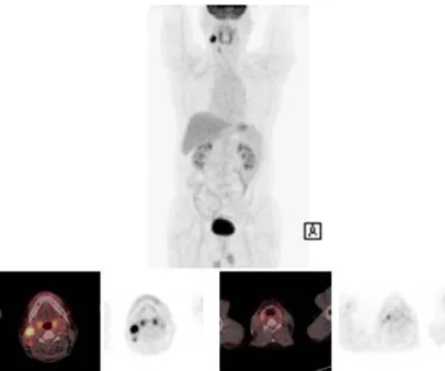
Fig. 1. Left nasopharyngeal cancer with left cervical lymphadenopathy. On PET/CT image there is another focal FDG uptake lesion in the liver, suggesting distant metastasis.
존의 CT나 MRI에 비해 더 우수하거나 유사한 성적을 보이 는 것으로 알려져 있다.5-8 Schoder와 Yeung 등이 조사한 바에 의하면 FDG PET의 림프절전이 진단의 예민도 87~90
%, 특이도 80~93%를 보였으며,9 MRI와 CT는 민감도 61~
97%, 특이도 21~100%를 보여 PET에서 더 고른 성적을 보 였다.10 최근 보고에 따르면 CT, MRI, 초음파검사와 PET/
CT검사결과를 비교하면 각 검사간에 통계적으로 유의한 차이는 없지만, 상기 검사들을 모두 시행한 결과를 비교하 는 것이 진단의 특이도와 정확도를 떨어뜨리지 않으면서 예민도를 높인다고 하였다.11
이전 연구에 따르면 수술 전 임상적으로 림프절전이가 없었던 N0 병기의 환자 19명의 FDG PET, 감시림프절 영 상 및 조직검사결과 PET에서 위음성을 보인 환자 4명 중 3 명의 감시림프절에서 양성으로 확인되었으며 위음성을 보 인 림프절의 크기는 1 cm내외였다.12, 13 또 다른 연구에서 는 FDG PET/CT 결과는 31명의 N0 병기 환자중 3명의 환 자에서 위음성을 나타냈으며 림프절크기는 5 mm 이하였 다.14 이와 같이 임상적으로 N0 병기의 환자에서 시행한
PET, PET/CT검사에서 크기가 작은 병변은 공간해상력의 한계로 인한 위음성결과를 보일 수 있지만 향후 추적관찰 을 위해서 PET/CT를 초기병기설정시 시행하는 것이 좋다.
특히 치료 반응 평가를 위해 시행하는 FDG PET에서 인두 의 근육, 타액선, 림프조직 등은 정상적으로도 FDG 섭취가 증가할 수 있는 곳이므로 FDG 섭취가 증가한 곳이 정상 조 직의 생리적 섭취 증가인지 병변인지 여부를 확인하기 위 해서는 초기병기설정 시 PET/CT를 시행하는 것이 유용하 다. 지금까지의 보고된 논문들은 주로 FDG PET의 성적이 주를 이루고 있으므로 PET/CT의 결과는 더 좋을 것으로 기대된다.
또한 FDG PET/CT 검사시 종격동이나 액와부와 같이 예 상치 못했던 림프절 전이를 진단할 수 있으며 전신영상을 얻음으로써 고식적인 검사에서 발견할 수 없는 원격전이를 진단하는데 중요한 역할을 한다.(Fig. 1) 몇몇 연구결과에 따르면 두경부 국소 진행암 환자의 약 10%에서 PET검사를 통해 숨은 전이암을 발견할 수 있었으며,4, 5, 7, 9 Goyal 등 은 수술전 PET을 시행한 환자의 13%에서 치료계획을 바꾸
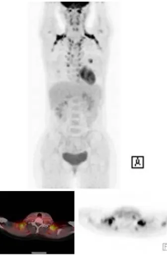
Fig. 2. Right vocal cord tumor with right supraclavicular lymph node metastasis.
Another second primary malignancy is noted in esophagus.
었다고 하였다.15
두경부편평세포암 환자는 특히 상부 호흡소화관을 포함 하여 신체 다른 부위에 동시에 발생한(synchronous) 악성 종양의 발생율이 2~16% 정도로 알려져 있으며, 그 빈도가 높은 종양으로는 폐암과 식도암이 있고, 그외에 대장암, 췌 장암, 방광암 등이 드물게 발생하는 것으로 알려져 있다.
(Fig. 2) 이러한 경우 PET/CT는 전신을 평가하므로 한번의 검사로 원격전이와 병발암을 진단하는데 매우 유용한 검사 이다.16-18
2. 원발불분명 경부림프절전이암 (Fig. 3)
일반적으로 경부종물의 조직검사에서 편평세포암 전이 림프절로 진단된 후, 임상 소견, 방사선학적, 내시경적 검 사 및 종양이 숨어있을 만한 부위의 조직검사 등에서 원발
종양을 찾을 수 없는 경우가 10%가량 된다. 대개 종양의 크 기가 0.5 cm 이하로 매우 작거나 구개편도 깊숙이 위치한 종양, 설편도(lingual tonsil) 조직처럼 주변 조직과 종양의 구분이 어려운 위치에 있는 작은 종양의 경우 방사선학적 및 내시경적 검사에서 찾기 어려울 수 있다. 두경부 편평세 포암은 수술로 제거하거나 직접적인 방사선조사로 치료하 는 것이 원칙이므로 경부림프절 전이암의 발견시 원발종양 의 위치를 알아내는 것이 중요하며, 만일 원발종양의 위치 를 찾아내지 못하는 경우에는 상부호흡소화관 점막 전체 범위에 걸친 방사선치료를 시행하여야 하므로 구강건조증, 상부호흡소화관 협착증 등의 심각한 부작용을 동반할 수 있다. 원발종양을 찾기 위한 여러 연구 결과 PET검사의 민 감도는 25~35%로 나타났으며,19-26 PET/CT검사의 예비분 석에 따르면 33~57%로 PET검사에 비하여 비교적 높은 예
Fig. 3. Metastatic squamous cell carcinoma of right cervical lymphadenopathy with unknown primary tumor. Focal FDG uptake was detected in right pyriform sinus on PET/CT, which was proven to be primary malignancy focus.
Fig. 4. False positive lymphadenopathy on FDG PET/CT in a patient with left vocal cord cancer, which was removed 10 days before. The patient didn’t get treated, and followed FDG PET/CT one year later shows complete disappearance of hypermetabolic lesions.
민도를 나타냈다.27, 28 그러나 PET/CT로 경부림프절전이 암의 원발종양을 찾는데 있어 제한점은 구개편도, 설편도 등 원발종양이 숨어있을 만한 부위에 FDG의 생리적 섭취 가 다양한 정도로 나타나며 때로는 비대칭적으로 나타나기 때문에 종양과의 구분이 어렵고 때로는 종양으로 오인할 수도 있다는 점이다. 그렇지만 원발종양을 진단하기 위해 시행하는 PET/CT 검사에서는 보다 예민도를 높여 판독함 으로써 조직검사결과에서 음성으로 나오더라도 종양이 있 을만한 위치를 알려주는 것이 중요하다.
3. 방사선치료/화학방사선치료에 대한 반응평가 두경부편평세포암에서 치료에 대한 반응을 평가하는 방 법으로 CT나 MRI 등 해부학적 영상과 FDG PET을 이용한 기능적 영상을 얻는 것이 일반적이다. 완치목적으로 약물 또는 방사선치료를 받은 후의 FDG PET/CT는 경부림프절 등에 잔존하는 종양 있는지 여부를 알아보는데 중요하다.
Yao 등은 53명의 환자의 치료 후 평균 15주 후에 시행한 PET 또는 PET/CT에서 예민도 100%, 특이도 94%, 양성예 측도 43%, 음성예측도 100%의 결과를 보였다.29 Ong 등은 65명의 환자에서 화학방사요법 후 적어도 8주 후에 시행한 FDG PET/CT에서 예민도 71%, 특이도 89%, 양성예측도 38%, 음성예측도 97%를 보였다.30 이들은 위양성을 보인
림프절들은 대개 염증성 또는 육아종성변화를 보인 림프절 이었으며, 해부학적영상만 시행했을 때보다 PET/CT를 함 께 시행했을 때 위양성이 27%에서 10%로 감소하였다고 보고했다.(Fig. 4) 방사선치료 또는 화학방사선치료 후의 환자에서 FDG PET의 음성예측도가 매우 높기 때문에 조 직검사나 planned neck dissection 없이 임상 추적 또는 주기적 영상검사로 추적할 수 있다. 화학방사선치료후의
Fig. 5. Physiologic brown fat activity in bilateral neck should not be misinterpreted as pathologic cervical lymph nodes.
PET의 양성예측도는 비교적 낮지만, 지속적으로 강한 FDG 섭취증가를 보이는 경우에는 치료 결과가 나쁘거나 치료에 따른 합병증을 시사하는 소견으로 간주할 수 있다. 화학방 사선치료 후 어느 정도 시간이 지나야 치료에 따른 염증성 변화가 회복되면서 위양성 결과를 줄일 수 있으므로 치료 후 검사시기는 10~12주 가량 지나서 검사를 하는 것이 권 장되며31 6개월 이상 지난 경우에는 위양성율이 낮은 것으 로 알려져 있다. 그러나 다른 한편으로는 치료 후 4~8주 이 내에 시행한 PET에서는 잔류종양의 부피가 매우 작아서 위 음성율이 높을 수 있다는 보고도 있다.32, 33 상기 환자들의 PET판독에 있어 특정한 SUV 값으로 잔류암과 염증을 감별 할 수 없으며, 오히려 오랜 경험에서 얻어진 판독기술과 판 단력이 더 정확한 결과를 보여줄 수 있다. 또한 PET보다는 PET/CT를 시행하는 것이 진단의 정확도를 높이고 정상조 직의 섭취와 림프절을 쉽게 구분하게 하고 조직검사할 위 치를 알려줄 수 있고, 방사선치료계획 등에도 도움줄 수 있 다.(Fig. 5)
4. 방사선치료계획
FDG PET/CT는 해부학적 정보와 기능적 정보를 동시에 제공하는 검사로 방사선치료 계획 수립에 이상적인 검사방
법이다.34, 35 방사선치료시 조사영역은 CT나 MRI와 같은
해부학적 영상에 기초하여 정하게 되는데, PET/CT를 이용 하면 종양의 윤곽을 보다 정확하게 그릴 수 있다. 강도변조 방사선치료(IMRT)를 이용한 치료법이 출현함에 따라, 주 변조직의 손상을 최소화하면서 종양의 방사선조사량을 증 가시키기 위해서는 종양의 위치를 정확히 정하는 것이 필 수적이다. 몇몇 보고에 의하면 CT만을 사용하여 방사선치 료 계획을 세웠을 때와 비교하여 PET/CT를 사용하였을 때 많은 경우 20%의 증례에서 표적용적(target volume)을 변 화시켜야 했다고 한다. 그러나 아직 PET/CT는 보조적인 검사로 활용되며 방사선치료의 표적 설정에는 공간해상력 이 뛰어난 CT와 MRI가 기본으로 활용되고 있다. 그러나 혀 저부의 종양처럼 해부학적 영상검사법으로는 원발 종양의 형태를 구분하기 어려울 때에는 PET/CT가 도움 될 수 있 다. 아직까지는 PET/CT를 이용한 표적용적 결정의 치료실 패와의 관련성에 대한 장기간의 분석이 없으며, 방사선치 료계획 수립에 PET/CT를 일상적으로 사용할 수 있으려면 이에 대한 표준화된 방법이 정의되어야 할 것이다.
5. 예후예측
두경부이외의 종양에서 FDG PET의 SUV가 환자의 예후 와 관련있는 것으로 알려져 있으며, 최근 보고된 바에 의하 면 두경부편평세포암에서도 SUV는 종양의 국소제어 및 무 병생존과 관계있으므로 종양의 FDG섭취가 높은 환자는 보다 적극적인 치료를 고려하는 것이 좋겠다고 하였으나, 아직까지는 보다 많은 연구가 필요하다.36-39
6. FDG이외에 두경부종양 PET/CT에 사용될 수 있는 방사성의약품
1) F-18 Fluorothymidine (FLT)
FLT는 DNA의 합성원료인 티미딘(thymidine)유사체로 세포증식이 활발한 종양세포에 더 많은 섭취를 보이게 된
다. 즉, 세포주기중 S-상에 있는 세포분획 및 thymidine kinase-1의 활성도에 비례하여 세포내 FLT 섭취증가를 보
이며,40, 41 여러가지 항암화학요법제제나 방사선조사에 의
하여 thymidine kinase-1의 활성도가 억제되고 방사선조 사를 받은 종양세포가 S-상에 잔류하고 분획이 감소하면 세 포내 FLT섭취가 감소하게 된다. 두경부종양에서 FLT의 이 용에 대해서는 아직 연구과정에 있으나, 다른 종양에서와 마찬가지로 FDG나 CT/MRI에 비하여 치료에 대한 반응을 보다 조기에 정확히 평가할 수 있는 가능성을 가지고 있다.
그러나 FLT가 약물치료 후 림프절에 섭취되어 위양성 소견 을 보일 수 있다고 되어 있으며, 그 빈도는 아직 알려져 있 지 않지만 만일 흔히 발생하는 소견이라면 치료반응평가에 이용하기는 어려울 것으로 보인다.
2) F-18 Fluoromisonidazole (FMISO)
종양내 저산소세포는 항함화학요법이나 방사선치료의 세포독성에 대한 저항성을 가지므로, 다른 정상적 산소공 급을 받는 세포와 비슷한 정도의 치료효과를 위해서는 3배 이상의 방사선량을 조사하여야 한다. 두경부종양에서는 종 양내 저산소상태가 흔하며, 이러한 저산소상태로 인하여 종양의 병기나 치료방법에 무관하게 종양의 재발이나 원격 전이를 일으킬 수 있는 것으로 보고되고 있다.42 FMISO는 종양의 저산소상태를 찾아내는데 사용되는 방사성의약품 으로, 두경부종양에서 치료 전 저산소상태의 종양이 확인 되면 방사선치료시 방사선민감제, 혈관확장제, 저산소세포 의 cytotoxin등을 사용하여 방사선치료의 효과를 높일 수 있는 것으로 보고되고 있다.43
결 론
FDG PET/CT는 이미 다양한 악성종양의 진단검사로 널 리 이용되고 있으며 특히 두경부암의 진단과 치료계획 수 립, 치료 효과 판정과 추적관찰시에 표준 검사법으로 자리 잡아 검사 빈도가 증가하고 있다. 두경부편평세포암 진단 후 PET/CT는 국소림프절 전이와 원격 전이 여부 판단에 유용하며, 원발불분명 종양을 발견하거나 동시에 존재하는 다른 원발암의 발견에도 유용하다. 또한 화학방사선요법 시행 후에 치료 반응의 평가에 사용될 수 있으며 장기간의 추적관찰시 재발암이나 전이성암의 발견 민감도가 높아 임
상적으로 유용하며, 방사선치료 계획을 수립하는 데에도 PET/CT를 이용할 수 있다. 아직까지는 FDG PET/CT가 두 경부편평세포암에서 환자의 진단 및 치료에 중요한 영상수 단으로 이용되고 있으나, 장차 FLT나 FMISO 등 다양한 방 사성의약품의 개발에 따라 점차 PET/CT의 영역이 확장될 것으로 예측된다.
References
1. Kim JS, Lee JH. Clinical application of PET in head and neck. Hanyang Medical Review 2007;4:16-27
2. Ha PK, Hdeib A, Goldenberg D, Jacene H, Patel P, Koch W, Califano J, Cummings CW, Flint PW, Wahl R, Tufano RP. The role of positron emission tomography and computed tomography fusion in the management of early-stage and advanced-stage primary head and neck squamous cell carcinoma. Arch Otolaryngol Head Neck Surg 2006;132:12-6.
3. Paulus P, Sambon A, Vivegnis D, Hustinx R, Moreau P, Collignon J, Deneufbourg JM, Rigo P. 18FDG-PET for the assessment of primary head and neck tumors:
clinical, computed tomography, and histopathological correlation in 38 patients. Laryngoscope 1998;108:
1578-83.
4. Goerres GW, Schmid DT, Gratz KW, von Schulthess GK, Eyrich GK. Impact of whole body positron emission tomography on initial staging and therapy in patients with squamous cell carcinoma of the oral cavity. Oral Oncol 2003;39:547-51.
5. Schmid DT, Stoeckli SJ, Bandhauer F, Huguenin P, Shumid S, von Schlulthess GK, Goerres GW. Impact of positron emission tomography on the initial staging and therapy in locoregional advanced squamous cell carcinoma of the head and neck. Laryngoscope 2003;
113:888-91.
6. DiMartino E, Nowak B, Hassan HA, Hausmann R, Adam G, Buell U, Westhofen M. Diagnosis and staging of head and neck cancer: a comparison of modern imaging modalities (positron emission tomography, computed
tomography, color-coded duplex sonography) with panendoscopic and histopathologic findings. Arch Otolaryngol Head Neck Surg 2000;126:1457-61.
7. Schwartz DL, Rajendran J, Yueh B, Coltrera M, Anzai Y, Krohn K, Eary J. Staging of head and neck squamous cell cancer with extended-field FDG-PET. Arch Oto- laryngol Head Neck Surg 2003;129:1173-8.
8. Adams S, Baum RP, Stuckensen T, Bitter K, Hor G.
Prospective comparison of 18F-FDG PET with conven- tional imaging modalities (CT, MRI, US) in lymph node staging of head and neck cancer. Eur J Nucl Med 1998;25:1255-60.
9. Schoder H, Yeung HW. Positron emission imaging of head and neck cancer, including thyroid carcinoma.
Semin Nucl Med 2004;34:180-97.
10. Dammann F, Horger M, Mueller-Berg M, Schlemmer H, Claussen CD, Hoffman J, Eschmann S, Bares R. Ra- tional diagnosis of squamous cell carcinoma of the head and neck region: comparative evaluation of CT, MRI, and 18FDG PET. Am J Roentgenol 2005;184:1326-31.
11. Yoon DY, Hwang HS, Chang SK, Rho YS, Ahn HY, Kim JH, Lee IJ. CT, MR, US, F-18 FDG PET/CT, and their combined use for the assessment of cervical lymph node metastasis in squamous cell carcinoma of the head and neck. Eur Radiol 2009;19:634-42
12. Hyde NC, Prvulovich E, Newman L, Waddington WA, Visvikis D, Ell P. A new approach to pre-treatment assessment of the N0 neck in oral squamous cell car- cinoma: the role of sentinel node biopsy and positron emission tomography. Oral Oncol 2003;39:350-60.
13. Stoeckli SJ, Steinert H, Pfaltz M, Schmid S. Is there a role for positron emission tomography with 18F-fluoro- deoxyglucose in the initial staging of nodal negative oral and oropharyngeal squamous cell carcinoma.
Head Neck 2002;24:345-9.
14. Schoder H, Carlson DL, Kraus DH, Stambuk HE, Gonen M, Erdi YE, Yeung HW, Huvos AG, Shah JP, Larson SM, Wong RJ. 18F-FDG PET/CT for detecting nodal metastases in patients with oral cancer staged N0 by clinical examination and CT/MRI. J Nucl Med 2006;47:
755-62.
15. Goyal P, Hsu JM, Kellman RM. Effect of F-18 FDG PET on the management of patients with head and neck squamous cell carcinoma. J Otolaryngol Head Neck Surg 2008;37:694-9.
16. Wax MK, Myers LL, Gabalski EC, Husain S, Gona JM, Nabi H. Positron emission tomography in the evaluation of synchronous lung lesions in patients with untreated head and neck cancer. Arch Otolaryngol Head Neck Surg 2002;128:703-7.
17. Keyes JW Jr, Chen MY, Watson NE Jr, Greven KM, McGuirt WF, Williams DW III. FDG PET evaluation of head and neck cancer: value of imaging the thorax.
Head Neck 2000;22:105-10.
18. Perlow A, Bui C, Shreve P, Sundgren PC, Teknos TN, Mukherji SK. High incidence of chest malignancy de- tected by FDG PET in patients suspected of recurrent squamous cell carcinoma of the upper aerodigestive tract. J Comput Assist Tomogr 2004;28:704-9.
19. Regelink G, Brouwer J, de Bree R, Pruim J, van der Laan BF, Vaalburg W, Hoekstra OS, Comans EF, Vissink A, Leemans CR, Roodenburg JL. Detection of unkown primary tumours and distant metastases in patients with cervical metastases: value of FDG PET versus conventional modalities. Eur J Nucl Med Mol Imaging 2002;29:1024-30.
20. Nieder C, Gregoire V, Ang KK. Cervical lymph node metastases from occult squamous cell carcinoma: cut down a tree to get an apple? Int J Radiat Onocol Biol Phys 2001;50:727-33.
21. Jungehulsing M, Scheidhauer K, Damm M, Pietrzyk U, Eckel H, Schicha H, Sennert E. FDG PET is a sensitive tool for the detection of occult primary cancer (carcino- ma of unknown primary syndrome) with head and neck lymph node manifestation. Otolaryngol Head Neck Surg 2000;123:294-301.
22. Bohuslavizki KH, Klutmann S, Kroger S, Sonnermann U, Buchert R, Werner JA, Meser J, Clausen M. FDG PET detection of unknown primary tumors. J Nucl Med 2000;41;816-22.
23. Stokkel MP, Terhaard CH, Hordijk GJ, van Rijk PP. The detection of unknown primary tumors in patients with cervical metastases by dual-head positron emission tomography. Oral Oncol 1999;35:390-4.
24. Lassen U, Daugaard G, Eigtved A, Damgaard K, Friberg L. F-18 FDG whole body positron emission tomography (PET) in patients with unknown primary tumors (UTP).
Eur J Cancer 1999;35:1076-82.
25. Greven KM, Keye JW Jr, William DW 3rd, Watson N.
Occult primary tumors of the head and neck: lack of benefit from positron emission tomography imaging with FDG. Cancer 1999;86:114-8.
26. Braams JW, Pruim J, Kole AC, Nikkels PG, Vaalburg W, Vermey A, Roodenburg JL. Detection of unknown pri- mary head and neck tumors by positron emission tomography. Int J Oral Maxillofac Surg 1997;26:112-5.
27. Paul SA, Stoeckli SJ, von Schulthess GK, Goerres GW.
FDG PET and PET/CT for detection of the primary tumor in patients with cervical nonsquamous cell car- cinoma metastasis of an unknown primary. Eur Arch Otorhinolaryngol 2007;264:2189-95.
28. Freudenberg LS, Fischer M, Antoch G, Jentzen W, Gutzeit A, Rosenbaum SJ, Bockisch A Egelhof T. Dual modality of FDG PET/CT in patients with cervical car- cinoma of unknown primary. Med Princ Pract 2005;14:
155-60.
29. Yao M, Smith RB, Graham MM, Hoffman HT, Tan H, Funk GF, Graham SM, Chang K, Dornfeld KJ, Menda Y, Buatti JM. The role of FDG PET in management of neck metastasis from head and neck cancer after de- finitive radiation treatment. Int J Radiat Oncol Biol Phys 2005;63:991-9.
30. Ong SC, Schoder H, Lee NY, Patel SG, Carlson D, Fury M, Pfister DG, Shah JP, Larson SM, Kraus DH. Clinical utility of FDG PET/CT in assessing the neck after con- current chemoradiotherapy for locoregional advanced head and neck cancer. J Nucl Med 2008;49:532-40.
31. Lonneus M, Lawson G, Ide C, Bausart R, Remacle M, Pauwels S. Positron emission tomography with FDG for suspected head and neck tumor recurrence in the
symptomatic patient. Laryngoscope 2000;110:1493-7.
32. Andrade RS, Heron DE, Degirmenci B, Filho PA, Bran- stetter BF, Seethala RR, Ferris RL, Avril N. Posttreatment assessment of response using FDG PET/CT for patients treated with definitive radiation therapy for head and neck cancers. Int J Radiat Oncol Biol Phys 2006;65:
1315-22.
33. Greven KM, Williams DW III, McGuirt WF Sr, D'Agostino RB Jr. Serial positron emission tomography scans fol- lowing radiation therapy of patients with head and neck cancer. Head Neck 2001;23:942-6.
34. Ciernik IF, Dizendorf E, Baumert BG, Reiner B, Burger C, Davis JB, Lutolf UM, Steinert HC, Von Schulthess GK. Radiation treatment planning with an integrated positron emission and computer tomography (PET/CT):
a feasibility study. Int J Radiat Oncol Biol Phys 2003;
57:853-63.
35. Heron DE, Andrade RS, Flickinger J, Johnson J, Agar- wala SS, Wu A, Kalnicki S, Avril N. Hybrid PET-CT si- mulation for radiation treatment planning in head-and- neck cancers: a brief technical report. Int J Radiat Oncol Biol Phys 2004;60:1419-24.
36. Torizuka T, Tanizaki Y, Kanno T, Futatsubashi M, Naitou K, Ueda Y, Ouchi Y. Prognostic value of FDG PET in patients with head and neck squamous cell cancer. Am J Roentgenol 2009;192:156-60.
37. Machtay M, Natwa M, Andrel J, Hyslop T, Anne PR, Lavarino J, Intenzo CM, Keane W. Pretreatment FDG PET standardized uptake value as a prognostic factor for outcome in head and neck cancer. Head Neck 2009;31:195-201.
38. Liao CT, Chang JT, Wang HM, Ng SH, Hsueh C, Lee LY, Lin CH, Chen IH, Hwang SF, Cheng AJ, Yen TC.
Preoperative FDG PET SUV of neck lymph nodes pre- dicts neck cancer control and survival rates in patients with oral cavity squamous cell carcinoma and patholo- gically positive lymph nodes. Int J Radiat Oncol Biol Phys. 2008 Dec 18. [Epub ahead of print]
39. Suzuki H, Hasegawa Y, Terada A Hyodo I, Nakashima T, Nishio M, Tamaki T. FDG PET predicts survival and
distant metastasis in oral squamous cell carcinoma.
2008 Sept 17.[Epub ahead of print]
40. Rasey JS, Grierson JR, Wiens LW, Kolb PD, Schwatz JL. Validation of FLT uptake as a measure of thymidine kinase-I activity in A549 carcinoma cells. J Nucl Med 2002;43:1210-7.
41. Schwatz JL Tamura Y, Jordan R, Grierson JR, Krohn KA. Effect of p53 activation on cell growth, thymidine kinase-I activity and 3-deoxy-3fluorothymidine uptake.
Nucl Med Biol 2004;31:419-23.
42. Nordsmark M, Bentzen SM, Rudat V, Brizel D, Lartigau E, Stadler P, Becker A, Adam M, Molls M, Dunst J, Terris DJ, Overgaard J. Prognostic value of tumor oxygenation
in 397 head and neck tumors after primary radiation therapy: an international multicenter study. Radiother Oncol 2005;77:18-24.
43. Rischin D, Hicks RJ, Fisher R, Binns D, Corry J, Porced- du S, Peters LJ, Trans-tasman Radiation oncology group study 98.02. Prognostic significance of F-18 Misonidazole positron emission tomography-detected tumor hypoxia in patients with advanced haead and neck cancer randomely assigned to chemoradiation with or without tirapazamine; a substudy of Trans- Tasman Radiation Oncology Groaup Study 98.02. J Clin Oncol 2006;24:2098-104.