Developmental salivary gland defect is a bone depression on the lingual side of the mandible containing salivary gland or fatty soft tissue. This defect has been widely known as Stafne’s bone defect since it was described by Stafne in 1942. Stafne reported 35 cases of asymptomatic unilateral radiolucent cavities in the posterior region of the mandible.1The lesions were located between the man- dibular angle and the third molar, below the inferior dental canal and above the mandibular base. The developmental salivary gland defect has no symptoms and is occasionally observed, in general, as a cyst-like oval radiolucent image with cortical bone thickening around the gonial angle of the mandible under the mandibular canal, on panoramic radiography.2,3 Although this defect appearing as a round or ovoid radiolucency is not uncommon, multilocular radio- lucency of this defect is rare.4This report presents a cone- beam computed tomographic (CBCT) scan, showing a developmental salivary gland defect with multilocular radiolucency occurring in a male patient.
Case Report
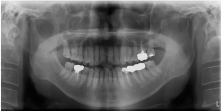
A 66-year-old healthy man was referred from a private dental clinic for evaluation of a multilocular radiolucency at the left molar region of the mandible. Panoramic radio- graph revealed a well-defined multilocular radiolucent area under the mandibular canal at the distal root of the second molar tooth (Fig. 1). The lesion showed three lobulated radiolucencies with radiopaque margins in the region of the submandibular gland fossa. On clinical examination, there was no sign of infection, fistula, or nerve damage.
─ 261 ─
Multilocular developmental salivary gland defect
Jin-Soo Kim
Department of Oral and Maxillofacial Radiology and Oral Biology Research Institute, School of Dentistry, Chosun University, Gwangju, Korea
ABSTRACT
Developmental salivary gland defect is a bone depression on the lingual surface of the mandible containing salivary gland or fatty soft tissue. The most common location is within the submandibular gland fossa and often close to the inferior border of the mandible. This defect is asymptomatic and generally discovered only incidentally during radio- graphic examination of the area. This defect also appears as a well-defined, corticated, unilocular radiolucency below the mandibular canal. Although it is not uncommon for this defect to appear as a round or ovoid radiolucency, multi- locular radiolucency of these defects is relatively rare. This report presents a case of a developmental salivary gland defect with multilocular radiolucency in a male patient. (Imaging Sci Dent 2012; 42 : 261-3)
KEY WORDS: Bone Cysts; Salivary Glands; Mandible; Cone-Beam Computed Tomography
*This study was supported by research funds from Chosun University, 2010.
Received April 22, 2012; Revised June 7, 2012; Accepted July 3, 2012 Correspondence to : Prof. Jin-Soo Kim
Department of Oral and Maxillofacial Radiology, School of Dentistry, Chosun University, 309 Pilmun-daero, Dong-gu, Gwangju 501-759, Korea
Tel) 82-62-220-3884, Fax) 82-62-227-0270, E-mail) [email protected]
Imaging Science in Dentistry 2012; 42 : 261-3 http://dx.doi.org/10.5624/isd.2012.42.4.261
Copyright ⓒ 2012 by Korean Academy of Oral and Maxillofacial Radiology
This is an Open Access article distributed under the terms of the Creative Commons Attribution Non-Commercial License (http://creativecommons.org/licenses/by-nc/3.0) which permits unrestricted non-commercial use, distribution, and reproduction in any medium, provided the original work is properly cited.
Imaging Science in Dentistry∙pISSN 2233-7822 eISSN 2233-7830
Fig. 1.Panoramic radiograph reveals a well-defined multilocular radiolucent area beneath the mandibular canal at the root of the second molar tooth.
No history of expansion on the left mandibular angle area was recorded. The patient had no systemic disease and no
specific medical history.
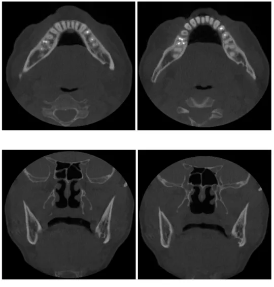
CBCT scan was recommended for further study. Axial CBCT images showed a lingual bony defect located at the lingual side of the mandibular canal (Fig. 2). Coronal CBCT images showed a lobulated bony defect below the mandi- bular canal (Fig. 3). The maximum mesiodistal length of the defect was 14.16 mm, and the height was 7.38 mm. The maximum buccolingual length of the defect was 4.44 mm, not showing the expansion or thinning of the buccal corti- cal bone.
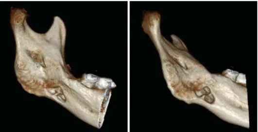
Parasagittal CBCT images showed three lobulated radio- lucencies with radiopaque margins between the mandibular canal and the inferior border of the mandible (Fig. 4). Three- dimensional, reformatted CBCT images revealed a bony defect extending from the medial surface of the mandible (Fig. 5).
The lesion was diagnosed as a developmental salivary gland defect from the radiographic features. The patient was informed about the lesion and was scheduled for fol- low-up.
─ 262 ─ Multilocular developmental salivary gland defect
Fig. 2.Axial CBCT images show a lingual bony defect located at the lingual side of the mandibular canal.
Fig. 4.Para-sagittal CBCT image shows three lobulated radiolu- cencies with radiopaque margins between the mandibular canal and the inferior border of the mandibular canal.
Fig. 3.Coronal CBCT images show a lobulated bony defect below the mandibular canal.
Discussion
Developmental salivary gland defect is typically asymp- tomatic and next to impossible to palpate, and is generally found incidentally during radiographic examination of the area. In a recent review of a large number of cases, males predominated over females at a ratio of 6.1 : 1, with a peak incidence in the fifth and sixth decades of life.4Many other terms have been used for this defect including lingual sali- vary gland depression, Stafne bone cavity, Stafne cyst, static bone cavity, and idiopathic bone cavity.2,5-8
On panoramic radiograph, the defect appears as a cir- cular or ovoid, well-defined radiolucency that ranges in diameter from 1 to 3 cm. The margins of the radiolucent defect are well-defined by a dense sclerotic radiopaque margin of variable width, which is usually thicker on the superior aspect. Characteristically, it is situated just above or at the inferior border of the mandible, between the area of the first molar and the mandibular angle, and always inferior to the mandibular canal. In this case, the defect appeared as a multilocular radiolucency with a radiopaque margin. The defect, however, was located at the character- istic region.
In recent years, computed tomography (CT), CBCT, and magnetic resonance imaging (MRI) have been shown to be diagnostically valuable in addition to panoramic radio- graphs. MRI, with its enhanced definition of adjacent soft tissue, may reveal the occurrence of soft tissue extending into the defect.2
Advanced radiological assessment using CBCT is usually sufficient to achieve a final diagnosis of a developmental salivary gland defect and to avoid surgical treatment, which
would be an unnecessary option in the management of a developmental salivary gland defect except for symptomatic or other concomitant pathologies.
Individual developmental salivary gland defects having been followed for some years with periodic radiographic examinations showed no changes in size or shape of the defects over time. Periodic radiographic examination is recommended to document the static nature of the lesion.
References
1. Stafne EC. Bone cavities situated near the angle of the mandi- ble. J Am Dent Assoc 1942; 29 : 1969-72.
2. Philipsen HP, Takata T, Reichart PA, Sato S, Suei Y. Lingual and buccal mandibular bone depressions: a review based on 583 cases from a world-wide literature survey, including 69 new cases from Japan. Dentomaxillofac Radiol 2002; 31 : 281- 90.
3. Kim HK, Kim JS, Kim JD. Developmental salivary gland defect.
Literatures review and case analysis of 12 cases. Korean J Oral Maxillofac Radiol 2006; 36 : 81-8.
4. Etöz M, Etöz OA, Sahman H, Sekerci AE, Polat HB. An un- usual case of multilocular Stafne bone cavity. Dentomaxillofac Radiol 2012; 41 : 75-8.
5. White SC, Pharoah MJ. Oral Radiology; principles and Inter- pretation. 6th ed. St. Louis: Mosby Inc; 2009. p. 574.
6. Nah KS, Jung YH, Cho BH. Unusual Stafne bone cavity mi- micking infected cyst or neural origin tumor. Korean J Oral Maxillofac Radiol 2007; 37 : 221-3.
7. Shimizu M, Osa N, Okamura K, Yoshiura K. CT analysis of the Stafne’s bone defects of the mandible. Dentomaxillofac Radiol 2006; 35 : 95-102.
8. Minowa K, Inoue N, Sawamura T, Matsuda A, Totsuka Y, Nakamura M. Evaluation of static bone cavities with CT and MRI. Dentomaxillofac Radiol 2003; 32 : 2-7.
─ 263 ─
Jin-Soo Kim
Fig. 5.Three-dimensional CBCT images reveal a bony defect extend- ing from the medial surface of the mandible.