• 검색 결과가 없습니다.

Distribution of Oncogenic Human Papillomavirus Genotypes at High Grade Cervical Lesions above CIN 2 Grade with Histological Diagnosis

N/A
N/A
Protected

Academic year: 2021

Share "Distribution of Oncogenic Human Papillomavirus Genotypes at High Grade Cervical Lesions above CIN 2 Grade with Histological Diagnosis"

Copied!
9
0
0

로드 중.... (전체 텍스트 보기)

전체 글

(1)

Biomedical Science Letters 2016, 22(2): 37~45 http://dx.doi.org/10.15616/BSL.2016.22.2.37 eISSN : 2288-7415

Distribution of Oncogenic Human Papillomavirus Genotypes at High Grade Cervical Lesions above CIN 2 Grade with Histological Diagnosis

Geehyuk Kim 1 , Sungyoung Park 1 , Hye-young Wang 2 , Sunghyun Kim 3 , Sangjung Park 4 , Kwangmin Yu 1 , Boohyung Lee 7 , Seung-Ju Ahn 5 , Eun-Joong Kim 6 and Dongsup Lee 7,†

1

Department of Biomedical Laboratory Science, College of Health Sciences, Yonsei University, Wonju, Gangwon 26493, Korea

2

M&D, Inc., Wonju Eco Environmental Technology Center, Wonju, Gangwon 26493, Korea

3

Department of Clinical Laboratory Science, College of Health Sciences, Catholic University of Pusan, Pusan 46252, Korea

4

Department of Biomedical Science, College of Life and Health Sciences, Hoseo University, Asan, Chungcheong 31499, Korea

5

Department of Clinical Laboratory Science, Daegu Health & Science College, Daegu 41453, Korea

6

Department of Clinical Laboratory Science, Chungbuk Health & Science University, Chungju 28250, Korea

7

Department of Clinical Laboratory Science, Hyejeon College, Choongchung, Hongseoung 32244, Korea

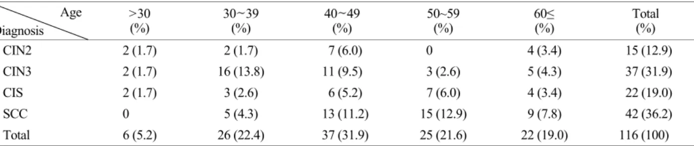
High risk human papillomavirus (HR-HPV) is major risk factor for uterine cervical cancer. There are approximately 15 types of HR-HPV. Liquid based cytology samples (116 samples) with high grade cervical lesions belonging to cervical intraepithelial neoplasia (CIN) 2, CIN 3, carcinoma in situ (CIS) and squamous cell carcinoma (SCC) were used after histologic confirmation. HR-HPV genotype assay was conducted using DNA chips. The HR-HPV infection rate was 81.9% with SCC samples showing the highest HR-HPV infection rate of 31%. CIN 3, CIS and CIN 2 showed infection rates of 25%, 16.4% and 9.5%, respectively. According to age with HR HPV infection rate, the 30~39 years-old group showed the highest infection rate by 92.3%. According to distribution with HR HPV genotyping, HPV 16 showed the highest infection rate by 42.3% whereas HPV 33 and HPV 58 showed infection rates of 11.7% and 10.8%, respectively.

HPV 18 which is the second most common infected HPV genotype in the world showed 3.6%. Of the three most common oncogenic HR-HPV genotypes in CIN 2, we detected HPV 16, 35, 58; CIN 3 was HPV 16, 33, 58; CIS was HPV 16, 58, 33 (35/52); and SCC was HPV 16, 33, and 18 (31/52/58). Among the HPV 18, CIN 2, CIN 3, CIS and SCC showed 0.9%, 0.9%, 0% and 1.8%, respectively. The most often used preventive vaccines for cervical cancers use HPV 16 and HPV 18 as targets. However, results derived from this study suggest that a preventive vaccine against HPV 16 and HPV 18 would not be optimal for populations in this study.

Key Words: Human papillomavirus, Uterine cervix cancer, Cervical intraepithelial neoplasia, Carcinoma in situ, Squamous cell carcinoma, DNA chip

Original Article

*

Received: May 18, 2016 / Revised: June 28, 2016 / Accepted: June 28, 2016

Corresponding author: Dongsup Lee. Department of Clinical Laboratory Science, Hyejeon College, Hongsung, Chungnam 32244, Korea.

Tel: +82-41-630-5342, Fax: +82-41-630-5364, e-mail: [email protected]

C

The Korean Society for Biomedical Laboratory Sciences. All rights reserved.

CC

This is an Open Access article distributed under the terms of the Creative Commons Attribution Non-Commercial License (http://creativecommons.org/licenses/by-nc/3.0/) which

permits unrestricted non-commercial use, distribution, and reproduction in any medium, provided the original work is properly cited.

(2)

INTRODUCTION

Uterine cervical cancer is the third most common malig- nancy among women worldwide (Jemal et al., 2011; Bosch et al., 1995). Every year, approximately 500,000 new patients are diagnosed and 270,000 women die from uterine cervical cancer (Parkin et al., 1999). The Korean National Cancer information Center reported 111,792 cases of cervical cancer in 2012. Among these, an incidence rate showed 14.2%

(about 3,200/100,000 cases) which is seventh after thyroid cancer, breast cancer, colon cancer, stomach cancer, liver cancer and lung cancer in women.

Human papillomavirus (HPV) is considered to be the main contributing factor for causing cervical cancer (Vizcaino et al., 1998). HPV is the most common sexually transmitted pathogen between women and men. Approximately 60 HPV genotypes are known to infect the genital tract (Ergünay et al., 2007) and cervical cancer is caused by persistent infection with oncogenic high risk (HR) HPV genotypes that belong to a few phylogenetically related HR species. Oncogenic HPV genotypes cause squamous cell carcinoma (SCC) or cervical intraepithelial neoplasia (CIN) upon infection of cervical epithelial cells (zur Hausen et al., 2009). Especially, the oncogenic HR-HPV genotypes have been closely asso- ciated with development of high grade cervical lesion in- cluding cervical cancer (Gaarenstroom et al., 1994).

Much is known about the mechanisms by which HPV causes cervical cancer. After HR-HPV infection, HPV E6 and E7 viral oncoproteins induce degradation and inactivation of the tumor suppressor protein p53 and retinoblastoma protein (pRb), respectively, resulting in deregulation of the cell cycle checkpoints (Levin et al., 1991; Kim et al., 2006).

These proteins are also responsible for the transformation of epithelial cells and maintenance of malignancy (Yugawa et al., 2009; Andersson et al., 2011). This results in immort- alization of the uterine cervix squamous epithelium and inhibition of apoptosis (Dyson et al., 1989; Scheffner et al., 1990). Increased expression of E6/E7 transcripts has been described for both high-grade cervical lesions and malig- nancy (Cattani et al., 2009).

There are currently 13 high-risk HPV types classified as

Group 1 carcinogens (i.e., highly carcinogenic to humans) (HPV 16, 18, 31, 33, 35, 39, 45, 51, 52, 56, 58, 59 and 68) and Group 2A carcinogens (i.e., probably carcinogenic) (HPV 26, 30, 34, 53, 66, 67, 69, 70, 73, 82, 85 and 97) (IARC, 2012) (Bouvard et al., 2009). HPV types 16 and 18 cause approximately 70% of cervical cancers and types 31, 33, 35, 45, 52, and 58 accounts for an additional 20% of cases (Clifford et al., 2005).

HPV genotypes distribution is various depending on the country, province and substantial geographical variation (Bruni et al., 2010). In previous studies, HPV 16, 18, 31, 33, and 45 were reported to be the most prevalent HPV genotypes associated with cervical cancer in Europe; HPV 16, 18, 45, 31, and 33 were the most common HPV geno- types in North America; and HPV 16, 18, 45, 33, and 31 were the most common in Western and Central Asia, whereas HPV 16, 18, 58, 52, and 33 were the most prevalent HPV genotypes in Eastern Asia (Korea, China, and Japan) (Im et al., 2005; Li et al., 2011; Kim et al., 2012; Lee et al., 2015).

Therefore, the HPV genotype-distribution between Eastern Asia and other continents differs greatly. In Korea, the most common HPV genotypes were HPV 16, 18, 33, 53, 56, and 58 (Hong et al., 2009; Kim et al., 2012; Lee et al., 2011).

Utilization of the Pap test has been a key reason for the decrease in cervical cancer incidence rate and accompanying mortality (Okadome et al., 2014). Although the Pap test has been used widely as a cervical screening test due to its low cost and simple procedure, one main drawback is the false negative error rate of 20~50% (Im et al., 2005). In addition, the sensitivity for uterine cervix high-grade squamous intra- epithelial lesion detection and predictive value has been questioned by previous reports (Vizcaino et al., 1998). For these reasons, this research was conducted with liquid based cytology specimen diagnosed with cervical intraepithelial neoplasia (CIN) and analyzed HPV genotypes. A diagnosis of CIN refers to changes in the cervix which then subsequently can lead to uterine cervical cancer. The progression of pre- cancerous lesion to invasive carcinoma in uterine cervical cancer occurs slowly. Therefore, early HPV molecular diag- nosis test to stratify HR-HPV groups is the most effective way to prevent uterine cancers.

Several PCR-based multiplex detection methodologies

(3)

have been shown to be effective in detecting HPV infections (Jennifer, 2013). For uterine cervical cancer diagnosis, most molecular diagnostics are currently based on HPV DNA test with Pap test in Korea. As a result, testing for oncogenic HPV infection in cervical lesions could serve as an accurate means of identifying women who are at risk for developing cervical cancer. In this study, as an effort to characterize HR- HPV genotypes that are prevalent in Korean women, the HPV L1 gene DNA based genotype analysis was performed.

Cervical liquid-based cytology specimens (116 samples) with high grade cervical lesion were used. CIN grade 2, CIN grade 3, carcinoma in situ (CIS) and squamous cell carcinoma (SCC) histologically belong to high grade cervical lesion.

MATERIALS AND METHODS Clinical samples

Liquid-based cytology samples were obtained with Thin- Prep Pap test (Hologic Inc., Acton, MA, USA) from 116 women between the ages of 21 and 84. The clinical sam- ples were archived in the Department of Pathology, Yonsei University Wonju Severance Christian Hospital between September 2010 and July 2011. All subjects were confirmed above CIN grade 2 (CIN 2, CIN 3, CIS and SCC) with histological diagnosis through punch biopsy and cone biopsy.

The prepared slides were stained by the hematoxylin and eosin stain, and evaluated according to the histological CIN grades diagnosis by pathologists. This study was approved by the Institutional Ethics Committee at Yonsei University Wonju College of Medicine (approval number YWMR-12- 4-010) and all subjects provided written informed consent.

The remaining fluid samples were stored at 4℃ after cytology slide preparation and prior to DNA extraction.

DNA extraction

Clinical specimens were collected and vortexed for 1 min.

The volume was adjusted to 40 μL with PBS (pH 7.2) and centrifuged at 8,000×g for 5 min. The supernatant was discarded, and 300~500 μL of washing buffer were added to the pellet. DNA extraction was performed using a QIAamp DNA Mini Kit (Qiagen, Amsterdam, Netherlands) according

to the manufacturer's instructions. Then the supernatant (3 to 5 μL) was used as a template for PCR.

HPV DNA chip test

The HPV genotyping assay was performed using the Goodgene HPV chip (Goodgene Inc., Seoul, Republic of Korea) following the manufacturer's recommendations. The Goodgene HPV chip is designed to detect 15 HR HPV genotypes (16, 18, 31, 33, 35, 39, 45, 51, 52, 56, 58, 59, 68, 69, and 70) together with 7 low-risk (LR) HPV genotypes (6, 11, 34, 40, 42, 43, and 44). The genotyping method in- volves nested PCR to amplify the target region using MY11 and MY9 primers, followed by amplification with the GP5 and GP6 primer pair. Nested PCR conditions consist of an initial denaturation step for 5 min at 94℃ followed by 15 cycles of denaturation for 30 s at 94℃ and extension for 30 s at 65℃. The second amplification involved 45 cycles of 30 s at 94℃, and 30 s at 54℃. A final extension step was performed at 72℃ for 7 min. After PCR amplification of the target region, subsequent steps were performed according to the manufacturer's recommendations. For the Goodgene HPV chip, PCR products were loaded onto the probe- labeled glass chip and the resulting signal was read using a GenePix Pro6.0 (Axon Instruments, Foster City, CA) scanner.

Statistical analysis

All statistical analyses were performed using Prism 5 software (GraphPad, La Jolla, CA) and SPSS statistics soft- ware version 21.0 (IBM, Armonk, NY, USA). Sensitivity, specificity, correlation P value, and 95% confidence interval (CI) of the predictive ability for each HPV DNA chip were estimated with respect to histological diagnosis.

RESULTS

Histological diagnosis for cervical specimens

A total of 116 histological samples were used in this study.

Also, the samples were previously evaluated by histological

and cytological slides were evaluated by cytopathologists

and pathologists. CIN 3 normally belongs to CIS, but in

this study, CIN 3 and CIS are distinguished for segmented

analysis. CIN 2, CIN 3, CIS and SCC (including invasive

(4)

squamous cell carcinoma) are 15 (12.9%), 37 (31.9%), 22 (19%) and 42 (36.2%), respectively (Table 1). Five age groups, 5 (5.2%, mean 25.7 age) were >30 years group, 26 (22.4%, mean 34.9 age) were within the 30~39 years group, 37 (31.9%, mean 44.1 age) were within the 40~49 years group, 25 (21.6%, mean 54.2 age) were within the 50~59 years group and 22 (19%, mean 73.2 age) were within the 60≤ years group.

Total HPV infection patterns

The samples were previously evaluated by liquid-based cytology test. The total HPV infection rates were 108 (93.1%) and HPV non- infection rates were 8 (6.9%) by the DNA chip assay. HR-HPV infection rate of the HPV positive samples was 100 (86.2%) and other type infection rates were 8 (6.9%). Other types are not detected with DNA chip and included neither HR-HPV nor LR-HPV. When infected LR-HPV genotypes, there is no single infection. Multiple infections with HR-HPV are 5 (4.3%) cases (Table 2).

In high grade cervical lesions, overall rate of oncogenic

HR-HPV is 81.9%. SCC shows 31% which is the highest and CIN 3, CIS and CIN 2 shows 25%, 19% and 9.5%

respectively in order. LR-HPV infection was totally absent in high grade cervical lesion. Multiple infection which means HR-HPV and LR-HPV infection represent at the same time was found by 4.3% (Table 2).

Oncogenic HR-HPV infection rate according to the age To inspection of correlation between high grade cervical lesions and age, LR-HPV infection was excluded and re- searcher assessed the oncogenic HR-HPV infection rate according to five age groups. We evaluated the oncogenic HR HPV infection rate in each age group using the DNA chip assay. Oncogenic HR HPV infection rate according to age, >30 years group was 5/6 (83.3%), 30~39 years group was 24/26 (92.3%), 40~49 years group was 31/37 (83.8%), 50~59 years group was 23/25 (92%) and 60≤ years group was 17/22 (77.3%), respectively. In all age groups, the oncogenic HR-HPV infection rates was 100/116 (86.2%) by the DNA chip test (Table 3). HR HPV infection rate shows Table 2. Total HPV infection patterns by histological diagnosis

Infection pattern Histological diagnosis

CIN2 (%) CIN3 (%) CIS (%) SCC (%) Total (%)

High risk infection 11 (9.5) 29 (25.0) 19 (16.4) 36 (31.0) 95 (81.9)

High & low infection 0 (0) 4 (3.5) 1 (0.9) 0 (0) 5 (4.3)

Low risk infection 0 (0) 0 (0) 0 (0) 0 (0) 0 (0)

Other type infection 4 (3.5) 0 (0) 1 (0.9) 3 (2.6) 8 (6.9)

HPV non-infection 0 (0) 4 (3.5) 1 (0.9) 3 (2.6) 8 (6.9)

Total 15 (12.9) 37 (31.9) 22 (19.0) 42 (36.2) 116 (100)

Abbreviations; CIN, cervical intraepithelial neoplasia; CIS, carcinoma in situ; SCC, squamous cell carcinoma.

Table 1. Histological diagnosis according to age Age

Diagnosis

>30

(%) 30~39

(%) 40~49

(%) 50~59

(%) 60≤

(%) Total

(%)

CIN2 2 (1.7) 2 (1.7) 7 (6.0) 0 4 (3.4) 15 (12.9)

CIN3 2 (1.7) 16 (13.8) 11 (9.5) 3 (2.6) 5 (4.3) 37 (31.9)

CIS 2 (1.7) 3 (2.6) 6 (5.2) 7 (6.0) 4 (3.4) 22 (19.0)

SCC 0 5 (4.3) 13 (11.2) 15 (12.9) 9 (7.8) 42 (36.2)

Total 6 (5.2) 26 (22.4) 37 (31.9) 25 (21.6) 22 (19.0) 116 (100)

Abbreviations; CIN, cervical intraepithelial neoplasia; CIS, carcinoma in situ; SCC, squamous cell carcinoma.

(5)

the highest in 30~39 years group.

Oncogenic HR-HPV genotypes infection according to age

The overall oncogenic HR-HPV genotype infection dis- tribution was recognized by the DNA chip. Most common oncogenic HR-HPV genotypes were HPV 16 by 47 cases (42.3%) showing the highest. Sequentially, HPV 33 was 13 cases (11.7%) and HPV 58 was 12 cases (10.8%). Combi- nation of these most common three types shows 64.9%.

HPV 18, the second most common infected HPV genotypes around the world, was 4 cases (3.6%) and ranked 7

th

.

Oncogenic HR-HPV genotypes were classified according to age (Table 3). Of the most common oncogenic HR-HPV genotypes HPV 16 shows 12.6% in 40~49 year groups and 50~59 year groups. In >30 year group, it is detected by 0.9%. HPV 33 representing the second most common HPV genotypes in this study shows 3.6% in 40~49 year groups and 60≤ year group. In >30 group, it is not detected. HPV 58 representing the third most common HPV genotypes in this study shows 3.6% in 30~39 year groups and 60≤ year group. In 50~59 year group, it is not detected. HPV 18, the

second most common infected HPV genotypes around the world, is not detected in >30 years group and 30~39 years group. Overall rate is relatively low by 3.6%.

Oncogenic HR-HPV prevalence and genotype-distribution by histological diagnosis

We evaluated the oncogenic HR-HPV prevalence and distribution in the histological diagnosis as assessed by CIN 2, CIN 3, CIS and SCC (Table 4). By the DNA chip test the total infection was 116 cases the total oncogenic HR-HPV infection was 111 cases. In the most frequently infected HR-HPV 16 group, in CIN 2 samples, HPV 16 was detected in 4.5% (5 cases). In CIN 3 samples, HPV 16 was detected in 9.9% (11 cases). In CIS samples, HPV 16 was detected in 8.1% (9 cases). In SCC samples, HPV 16 was detected in 19.8% (22 cases) as determined by the DNA chip.

Of the three most common oncogenic HR-HPV genotypes in CIN 2, the DNA chip test detected HPV 16, 35, and 58, CIN 3 was HPV 16, 33, and 58, CIS was HPV 16, 58, and 33/35/52, SCC was HPV 16, 33, and 18/31/52/58. In case of HPV 18, the second most common infected HPV geno- types around the world, infection rate is relatively low in Table 3. Oncogenic HR-HPV genotypes infection according to age

Oncogenic HR-HPV

Groups of according to age

Total (%)

>30 (%) 30~39 (%) 40~49 (%) 50~59 (%) 60≤ (%)

HPV 16 1 (0.9) 11 (9.9) 14 (12.6) 14 (12.6) 7 (6.3) 47 (42.3)

HPV 18 0 0 2 (1.8) 1 (0.9) 1 (0.9) 4 (3.6)

HPV 31 0 4 (3.6) 2 (1.8) 0 1 (0.9) 7 (6.3)

HPV 33 0 2 (1.8) 4 (3.6) 3 (2.7) 4 (3.6) 13 (11.7)

HPV 35 1 (0.9) 0 2 (1.8) 3 (2.7) 1 (0.9) 7 (6.3)

HPV 39 0 0 1 (0.9) 0 0 1 (0.9)

HPV 45 0 0 0 1 (0.9) 0 1 (0.9)

HPV 51 1 (0.9) 2 (1.8) 0 0 1 (0.9) 4 (3.6)

HPV 52 0 3 (2.7) 2 (1.8) 3 (2.7) 0 8 (7.2)

HPV 56 0 1 (0.9) 0 0 0 1 (0.9)

HPV 58 1 (0.9) 4 (3.6) 3 (2.7) 0 4 (3.6) 12 (10.8)

HPV 66 0 1 (0.9) 1 (0.9) 0 0 2 (1.8)

HPV 68 2 (1.8) 0 2 (1.8) 0 0 4 (3.6)

Total 6 (5.4) 28 (25.2) 33 (29.7) 25 (22.5) 19 (17.1) 111 (100)

Abbreviations; CIN, cervical intraepithelial neoplasia; CIS, carcinoma in situ; SCC, squamous cell carcinoma.

(6)

CIN 2, CIN 3, CIS and SCC representing 0.9%, 0.9%, 0%

and 1.8%, respectively.

DISCUSSION

Uterine cervical cancer progresses from precancerous lesion for 5~20 years and is limited to the abnormal cancer cells from the cervix. As the mechanism of uterine cervical cancer is being elucidated, therapy is effective when the disease is detected during the precancerous lesion or early invasive cancer stage. However, once the disease has pro- gressed to the metastatic cervical cancer stage therapeutic options are often limited.

Goodgene HPV DNA Chip is commercially utilized techniques and have been used in other studies to clinically evaluate HPV genotyping analysis (Kim et al., 2012; Lee et al., 2011; Kim et al., 2012). In this study, DNA chip were used to analyze HPV genotype distribution with specimen liquid-based cytological specimens from Korean subjects (Gangwon Province). This subjects were confirmed above CIN grade 2 (CIN 2, CIN 3, CIS and SCC) with histological diagnosis through punch biopsy and cone biopsy.

HPV genotype-distribution data could provide useful in-

formation for establishing the appropriate vaccination pro- gram, and creating a diagnostic and treatment strategy for cervical cancer. The present study examined the correlation between HPV prevalence and age, as well as the relationship between HPV distribution and age. According to the Korea National Cancer Center report from 2008, HPV infection rate is 29.2% in the 40~49 year group, 20.0% in the 50~

59 year group and 18% in the 60≤ year group. The most common therapy is removal of HPV infected tissues but this is insufficient to completely remove HPV. The presence of high-grade cervical lesions based on histological diagnosis positively correlated with increased age. The prevalence of SCC was also higher in the higher aged patients. >30 year group show low rate relatively in CIN 2, CIN 3 and CIS by 1.7%. In 30~39 year group, CIN3 accounts for the highest proportion. Also, in 50~59 years group, SCC accounts for the highest proportion and so does in 60≤ year group. High grade cervical lesion rate of 60≤ year group is higher than that of >30 year group over all histological diagnosis. In terms of age, the median age of this subject was 48.8 years.

SCC and CIN 3 are associated with a high-risk of cancer and are most commonly found in women in their 40 years old and 50 years old, whereas CIN 1 is associated with a Table 4. Oncogenic HR-HPV genotypes according to histologic diagnosis

Oncogenic HR HPV

Histological diagnosis

Total (%)

CIN2 (%) CIN3 (%) CIS (%) SCC (%)

HPV 16 5 (4.5) 11 (9.9) 9 (8.1) 22 (19.8) 47 (42.3)

HPV 18 1 (0.9) 1 (0.9) 0 2 (1.8) 4 (3.6)

HPV 31 1 (0.9) 4 (3.6) 0 2 (1.8) 7 (6.3)

HPV 33 0 5 (4.5) 2 (1.8) 6 (5.4) 13 (11.7)

HPV 35 3 (2.7) 2 (1.8) 2 (1.8) 0 7 (6.3)

HPV 39 0 1 (0.9) 0 0 1 (0.9)

HPV 45 0 1 (0.9) 0 0 1 (0.9)

HPV 51 1 (0.9) 2 (1.8) 1 (0.9) 0 4 (3.6)

HPV 52 0 4 (3.6) 2 (1.8) 2 (1.8) 8 (7.2)

HPV 56 0 1 (0.9) 0 0 1 (0.9)

HPV 58 2 (1.8) 5 (4.5) 3 (2.7) 2 (1.8) 12 (10.8)

HPV 66 0 1 (0.9) 0 1 (0.9) 2 (1.8)

HPV 68 1 (0.9) 1 (0.9) 1 (0.9) 1 (0.9) 4 (3.6)

Total 14 (12.6) 39 (35.1) 20 (18.0) 38 (34.2) 111 (100)

(7)

low-risk of wart and are most commonly found in women in their 30 years old. These observations imply that oncogenic HR-HPV and immunological status both contributes to cer- vical cancer. These results indicate that persistent oncogenic HR-HPV infection can induce cervical cancer.

Oncogenic HR-HPV single infection shows greatly high in each of high grade cervical lesion (CIN 2, CIN 3, CIS and SCC). Especially HR-HPV single infection rates were highest for women 60≤ years old. In Table 2, high-grade cervical lesions are found in older individuals and with single infection with HR-HPV. In overall histological diag- nosis were no LR HPV single-infections.

In HPV DNA test, in all age groups, the five most com- mon oncogenic HR-HPV genotypes were HPV 16, 33, 58, 52, and 31/35 from this subjects (Table 3). In all age groups, HPV genotype 16 was the most common. In contrast, in case of HPV 18, it is not detected in >30 year group and 30~39 yeas group. In case of 50~59 year group and 60≤

year group, only 1 case is detected. These results show the gap with prevalence over the world. To review oncogenic HR-HPV genotypes distribution according to histological diagnosis, HPV 16 is detected in CIN 2, CIN 3, CIS and SCC by 4.5%, 9.9%, 8.1% and 19.8%, respectively. HPV 58 is detected in CIN 2, CIN 3, CIS and SCC by 1.8%, 4.5%, 2.7% and 1.8%, respectively. In contrast, HPV 18 is detected in CIN 2, CIN 3, CIS and SCC by 0.9%, 0.9%, 0% and 1.8%, respectively.

Among women with high-grade intraepithelial neoplasia, HPV 16 is still the most prevalent genotype worldwide, but greater geographic variability has been observed for other HPV genotypes. Compared to Western country (North America, Europe), major types causing high grade cervical lesions in Korean was shown different types. HPV 18 was rarely detected from Korean. Instead, HPV 33, 58, and 52 were frequently detected from Korea. Therefore, differences in HPV genotype distribution across studies can be explained by differences in the quality and type of uterine cervical histological samples.

HPV genotyping is important for monitoring the efficacy of HPV vaccination. The findings of this study suggest that a preventive vaccine against HPV 16 and 18 is not optimal for populations in Korea. Other meta-analysis studies widely

support this result (Bao et al., 2008). If a preventive vaccine included HPV 33 and 58, the vaccine efficacy could be increased in Korea, based on the results of this study.

The purposes of the present study were to identify the distribution of type-specific oncogenic HR-HPV genotypes based on the DNA chip and the association of different HPV genotypes with cervical dysplasia. In conclusion, the vaccine-targeted HPV 16 and 18 are the most frequent HPV genotypes worldwide and have also been shown to be asso- ciated with uterine cervical cancer, the extent to which vaccines directed against HPV 16 and 18 would prevent disease associated with other HPV genotypes is not yet clear.

However, the findings of this study suggest that a preventive vaccine against HPV 16 and HPV 18 is not optimal for populations in Korea. If a preventive vaccine included HPV 33 and HPV 58, the vaccine efficacy could be increased in this province, based on the results of this study. These data may provide guidance for national or regional vaccination programs in this province to substantially reduce the burden of cervical lesions and HPV infections. The differences in HPV prevalence and genotypes distribution identified in this study have a potential influence on the effectiveness of HPV vaccines for cervical cancer and the development of screening programs, which should be investigated in future studies.

Conflict of interest None.

REFERENCES

Andersson E, Kärrberg C, Rådberg T, Blomqvist L, Zetterqvist BM, Ryd W, Lindh M, Horal P. Type-specific human papilloma- virus E6/E7 mRNA detection by real-time PCR improves identification of cervical neoplasia. J Clin Microbiol. 2011.

49: 3794-3799.

Bao YP, Li N, Smith JS, Qiao YL. Human papillomavirus type dis- tribution in women from Asia: a meta-analysis. Int J Gynecol Cancer. 2008. 18: 71-79.

Bosch FX, Manos MM, Muñoz N, Sherman M, Jansen AM, Peto

J, Schiffman MH, Moreno V, Kurman R, Shah KV. Prevalence

of human papillomavirus in cervical cancer: a worldwide

perspective. International biological study on cervical cancer

(8)

(IBSCC) study group. J Natl Cancer Inst. 1995. 87: 796-802.

Bouvard V, Baan R, Straif K, Grosse Y, Secretan B, El Ghissassi F, Benbrahim-Tallaa L, Guha N, Freeman C, Galichet L, Cogliano V. A review of human carcinogens―part B: biological agents.

Lancet Oncol. 2009.10: 321-322.

Bruni L, Diaz M, Castellsagué X, Ferrer E, Bosch FX, de Sanjosé S. Cervical human papillomavirus prevalence in 5 continents:

meta-analysis of 1 million women with normal cytological findings. J Infect Dis. 2010. 202: 1789-1799.

Cattani P, Siddu A, D'Onghia S, Marchetti S, Santangelo R, Vellone VG, Zannoni GF, Fadda G. RNA (E6 and E7) assays versus DNA (E6 and E7) assays for risk evaluation for women in- fected with human papillomavirus. J Clin Microbiol. 2009.

47: 2136-2141.

Clifford GM, Gallus S, Herrero R, Munoz N, Snijders PJ, Vaccarella S, Anh PT, Ferreccio C, Hieu NT, Matos E, Molano M, Rajkumar R, Ronco G, de Sanjose S, Shin HR, Sukvirach S, Thomas JO, Tunsakul S, Meijer CJ, Franceschi S. Worldwide distribution of human papillomavirus types in cytologically normal women in the International Agency for Research on Cancer HPV prevalence surveys: a pooled analysis. Lancet.

2005. 366: 991-998.

Dyson N, Howley PM, Münger K, Harlow E. The human papillomavirus-16 E7 oncoprotein is able to bind to the retino- blastoma gene product. Science. 1989. 243: 934-940.

Ergünay K, Misirlioğlu M, Pinar F, Tuncer ZS, Tuncer S, Ustaçelebi S. Human papilloma virus DNA in cervical samples with cytological abnormalities and typing of the virus. Mikrobiyol Bul. 2007. 41: 219-226.

Gaarenstroom KN, Melkert P, Walboomers JM, Van Den Brule AJ, Van Bommel PF, Meyer CJ, Voorhorst FJ, Kenemans P, Helmerhorst TJ. Human papillomavirus DNA and genotypes:

prognostic factors for progression of cervical intraepithelial neoplasia. Int J Gynecol Cancer. 1994. 4: 73-78.

Hong SR, Kim IS, Kim DW, Kim MJ, Kim AR, Kim YO, Kim HS, Rha SH, Park GS, Park YK, Park YS, Park HS, Suh KS, Sohn JH, Shin MK, Oh HK, Yun KJ, Yoon HK, Lee SN, Lee AW, Lee HJ, Cho HY, Choi C, Jung WW. Prevalence and genotype distribution of cervical human papillomavirus DNA in Korean women: a multicenter study. Korean J Pathol. 2009.

43: 434-342.

Im JE, Shim MJ, Ryang YS, Lee DC. Human papillomavirus testing with hybrid capture II and DNA chip. J Exp Biomed Sci. 2005. 11: 51-56.

Jemal A, Bray F, Center MM, Ferlay J, Ward E, Forman D. Global

cancer statistics. CA Cancer J Clin. 2011. 61: 69-90.

Laudadio J. Human papillomavirus detection: testing methodologies and their clinical utility in cervical cancer screening. Adv Anat Pathol. 2013. 20: 158-167.

Levin AJ, Momandd J, Finlay CA. The p53 tumor supressor gene.

Nature. 1991. 351: 453-456.

Kim YK, Park YP. Analysis of telomerase activity by HPV E6/E7 expression in SW13. J Exp Biomed Sci. 2006. 12: 399-403.

Kim SH, Lee DS, Kim Y, Kim GH, Park SJ, Choi YI, Kim TU, Park KH, Lee HY. Clinical evaluation of human papilloma- virus DNA genotyping assay to diagnose women cervical cancer. J Exp Biomed Sci. 2012. 18: 123-130.

Kim SH, Lee DS, Park SJ, Kim TU, Jeon BY, Park KH, Lee HY.

REBA HPV-ID

®

for efficient genotyping of human papilloma- virus in clinical samples from Korean patients. J Med Virol.

2012. 84: 1248-1253.

Lee DS, Kim SH, Park SJ, Jin HW, Kim TU, Park KH, Lee HY.

Human papillomavirus prevalence in Gangwon province using reverse blot hybridization assay. Korean J Pathol. 2011. 45:

348-353.

Im JA, Shim MJ, Ryang YS, Lee DC. Human papillomavirus testing with Hybrid Capture II and DNA chip. J Exp Biomed Sci. 2005. 11: 51-56.

Li N, Franceschi S, Howell-Jones R, Snijders PJ, Clifford GM.

Human papillomavirus type distribution in 30,848 invasive cervical cancers worldwide: Variation by geographical region, histological type and years of publication. Int J Cancer. 2011.

128: 927-935.

Lee DS, Kim GH, Kim SH, Park SY, Wang HY, Park SJ, Han Lin, Yubo Ren, Li Yingxue, Park KH, Lee HY. Human papilloma- virus distribution among women in western Shandong province, East China using reverse blot hybridization assay. Biomed Sci Lett. 2015. 21: 69-76.

Okadome M, Saito T, Tanaka H, Nogawa T, Furuta R, Watanabe K, Kita T, Yamamoto K, Mikami M, Takizawa K. Potential impact of combined high- and low-risk human papilloma- virus infection on the progression of cervical intraepithelial neoplasia 2. J Obstet Gynaecol Res. 2014. 40: 561-569.

Parkin DM, Pisani P, Ferlay J. Global cancer statistics. CA Cancer J Clin. 1999. 49: 33-64.

Scheffner M, Werness BA, Huibregtse JM, Levine AJ, Howley PM. The E6 oncoprotein encoded by human papillomavirus types 16 and 18 promotes the degradation of p53. Cell. 1990.

63: 1129-1136.

Vizcaino AP, Moreno V, Bosch FX, Muñoz N, Barros-Dios XM,

(9)

Parkin DM. International trends in the incidence of cervical cancer: I. Adenocarcinoma and adenosquamous cell carcinomas.

Int J Cancer. 1998. 75: 536-545.

Yugawa T, Kiyono T. Molecular mechanisms of cervical carcino-

genesis by high-risk human papillomaviruses: novel functions of E6 and E7 oncoproteins. Rev Med Virol. 2009. 19: 97-113.

zur Hausen H. Papillomaviruses in the causation of human cancers

- a brief historical account. Virology. 2009. 384: 260-265.

수치

Table 1. Histological diagnosis according to age  Age  Diagnosis  >30 (%)  30~39 (%)  40~49 (%)  50~59 (%)  60≤ (%)  Total (%)  CIN2  2 (1.7)  2 (1.7)    7 (6.0)  0  4 (3.4)    15 (12.9)  CIN3  2 (1.7)  16 (13.8)  11 (9.5)  3 (2.6)  5 (4.3)    37 (31.9)

참조

관련 문서

(Taekwondo, Weight Lifting Players) (90 min × 6 days/week) Warming

Therefore, this study aims to investigate the relationship between explicit grammar instruction and English speaking of lower-grade students in third-grade

[r]

The Eschar Distribution According to Gender and Orientia tsutsugamushi Genotypes of

(A) Before repair of RCT shows fatty degeneration of supraspinatus (stage 2) and infraspinatus (stage 3) muscle and atrophy (Grade 2) (B) After 18months later

회원국의 영토밖에서 다른 회원국의 , 영토내에서 회원국의 서비스 소비자에게

As a result of 1 st clean flotation, it found that various bubble size distribution increases its recovery and increasing of bias increases its grade,

[r]