Received: April 24, 2012, Revised: August 27, 2012, Accepted: August 28, 2012 ISSN 1598-4478 (Print) / ISSN 2233-7679 (Online)
†Correspondence to: Youn-Soo Shim
Department of Dental Hygiene, College of Health Sciences, Cheongju University, 298, Daeseong-ro, Sangdang-gu, Cheongju 360-764, Korea Tel: +82-43-229-8351, Fax: +82-43-229-8969, E-mail: [email protected]
Copyright © 2012 by the Korean Society of Dental Hygiene Science
Dicalcium Phosphate Dihydrate를 함유한 3.5%
과산화수소가 치아표면에 미치는 영향
심연수†
청주대학교 치위생학과
Effects of 3.5% Hydrogen Peroxide Containing Dicalcium Phosphate Dihydrate on the Tooth Enamel Surface
Youn-Soo Shim
†Department of Dental Hygiene, College of Health Sciences, Cheongju University, Cheongju 360-764, Korea
The purpose of this study was to evaluate the tooth whitening and properties of an enamel surface after treatments with tooth bleaching agents that contained dicalcium phosphate dihydrate (DCPD) and hydrogen peroxide (HP). Thirty specimens were obtained from fifteen premolar and were randomly divided into three groups (n=10): 1, 3.5% HP + 0 g DCPD; 2, 3.5% HP + 0.1 g DCPD; 3. 3.5% HP + 1 g DCPD. All groups were bleached 8 hours per day for 14 days. With increasing DCPD concentration, the pH values in the agents increased, making it less acidic. However, there was no statistically significant difference (p>0.05). As the concentration of DCPD was increased, the concentration of Ca and P was also increased.
In all groups, after the tooth whitening, the tooth color was found to have a value of L* (p<0.05). All groups showed significantly decreased enamel microhardness compared to their baseline (p<0.05). The percentage microhardness loss (PML) of the group A1 and A2 were significantly lower than that of group A3. The obvious variation of morphology was observed on enamel surfaces in group A1. Following an analysis of the constituents of enamel surface after bleaching, as DCPD content was increased, the amount of Ca and P was increased. In this study, the experimental results suggest that DCPD/HP agent less demineralization changes such as the erosion morphology and hardness loss without compromising whitening efficiency.
Key Words: Dicalcium phosphate dihydrate, Microhardness, pH, Tooth bleaching
Introduction
Recently, there has been an increased demand for tooth bleaching in order to improve the esthetic appearance of patients, because it is an easy, conservative method
1).
Haywood and Heymann
2)published the first clinical study on tooth whitening using the Proxigel in vacuum- formed custom trays. This is the technique known as
‘night guard vital bleaching’ now commonly used. Vari-
ous chemical combinations have been tried. Currently available peroxide-containing tooth- whitening materials include professionally dispensed and supervised products for use by patients at home, professional-use in-office products, and over-the-counter products for sale directly to consumers.
Hydrogen peroxide (HP) acts as a strong oxidizing
agent through the formation of free radicals, reactive
oxygen molecules and HP anions
3). Free radical ions are
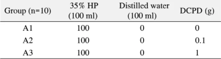
Table 1. The composition of bleaching agent containing DCPD Group (n=10) 35% HP
(100 ml)
Distilled water
(100 ml) DCPD (g)
A1 100 0 0
A2 100 0 0.1
A3 100 0 1
DCPD: dicalcium phosphate dihydrate, HP: hydrogen peroxide
unstable and immediately seek an available target with
which react
3). The bleaching effect is due to the degradation of complex organic molecules that are responsible for the color of teeth to the less complex molecules and result in a reduction or elimination of discoloration
4). HP is a main component of tooth blea- ching. CP is a chemical adduct of urea and HP, which upon dissolving in water or saliva disassociates back into HP and urea
3).
As the whitening process often involves direct contact of the whitening product with the surface of the teeth for an extended period of time, numerous studies have evaluated the effects of products that contain peroxide on the physical and chemical properties of tooth enamel
5,6). Some studies found that tooth bleaching led to tooth sensitivity or changes in chemical composition, surface morphology, or surface roughness some also found a reduction in microhardness
2,7-13). On the other hand, several studies did not find any significant changes in the surface properties of tooth enamel
14-16). Therefore, the effect of bleaching treatment on dental enamel is not yet clear and remains controversial. Although, no clinical studies or case reports in the literature have documented macroscopically or clinically visible demage due to vital bleaching or clinically relevant tissue destruction, It is important to minimize the risk of even minor damage in order to ensure life-long integrity of dental hard tissue
17).
To minimize damage, ingredients like fluoride, calcium, sodium nitrate, amorphous calcium phosphate, or syn- thetic hydroxyapatite (HA) have been added to the blea- ching agent by the manufactures. However, the final results may be unpredictable
18).
Dicalcium phosphate dihydrate (DCPD), also known as brushite, is a hydrated calcium phosphate mineral with the composition CaHPO
4ㆍ2H
2O. It has been shown that DCPD incorporated into snacks
19)and chewing gums
20)reduced the incidence of caries in children. Dentifrices containing DCPD as the abrasive are very popular and are sold throughout the world. There is evidence from intra-oral remineralization studies that indicate that DCPD works with fluoride to provide improved remineralization benefit above and beyond that of fluoride alone. For DCPD to work with fluoride, DCPD must be hydrolyzed
in the oral environment to release calcium and phosphate ions. According to the study, it can be seen that the use of saturated DCPD remineralization in the parts of the artificial caries lesions
21).
The effects of DCPD/HP agent with different pH on the enamel surface microhardness, morphology, composition, as well as their potential for affecting tooth color change, have also been questioned. The aim of this study was to evaluate the effects of 3.5% HP containing the DCPD on the tooth whitening and enamel surface properties.
Materials and Methods
1. Materials
This study used dicalcium phosphate dehydrate (Junsei Chemical Co. Ltd, Tokyo, Japan) powder. The 35% HP (Sigma-Aldrich, St. Louis, USA) solution was diluted with distilled water to obtain a 3.5% solution.
2. Methods
1) Tooth bleaching agents containing DCPD
The tooth bleaching agents were prepared by adding 0 g for controls, 0.1 and 1 g of DCPD powder to 100 ml of 3.5% HP solution (Table 1). The DCPD/HP agent was stirred using a magnetic stir-bar at room temperature for 24 hours. In the pilot study, surface properties were no difference on concentration over the 1 g DCPD. Thus, this study selected 0.1 and 1 g of DCPD concentrations.
2) pH-measurements
After stirring, the pH value of each solution was
measured with a pH meter (Thermo Fisher Scientific,
USA).
3) Mineral measurements
An inductively coupled plasma-atomic emission spec- trometer (ICP-AES, OPTIMA 4300DV, Perkin-Elmer, USA) was used for quantitative analysis of the elements dissolved in the DCPD / HP agents.
4) Preparation of the specimens
15 human premolars, extracted for orthodontic reasons, were stored in distilled water. Teeth exhibiting any visible cracks, dentin exposures or hypoplastic defects were excluded. The enamel surfaces were then cleaned with a prophylaxis paste to remove any extrinsic stain. The roots were removed and immersed into a black tea solution using the method described by Sulieman et al
22). After staining was complete, the teeth were rinsed with distilled water. Each tooth was sectioned into 2 fragments (4 mm×
4 mm×3 mm) with a low-speed saw (NTI-kahla GmbH, Germany). The 30 enamel specimens selected. The enamel surfaces of the specimens were ground flat and polished using a polishing machine (Polisher DP-1, Dae Heung Science, Korea) with 600, 1200, 1800, and 2400 grits silicon carbide papers (ALLIED, High tech products.
Inc, CA). The polished tooth were cleaned ultrasonically in distilled water for 5 minutes. The specimens were embedded in plastic moulds with the enamel surface exposed for bleaching.
5) Bleaching procedure
The resulting 30 specimens were randomly divided into three groups (n=10): 1, 3.5% HP+0 g DCPD; 2, 3.5% HP+
0.1 g DCPD; 3, 3.5% HP+1 g DCPD. All Group received applications of 0, 0.1, and 1 g DCPD combined with 3.5%
HP on sectioned enamel surfaces for 8 hours per day for 14 days. After eight hours, the enamel surfaces were rin- sed under distilled running water for 30 seconds. During the remaining time (16 hours per day), the specimens were maintained in individual vials filled with 1 ml of distilled water at 37
oC. Distilled water was replaced every day.
6) Color measurements
Color measurements were carried out before and after bleaching with a spectrophotometer (MINOLTA CM- 3500d, Osaka, Japan) in the L
*a
*b
*mode described by the
Commission Internationale de l'Eclairage (International Commission on Illumination, CIELab). In this mode, the L
*represents the light value(brightness), the a
*represents either green (-a
*) or red (+a
*), and the b
*represents either blue (-b
*) or yellow (+b
*). Prior to the measurements, the spectrophotometer was calibrated with white and black reflectance standards supplied by the manufacturer. The L
*a
*b
*values were estimated from the middle of the buccal side of each tooth. The difference between the color coordinates was calculated as :
△ E={(△ L
*)
2+(△ a
*)
2+(△ b
*)
2}
1/27) Microhardness measurements
Each specimen from the distilled water was dried in air.
Microhardness was measured on a hardness testing machine (Micro-hardness tester, Tokyo, Japan) with a load of 200 g for 10 s intervals. The mean of four indentations was calculated. Before measurements, the enamel surface of the specimen was wiped to remove moisture. The average baseline microhardness was designated VHN (B), and the average after microhardness was designated VHN (A). The percentage microhardness loss (PML) was calculated with the following calculation:
PML (%)= VHN(B)
VHN(B)-VHN(A) ×100
8) SEM observation
To examine the changes in surface morphology after bleaching, the specimens were selected and cleaned ultra- sonically in distilled water. Dried teeth were gold sputter- coated and examined with a field-emission scanning electron microscope (FE-SEM, S-4700, Hitachi, Tokyo, Japan).
9) EDX analysis
Enamel surface concentrations of calcium and phos- phorus were measured with energy dispersive X-ray spectroscopy (EDX, EMAX-350, Hitachi, Tokyo, Japan).
10) Statistical analysis
The data from pH, color, microhardness and EDX mea-
surements were analyzed using repeated measures ANOVA
and one-way ANOVA. Tukey's test was used for the post
Table 2. The pH of bleaching agents
Group pH p
A1 6.4
>0.05
A2 6.8
A3 6.9
p-values are determined by ANOVA
Table 3. Concentrations of minerals in bleaching agents (ppm)
Group Ca P
A1 0 0
A2 252.1 208.7
A3 2419.7 2150.1
Table 4. Mean values of baseline and final L*, a* and b* for each group and ∆E
Group L* a* b*
Baseline Final Baseline Final Baseline Final ∆E
A1 50.3±2.0 60.6±0.4† 0.2±1.6 -2.9±0.1† 5.1±1.1 -1.6±0.2† 12.3±1.5b
A2 51.4±0.7 59.9±1.0† 0.1±0.2 -2.5±0.2† 4.4±0.5 -0.2±0.3† 9.9±0.6a
A3 49.7±1.6 60.6±0.8† 0.1±0.2 -2.1±0.4† 5.0±1.0 -0.2±0.5† 11.6±0.6b
Values are reported as the mean±standard deviation.
a,bThe same letter indicates no significance different at α=0.05 by Tukey's test.
†p-values are determined by repeated measures ANOVA and denote the significance between baseline and final bleaching.
hoc analysis (p<0.05 and 95% confidence level were considered significant).
Results
1. pH-measurements
With increasing DCPD concentration, the pH values in the agents increased, making it less acidic (Table 2). It can be seen that a change in pH from 6.4 to 6.9 causes a pro- nounced increase in the concentration of DCPD. However, there was no statistically significant difference (p>0.05).
2. Mineral measurements
The dissolved Ca and P ions increased in HP solutions and were also enhanced by increasing the concentration of DCPD (Table 3). The molar ratios of Ca and P ions from the DCPD were approximately 1.1∼1.2. The Ca ions was more prominent than the P ions in A2 and A3 groups.
3. Color measurements
Paired t-tests showed significant difference in color values of enamel before and after bleaching in all the groups (p<0.05) (Table 4). Results of the study from CIELab color parameters are shown: improvement of lightness (△L
*), reduction of redness (△a
*), reduction of yellowness (△b
*). Therefore, color change (△E) was statistically significant difference (p<0.05). There was no significant difference between control group (A1) and other groups (A2, A3).
4. Microhardness measurements
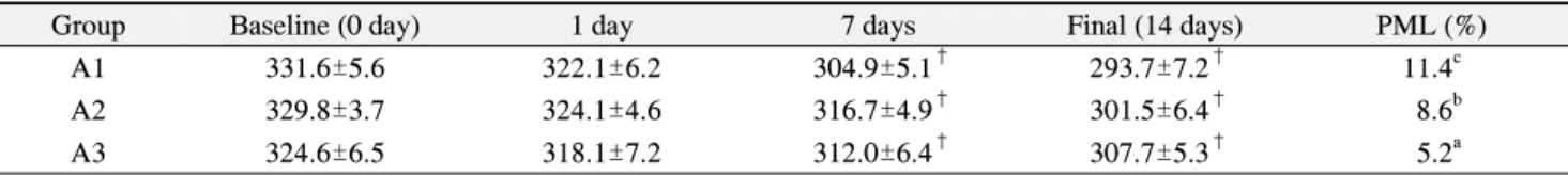
All groups showed significant difference (p<0.05) in mean microhardness values of enamel before and final bleaching (Table 5). However, the DCPD concentration increased in the bleaching agents, microhardness values less decreased (A3). Control group showed the lowest microhardness values (293.7 VHN) than that of groups A2 and A3 (Fig. 1). On the 1 day, microhardness values were similarly decreased at the groups A1, A2 and A3.
However, on the 7th and 14th days were statistical difference of microhardness values (p<0.05). The PML of group A3 was significantly lower than that of other groups (p<0.05).
5. SEM analysis
Control group showed increased surface porosities and erosion-like patterns around the enamel after bleaching.
Distinct variations of morphology were observed on the
enamel surfaces of the group A2 and A3 compared to the
control group (Fig. 2). With increasing DCPD concen-
trations, defective enamel surfaces became filled with a
particle. The substantial finding in this study was that
Table 5. Surface microhardness measurements and the percentage of microhardness loss (%)
Group Baseline (0 day) 1 day 7 days Final (14 days) PML (%)
A1 331.6±5.6 322.1±6.2 304.9±5.1† 293.7±7.2† 11.4c
A2 329.8±3.7 324.1±4.6 316.7±4.9† 301.5±6.4† 8.6b
A3 324.6±6.5 318.1±7.2 312.0±6.4† 307.7±5.3† 5.2a
Values are reported as the mean±standard deviation.
a∼cThe same letter indicates no significance different at α=0.05 by Tukey's test.
†p-values are determined by repeated measures ANOVA.
Fig. 1. Changes of enamel surface microhardness (VHN).
DCPD contained groups did not significantly change the enamel surface morphology.
6. EDX analysis
In the group A1 and A2, the amount of Ca and P ion found on the surfaces increased with increasing DCPD concentration in the DCPD/HP agent (Table 6). With increasing DCPD concentrations, Ca ions more increased than P ions. Control group showed the lower contents of Ca and P than that of groups A2 and A3.
Discussion
Enamel is the most dense, the mineral or inorganic phase content amounting to about 95 wt% (87 vol%). It is well known that mature human dental enamel crystals are carbonate-containing HA
23). DCPD has been known as a precursor phase to HA in the remineralization process.
Therefore, DCPD often coexists with HA in the dental hard tissues and interacts with saliva in the oral cavity at
the same time
19).
With increasing DCPD concentration, the pH values in the bleaching agents increased, making it less acidic. This can be explained by the increased supersaturation in the tooth bleaching agent containing DCPD. The low pH of some of the peroxide treatments caused severe changes in enamel topography, particularly the products with very low pH values
24). Low pH was probably the primary cause of demineralization
25).
The change in color of the enamel specimens in the all groups showed an increase in white (L
*), a decrease in red (a
*) and yellow (b
*). The directional change of these parameters confirm that tooth whitening has occurs. These results are in agreement with other studies, which found the L
*and b
*pre-treatment color results were shown to be affected by bleaching procedures
26,27). After the convert- sion of the mean △E values of all specimens to NBS units, it was determined that clinically detectable color changes occurred for all groups. Several authors have shown that color differences greater than 1 △E unit are visually detectable by 50% of human observers, although
△E values less than 3.3 are considered clinically insig- nificant
28). Most importantly, the data indicate that the addition of DCPD did not influence the whitening efficacy of the bleaching agent negatively. This study showed that the whitening effect of DCPD/HP agents were similar to that of HP alone.
The mean values of Vikers microhardness of enamel
before and after bleaching for the all groups showed a
tendency to decrease the microhardness. However, the
DCPD concentration increased in the bleaching, micro-
hardness values less decreased (A3). The variability of
hardness loss in different studies was contributed to
factors including the test methods, the immersion media
Table 6. Contents of calcium, phosphate and oxygen in enamel surface post bleaching
Group Ca (wt%) P (wt%) O (wt%)
A1 43.52±3.7 19.08±3.5 37.40±2.1
A2 46.83±5.3 19.93±3.1 33.90±3.6
A3 50.21±2.9* 20.52±2.1 29.37±3.2
Values and presented as mean±standard deviation.
*p<0.05.
Fig. 2. Field emission-SEM photographs of enamel specimens after bleaching with the following groups: (A1) Control, 0 g DCPD/3.5%
HP; (A2) 0.1 g DCPD/3.5% HP; (A3) 1 g DCPD/ 3.5% HP. SEM: scanning election microscopy, DCPD: dicalcium phosphate dihydrate, HP: hydrogen peroxide.
and exposure time. Microhardness after tooth bleaching was reduced by 5∼11% in all groups. Previous inve- stigations reported 12∼40% microhardness losses on 10% CP treated enamel using a hardness test
13). In this study, we found that the group A2 and A3 were still slight reduction in enamel microhardness. However, the group A3 that had 1 g DCPD in the agents showed less significantly loss of hardness than the control group.
DCPD seemed to enhance the microhardness of the enamel surface because the samples in that group were piled up with calcium and phosphate. Previously reported work to investigate the demineralization effects of blea- ching agents on tooth structures has primarily used microhardness technique. Traditionally, microhardness changes are related to a loss or gain of mineral (demi- neralization or remineralization) of the dental structure
29).
Scanning electron microscopy (SEM) is a rapid and convenient method for qualitatively analysing the surface morphology of enamel and dentin specimens following bleaching
30). A1 group showed increased surface poro-
sities and erosion-like patterns around the enamel prisms after bleaching. As in the current study, Several other studies have reported morphological changes including pitting, waviness and increased surface roughness
6,8,11,31). These results support the current findings. The obser- vations underline the findings mentioned above, that HP in combination with DCPD seems to affect the enamel surface more less than control group.
The EDX analysis showed that the release of Ca and P ions increased with increasing DCPD concentrations.
Mineral loss values obtained in this study are in corre- lation with Jiang et al's study
6). The release of Ca ions was consistently greater than release of phosphorous ions at all HP concentrations, for both enamel and dentin
32). Potocnik et al
33)using electron probe microanalysis showed lowered concentrations of Ca and P and with mean Ca/P value of all bleached samples decreasing after bleaching with 10%
CP. The liner relationship between the decrease in enamel
hardness and Ca and P loss shows that hardness measure-
ments can be used as an indication of the degree of enamel
mineralization, which relates to enamel caries
34). There-
fore, the remineralization supplement either during or after
bleaching is considered beneficial for preventing enamel
demineralization. The hydrogen peroxide can promote
chemical alterations in the composition of the tooth,
reducing in quantity of calcium and phosphate in enamel
and dentin
35,36). Calcium phosphate is the main organic
component of the tooth. Calcium phosphate minerals are
ideal candidates for obstructing dentin tubules and redu- cing sensitivity. High concentrations of Ca and P were used to increase the diffusion rates of the ions and, hence, the remineralization rates
37). 3.5% HP agents containing of 1 g DCPD were more assessible for ion diffusion, there- fore should be more susceptible to rapid remineralization.
Other factors that may contribute to the discrepancy in results among previous studies may be both the duration and storage medium of the post bleaching period before testing. Some in vitro studies used artificial saliva or fluoride products between of after the treatments, for these elements are known to be an important factor to simulate clinical situations. However, the aim of this study was to investigate the effects of DCPD on the enamel surface subjected to HP. This study did not involve these elements in order to prevent the influences of any other reminera- lization factors except DCPD. Nevertheless, it is necessary to involve these factors in the future studies to investigate the beneficial effects of DCPD under typical clinical conditions.
In this study, the experimental results suggest that DCPD/HP agent less demineralization changes such as the erosion morphology and hardness loss without compro- mising whitening efficiency.
Summary
The purpose of this study was to evaluate the tooth whitening and properties of an enamel surface after treatments with tooth bleaching agents that contained DCPD and 3.5% HP. 30 enamel specimens were obtained from fifteen human premolars and randomly divided into three groups (n=10): 1, 3.5% HP 0+g DCPD; 2, 3.5%
HP+0.1 g DCPD; 3, 3.5% HP+1 g DCPD. All groups were treated 8 hours per day for 14 days. pH and mineral composition (ICP-AES) in the DCPD/HP agent were measured. Tooth color, microhardness and surface charac- terization (SEM and EDX) of enamel surface were measured as well. The data were analyzed with ANOVA.
1. The tooth bleaching agents with DCPD showed a increase in pH as compared with the group A1. However, there was no statistically significant difference (p>0.05).
2. As the concentration of DCPD was increased, the
concentration of Ca and P was also increased. The ratio of Ca/P was found to be approximately 1.1∼1.2.
3. In all groups, after the tooth whitening, the tooth color was found to have a value of L
*which was significantly increased (p<0.05).
4. In all groups, the hardness of tooth after bleaching showed a significant decrease in the microhardness as compared to their baseline (p<0.05). PML of the group A1 and A2 were significantly lower than that of the group A3.
5. Following an analysis of the characteristics of enamel surface after bleaching, there were porosity and erosion in the control group. However, the group A3 was no surface change.
6. Following an analysis of the constituents of enamel surface after bleaching, as DCPD content was increased, the amount of Ca and P was increased.
Based on the above results, DCPD/HP agent was equally effective to the control group. By raising the pH and thereby effectively reducing the decalcification of tooth surface, a lower degree of the effects are given to the surface characteristics and constituent alterations of enamel. Thus, the commercial availability can be achieved for the constituents of tooth whitening materials.
요 약
본 연구는 3.5% 과산화수소(hydrogen peroxide, HP)에 dicalcium phosphate dihydrate (DCPD)를 함유한 치아미 백제가 치아 미백과 표면 특성에 미치는 영향에 대해 연구 하였다. 발치한 건전한 소구치 30개를 치아 시편으로 하였 고, 이 시편을 3군(A1, A2, A3)으로 나누었다(n=10). 3.5%
HP에 DCPD를 0 g (대조군, A1), 0.1 g (A2), 1 g (A3)을 함 유시켜 치아 미백제를 제조했다. 모든 군은 하루에 8시간 미 백하여 14일 동안 반복하였다. 미백제의 pH 측정, 유도 결 합 플라스마 원자 방출 분광기(ICP-AES)를 이용한 원소분 석을 실시하고, 법랑질의 색, 경도, 표면의 형태 및 무기질 성분을 측정하였고, ANOVA를 이용하여 분석하였다.
1. 치아미백제의 pH를 측정한 결과 DCPD를 함유한 치아 미백제의 pH는 함유하지 않은 치아미백제의 pH에 비해 증 가를 보였으나 통계적으로 유의한 차이는 없었다(p>0.05).
2. 치아미백제의 무기질 함량을 측정한 결과 DCPD 농도
가 증가할수록 칼슘과 인의 농도가 증가했으며, Ca/P 비는
1.1∼1.2 정도로 나타났다.
3. 모든 군에서 L
*값은 미백 후가 미백 전에 비해 통계적 으로 유의하게 증가했다(p<0.05).
4. 치아의 경도는 미백 후가 미백 전에 비해 통계적으로 유의하게 미세경도의 감소를 보였다(p<0.05). 하지만 DCPD 함유량이 증가할수록 대조군에 비해 실험군의 미세경도 감 소율이 적게 나타났다.
5. 미백 후 법랑질 표면의 형태는 대조군에서 표면의 다공 성과 침식 현상이 보였으나(A1), DCPD 함유량이 가장 높 은 군(A3)에서는 표면 변화가 나타나지 않았다.
6. 미백 후 법랑질 표면의 성분은 DCPD 함유량이 증가할 수록 Ca, P 함량이 증가하였다.
이상의 결과로부터 DCPD를 함유한 3.5% HP의 치아미 백제는 대조군과 동등한 치아미백 효과가 있고, pH를 상승 시켜서 치아 표면의 탈회를 감소시키며 법랑질의 표면 형태 와 성분 변화에 덜 영향을 줌으로써, 치아미백제의 구성성 분으로 실용화할 수 있을 것으로 생각된다.
References
1. Greenwall L: Bleaching techniques in restorative dentistry.
Martin Dunitz, London, pp.25-35, 2001.
2. Haywood VB, Heymann HO: Nightguard vital bleaching.
Quintessence Int 20(3): 173-176, 1989.
3. Dahl JE, Pallesen U: Tooth bleaching-a critical review of the biological aspects. Crit Rev Oral Biol Med 14(4): 292-304, 2003.
4. Reyto R: Laser tooth whitening. Dent Clin North Am 42(4):
755-762, 1998.
5. Yang Z et al.: Novel method to measure enamel surface porosity with hydrogen peroxide bleaching. Am J Dent 22(5):
283-289, 2009.
6. Jiang T et al.: Beneficial effects of hydroxyapatite on enamel subjected to 30% hydrogen peroxide. J Dent 36(11): 907-914, 2008.
7. Montgomery S: External cervical resorption after bleaching a pulpless tooth. Oral Surg Oral Med Oral Pathol 57(2):
203-206, 1984.
8. Akal N, Over H, Olmez A: Effect of carbamide peroxide containing bleaching agents on the morphology and subsurface hardness of enamel. J Clin Pediat Dent 25(4):
293-296, 2001.
9. Gu HJ, Song KB: The bleaching effect of plasma arc and
35% carbamaid peroxide and its influence on the enamel surface. J Dent Hyg Sci 9(5): 525-530, 2009.
10. Shim YS, Jung SH: Effects of bleaching agents containing fluoride and calcium on human enamel. J Dent Hyg Sci 10(4):
295-300, 2010.
11. Pinheiro HB, Cardoso PE: Influence of five home whitening gels and a remineralizing gel on the enamel and dentin ultrastructure and hardness. Am J Dent 24(3): 131-137, 2011.
12. Lee HJ et al.: 35% Hydrogen peroxide gel in the whitening effect and enamel changes. J Dent Hyg Sci 8(4): 255-260, 2008.
13. Zantner C et al.: Surface microhardness of enamel after different home bleaching procedures. Dent Mater 23(2): 243- 250, 2007.
14. Smidt A, Feuerstein O, Topel M: Mechanical, morphologic, and chemical effects of carbamide peroxide bleaching agents on human enamel in situ. Quintessence Int 42(5): 407-412, 2011.
15. Polydorou O, Hellwig E, Hahn P: The efficacy of three different in-office bleaching systems and their effect on enamel microhardness. Oper Dent 33(5): 579-586, 2008.
16. Duschner H et al.: Effects of hydrogen peroxide bleaching strips on tooth surface color, surface microhardness, surface and subsurface ultrastructure, and microchemical (Raman spectroscopic) composition. J Clin Dent 17(3): 72-78, 2006.
17. Attin T et al.: Potential of fluoridated carbamide peroxide gels to support post-bleaching enamel re-hardening. J Dent 35(9): 755-759, 2007.
18. Baratieri LN et al.: Non-vital tooth bleaching: guidelines for the clinician. Quintessence Int 26(9): 597-608, 1995.
19. Stralfors A: The effect of calcium phosphate on dental caries in school chidren. J Dent Res 43(6): 87-93, 1964.
20. Finn SB, Jamison HC: The effect of a dicalcium phosphate chewing gum on caries incidence in children: 30-month results. J Am Dent Assoc 74(5): 987-995, 1967.
21. Zhang YP et al.: Intra-oral remineralization of enamel with a MFP/DCPD and MFP/silica dentifrice using surface micro- hardness. J Clin Dent 6(2): 148-153, 1995.
22. Sulieman M et al.: The bleaching depth of a 35% hydrogen peroxide based in-office product: a study in vitro. J Dent 33(1): 33-40, 2005.
23. Sydney-Zax M, Mayer I, Deutsch D: Carbonate content in developing human and bovine enamel. J Dent Res 70(5):
913-916, 1991.
24. Joiner A: Review of the effects of peroxide on enamel and dentine properties. J Dent 35(12): 889-896, 2007.
25. Efeoglu N, Wood D, Efeoglu C: Microcomputerised tomo- graphy evaluation of 10% carbamide peroxide applied to enamel. J Dent 33(7): 561-567, 2005.
26. Luk K, Tam L, Hubert M: Effect of light energy on peroxide tooth bleaching. J Am Dent Assoc 135(2): 194-201, 2004.
27. Rosenstiel SF, Gegauff AG, Johnston WM: Duration of tooth color change after bleaching. J Am Dent Assoc 122(4):
54-59, 1991.
28. Ruyter IE, Nilner K, Moller B: Color stability of dental composite resin materials for crown and bridge veneers. Dent Mater 3(5): 246-251, 1987.
29. Featherstone JD et al.: Comparison of artificial caries-like lesions by quantitative microradiography and microhardness profiles. Caries Res 17(5): 385-391, 1983.
30. Joiner A, Thakker G: In vitro evaluation of a novel 6%
hydrogen peroxide tooth whitening product. J Dent 32(1):
19-25, 2004.
31. Sasaki RT et al.: Micromorphology and microhardness of
enamel after treatment with home-use bleaching agents containing 10% carbamide peroxide and 7.5% hydrogen peroxide. J Appl Oral Sci 17(6): 611-616, 2009.
32. Leonard RH Jr et al.: Nightguard vital bleaching of tetra- cycline-stained teeth: 90 months post treatment. J Esthet Restor Dent 15(3): 142-152, 2003.
33. Potocnik I, Kosec L, Gaspersic D: Effect of 10% carbamide peroxide bleaching gel on enamel microhardness, micro- structure, and mineral content. J Endod 26(4): 203-206, 2000.
34. Feagin F, Koulourides T, Pigman W: The characterization of enamel surface demineralization, remineralization, and asso- ciated hardness changes in human and bovine material. Arch Oral Biol 14(12): 1407-1417, 1969.
35. Rotstein I et al.: Histochemical analysis of dental hard tissues following bleaching. J Endod 22(1): 23-25, 1996.
36. Kawamoto K, Tsujimoto Y: Effects of the hydroxyl radical and hydrogen peroxide on tooth bleaching. J Endod 30(1):
45-50, 2004.
37. Tung MS et al.: Effects of calcium phosphate solutions on dentin permeability. J Endod 19(8): 383-387, 1993.