Introduction
Among foot structures, the arches are important for foot stability and resilience. In particular, the me- dial longitudinal arch (MLA), consisting of the first metatarsal, medial cuneiform, navicular, talus, and calcaneus bones, is a primary weight-bearing and shock-absorbing structure (Neumann, 2011). Pes pla- nus (or flatfoot) develops as the MLA decreases (Pandey et al, 2013) and is largely divided into rigid
type and flexible type. Rigid type includes states in which the MLA has dropped regardless of bearing weight, while flexible types occur when the MLA is formed without bearing weight but disappears during weight bearing (Kuhn et al, 1999).
A flexible pes planus is caused by tibialis posterior dysfunction, foot bone malformation, ligament loosen- ing, Achilles tendon shortening, and foot muscle weak- ness (Huang et al, 1993; Leung et al, 1998; Murley et al, 2009). These deformations lead to excessive prona- Corresponding author: Jong-duk Choi [email protected]
The Effects of Foot Intrinsic Muscle and Tibialis Posterior Strengthening Exercise on Plantar Pressure and Dynamic Balance in
Adults Flexible Pes Planus
Da-bee Lee1,2, BHSc, PT, Jong-duk Choi3, PhD, PT
1Rehabilitation Center, Chungnam University Hospital
2Dept. of Physical Therapy, The Graduate School, Daejeon University
3Dept. of Physical Therapy, College of Health & Medical Science, Daejeon University
Abstract
1)Background: In previous studies regarding flexible pes planus, Foot orthosis, special shoes have been used as interventions for correcting malalignment and intrinsic muscles strengthening exercise have been regarded as interventions for foot function and supporting medial longitudinal arch during walking.
However, some recent studies reported that strengthening extrinsic muscles as well as intrinsic muscles is more effective and active intervention for flexible pes planus. In particular, the tibialis posterior muscle of foot extrinsic muscles plays essential roles in maintaining the medial longitudinal arch during dynamic weight bearing and balance. In addition this muscle acts longer than other supination muscles during the stance phase in the gait cycle.
Objects: This study aimed to investigate the effect of foot intrinsic muscle and tibialis posterior muscle strengthening exercise for plantar pressure and dynamic balance in adults with flexible pes planus.
Methods: 16 young flexible pes planus adults (7 males, 9 females) were recruited and were randomized into two groups. The experimental group performed foot intrinsic muscle and tibialis posterior muscle strengthening training, the control group performed only foot intrinsic muscle strengthening training. All groups received strengthening training for 30 minutes five times a week for six weeks.
Results: The experimental group had significantly lower plantar pressure of medial heel area than the control group in stand (p<.05). The experimental group had significantly higher dynamic balance ability than control group (p<.05).
Conclusion: The results of this study provide evidence to suggest that foot intrinsic muscle and tibialis posterior muscle of extrinsic muscle strengthening exercises may improve plantar pressure distribution and dynamic balance ability in adults with flexible pes planus.
Key Words: Dynamic balance; Flexible pes planus; Plantar pressure; Tibialis posterior muscle.
tion of the foot during weight bearing and cause plan- tar flexion and adduction of the talus bone and the valgus of the calcaneus bone (Pandey et al, 2013).
Abnormal peripheral information from the foot affects muscle performance necessary for body posture and position control (Shumway-Cook and Horak, 1986) and stable maintenance on the base of support (Franco, 1987). Such abnormalities in the MLA leads to loss of the functional stability of the foot (Franco, 1987), which in turn causes balance problems (Hertel, 2002;
Hillstrom et al, 2013; Tsai et al, 2006).
Pes planus treatments are divided into surgical and conservative treatments. Conservative treatments in- clude taping, orthosis, special shoes, and foot muscle exercises. Among these treatments, foot muscle ex- ercises have been reported to reduce excessive prona- tion, strengthen the foot muscles, and improve foot functions (Panichawit et al, 2015) and have the advan- tages of helping restructure the foot and being simple to perform (Jung et al, 2011; Lynn et al, 2012). Foot muscles are subdivided into intrinsic and extrinsic muscles. Intrinsic foot muscles assist standing postures and balance during gait and support the MLA during push-off in the stance phase (Neumann, 2011).
Exercise interventions for intrinsic muscle strengthen- ing include toe curls (TC), shin curls, picking up ob- jects with the foot, unilateral balance activities, and short foot (SF) exercises (Anderson et al, 2004;
Prentice, 2009). Among these exercises, TC and SF exercises are most commonly recommended (Abdo and Iorio, 1994; Freiberger et al, 2007; Liebenson, 2001).
Extrinsic foot muscles, such as the tibialis posteri- or and peroneus longus muscles, provide dynamic support to the MLA during the stance phase of gait and contribute to stabilization of the intertarsal joints (Jung et al, 2011). In particular, the tibialis posterior muscle plays essential roles in maintaining the MLA during dynamic weight bearing and balance (Kamiya et al, 2012; Kohls-Gatzoulis et al, 2004). In addition, this muscle provides foot adduction, supination, and plantar flexion, assists in controlled flattening of the MLA through eccentric contractions during the stance
phase in the gait cycle, and acts longer than other supination muscles (Neumann, 2011). According to Prentice (2009), exercise interventions for selective strengthening of the tibialis posterior muscle include foot adduction, foot supination, and heel raises, with foot adduction and foot supination being the most ef- fective for selective strengthening (Kulig et al, 2004).
In prior studies, the malignment of the foot due to flattening of the MLA has been corrected and ex- cessive pronation of the subtalar joint has been ad- justed using the orthosis and special shoes(Brown et al, 1995; Johanson et al, 1994; Nigg et al, 1998).
Kelly et al (2014) stated that intrinsic foot muscles are important for foot arch postures while gait loads are applied, and other studies have reported that strengthening exercises targeting these muscles are necessary to maintain and enhance foot function.
Foot muscle exercise interventions for the height of the MLA have been limited to intrinsic foot muscle strengthening exercises without considering extrinsic foot muscle strengthening (Jam, 2006; Won and Lee, 2010). Recent studies suggested that to correct foot pronation inducing MLA flattening, increasing the strength of the intrinsic and extrinsic muscles is the most effective method (Panichawit et al, 2015;
Snyder et al, 2009). Currently, exercise interventions that combine both foot intrinsic muscle and foot ex- trinsic muscle are rare and studies of foot plantar pressure and dynamic balance in relation to the pes planus are lacking.
Therefore, the present study includes an examina- tion of the effects of strengthening the tibialis poste- rior muscle, which maintains foot supination for the longest time among the extrinsic muscles that main- tain the MLA, as well as effects on intrinsic muscles related to foot arch height, dynamic balance, and foot plantar pressure distribution in young adults with flexible pes planus. The hypothesis states that inter- vention methods combining foot intrinsic muscle strengthening exercise and tibialis posterior muscle strengthening exercise will affect flexible pes planus by decreasing foot arch height differences before and
after weight bearing, increasing dynamic balance, and decreasing plantar medial column pressure.
Methods
Subjects
This study included 16 young adults (7 males and 9 females) with flexible pes planus residing in Daejeon City of Korea. After hearing sufficient ex- planations of the study, three participants were ex- cluded during the selection process. The subjects were divided into an experimental group (foot in- trinsic muscle and tibialis posterior muscle strength- ening training; FTST) that performed intrinsic foot muscle and tibialis posterior muscle strengthening exercises and a control group that performed intrinsic foot muscle strengthening exercise (foot intrinsic muscle strengthening training; FST). The subjects were randomly assigned to the groups by having them draw a card indicating one of the two groups.
The general characteristics of the study subjects are provided in Table 1, and selection criteria included foot arch height differences before and after weight bearing exceeding 10 ㎜ (Cote et al, 2005), normal weight with a body mass index ranging from 18.5∼
23.0 (Chang et al, 2010), and no use of insoles or orthoses. The exclusion criteria for the study subjects were those that had other neurologic, orthopedic, or cardiorespiratory system diseases. The subjects in the present study signed a written agreement related to
the experiment and volunteered to participate in the study. The experimental procedure was approved by Daejeon University Institutional Review Board (approval number: 1040647-201506-HR-005-003).
Measurement tools
Foot arch heights measure
The navicular drop test (NDT) was used to select the subjects and measure foot arch heights before and after interventions (Shrader et al, 2005). This test is valuable for measuring and evaluating navicular heights with high reliability [Intraclass correlation co- efficient (ICC)>0.94] (Cote et al, 2005; Vicenzino et al, 2000) and can evaluate damage to and weakening of the musculoskeletal system that changes MLA height (Allen and Glasoe, 2000). In a sitting position, sub- jects were asked to bend the knee joints to 90° and place their feet flat on the ground with a neutral posture of the ankles. In both sitting and standing positions, a sheet marked with navicular bone tuber- osity was placed vertically on the floor, and the dis- tance from the floor to the navicular bone tuberosity was marked. The difference between the distance measured in the sitting position and the distance measured in a state of weight bearing was measured using a tape measure (Picciano et al, 1993).
Plantar foot pressure measure
A Gaitview AFA-50 system (alFOOTs, Seoul, Korea) was used to measure foot plantar pressure dis-
Characteristics Experimental groupa (n1=8) Contorl groupb (n2=8)
Gender (male/female) 4/4 3/5
Affected side (left/right) 6/2 3/5
Age (year) 24.9±2.9c 24.4±.7.0
Height (㎝) 169.5±7.0 170.5±5.9
Weight (㎏) 62.0±7.4 61.1±7.0
BMId (㎏/㎡) 21.5±1.1 21.0±1.6
afoot intrinsic muscle and tibialis posterior muscle strengthening training, bfoot intrinsic muscle strengthening training,
cmean±standard deviation, dbody mass index.
Table 1. General characteristics of the subjects (N=16)
tributions in standing positions. The Gaitview is a pressure pad in the form of a footboard consisting of 2,304 (48×48 ㎟) sensors placed in an area of 410×410 ㎜, and it is highly reliable (Kim and Lee, 2012). The foot plantar pressure measurements were divided into eight zones: hallux, the second through fifth toes, first metatarsal (M1), second through fourth metatarsals (M24), fifth metatarsal, midfoot (MF), medial heel (MH), and lateral heel. To meas- ure the foot plantar pressure distributions, the sub- jects were instructed to stand upright on the Gaitview for 30 seconds in a comfortable posture with eyes open and looking forward. The values were measured three times with a rest time of one minute after each measurement, and the average of the measured values was obtained. The measurement method was sufficiently explained to the subjects be- fore the measurement so that the subject fully un- derstood what was expected of them.
Dynamic balance test
The star excursion balance test (SEBT) was used to evaluate dynamic balance. This test evaluates bal- ance ability by measuring the distances of the sub- ject’s non-weight-bearing leg stretched in eight di- rections while the weight is being borne on the other leg (Gribble et al, 2004). The eight directions drawn at intervals of 45° are anterior (SEBT-A), ante- rior-lateral, lateral (SEBT-L), posterior-lateral, pos- terior, posterior-medial, medial (SEBT-M), and ante- rior-medial (SEBT-AM). Each subject was in- structed to place the leg with flexible pes planus on the center of a line and maximally stretch the other leg along the line. The distance from the center to the end of the big toe of the stretched leg was measured (Cote et al, 2005). During the measure- ments, subjects could only allow the stretched leg to slightly contact the bearing surface, which ensured that weight was not supported by the stretched leg.
After stretching the leg as far as possible, measured values were calculated as a percentage (%) of the subjects’ leg lengths (Gribble et al, 2004). Leg
lengths were measured in three times as the distance from the medial malleolus bone to the anterior supe- rior iliac spine (Beattie et al, 1990), at intervals of one minute, and the average of the measured values was obtained.
Intervention
The subjects in the experimental group performed selective tibialis posterior muscle strengthening ex- ercises along with the intrinsic foot muscle strength- ening exercise, while the subjects in the control group performed only the intrinsic foot muscle strengthening exercise. The interventions were im- plemented for 30 minutes per time, 5 times per week for 6 weeks.
Selected tibialis posterior muscle strengthening training
The selected tibialis posterior muscle strengthen- ing exercises consisted of foot adduction resistance and foot supination resistance exercises (Kulig et al, 2004). After the exercises and to prevent short- ening of the Achilles tendon, the subjects per- formed calf muscle stretching five times for ap- proximately 7 seconds with a relaxation period of approximately 3 seconds (Hyong et al, 2009). For the foot adduction resistance exercise, each subject placed his or her feet on the floor, forearm length apart, and sat with knee joints bent at a flexion angle of 80°. For leg stability, the subjects placed their forearms on opposite sides of the leg, which strengthened between the legs and the leg being strengthened. Elastic bands were provided depend- ing on each subject’s muscle strength (Theraband, GmbH, Hadamar, Germany), which were wound around the medial and lateral sides of each sub- ject’s foot, tied up, and pulled laterally at an angle of 45° in relation to the floor (Kulig et al, 2004).
During the exercise, the feet were maintained flat, in contact with the floor, and moved as it they were sweeping the floor. For the foot supination resistance exercise, each subject placed one leg on
and stood at the lateral end of the footboard with the knee joint of placed on the footboard main- tained a bend. The subject placed the medial part of the heel and foot at the base of the third meta- tarsal bones on the edge of the footboard to per- form foot supination (Figure 1).
Foot intrinsic muscle strengthening training
The intrinsic foot muscle strengthening exercises consisted of TCs and an SF exercise (Abdo and Iorio, 1994; Freiberger et al, 2007; Liebenson, 2001) recommended for intrinsic foot muscle strengthening.
For the TC, towels were prepared and placed be- low the feet of the subjects. While heels remained in contact, each subject bent the interphalangeal joint and metatarsophalangeal joint to hold the towel below the feet. During the SF exercise, without bending the toes, subjects shortened their feet in an anterior-posterior direction by moving the head of the M1 bone toward the heel. For TC and SF exercises in a sitting position, the subject sat on a chair to support the hip, knee, and ankle joints at 90° angles. The foot not being exercised was placed behind the foot being exercised. For TC and SF exercises in a one-leg- ged standing position, the subject maintained the legs shoulder-width apart and slightly bent the
knee joint. To maintain balance, the subject had the left and right index fingers gently come into contact with the wall.
All exercises were performed in a sequence from a sitting position, to a standing position, and finally a one-legged standing position (Jung et al, 2011). In addition, the researcher instructed the subjects to perform all exercises with maximum efforts.
Statistical analysis
The data collected from the experiments were an- alyzed using SPSS ver. 18.0 (SPSS Inc., Chicago, IL, USA) and the measured values were presented as means and standard deviations. Mann-Whitney U-tests were conducted to compare foot arch heights, foot plantar pressure and dynamic balance between the two groups, and Wilcoxon signed rank tests were conducted to compare foot arch heights, foot plantar pressure and dynamic bal- ance between before and after interventions. To analyze statistical significance, significance level α was set to .05.
Results
Foot arch heights
The foot arch heights of the FTST and FST
A B C D
Figure 1. Tibialis posterior strengthening training: foot adduction resistance exercise (A: start position, B: end position), foot supination resistance exercise (C: start position, D: end position).
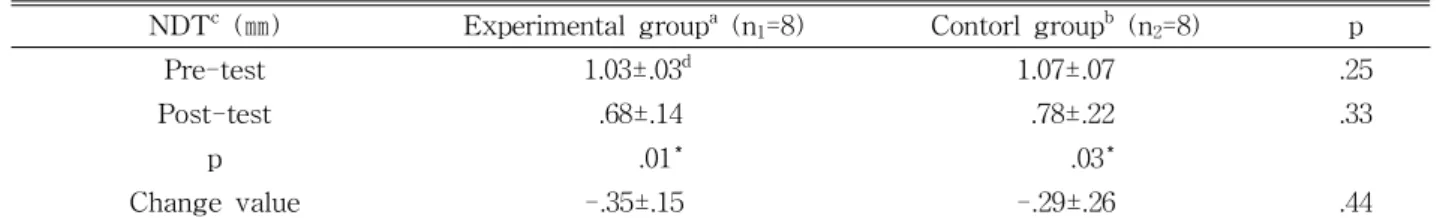
groups before and after interventions were compared (Table 2). Both groups showed significant differences in foot arch heights before and after interventions (p<.05), but the two groups did not show any differ- ence in foot arch heights (p>.05).
Plantar foot pressure
Plantar pressure distributions of the FTST and
FST groups before and after interventions were compared (Table 3). The FTST group showed significant differences before and after inter- ventions in the M1 bone, M24 bones, and MH (p<.05), while the FST group showed no sig- nificant difference (p>.05). In addition, the results from the two groups were significant different for the MH (p<.05).
NDTc (㎜) Experimental groupa (n1=8) Contorl groupb (n2=8) p
Pre-test 1.03±.03d 1.07±.07 .25
Post-test .68±.14 .78±.22 .33
p .01* .03*
Change value -.35±.15 -.29±.26 .44
afoot intrinsic muscle and tibialis posterior muscle strengthening training, bfoot intrinsic muscle strengthening training,
cnavicular drop test, dmean±standard deviation, *p<.05.
Table 2. Comparison of Foot medial longitudinal arch height outcomes within groups and between groups
Foot plantar pressure (㎪) Experimental groupa (n1=8) Contorl groupb (n2=8) p 1st metatarsal bone
Pre-test 108.45±24.93c 78.65±29.86 .06
t-test 60.74±41.84 65.10±39.25 .75
p .02* .09
Change value -47.71±40.46 -13.55±23.72 .17
2∼4th metatarsal bone
Pre-test 144.58±19.94 135.85±16.15 .53
Post-test 97.53±47.31 130.81±28.73 .25
p .04* .58
Change value -47.05±38.99 -5.04±25.90 .05
Midfoot
Pre-test 54.16±20.54 56.93±34.62 .80
Post-test 56.61±22.40 57.35±36.19 .92
p .77 .58
Change value 2.45±21.45 .43±26.24 1.00
Medial heel
Pre-test 141.58±13.01 156.08±10.37 .05
Post-test 140.5±15.19 157.06±12.10 .04*
p .03* 1.00
Change value -1.08±14.92 .99±8.45 .60
afoot intrinsic muscle and tibialis posterior muscle strengthening training, bfoot intrinsic muscle strengthening training,
cmean±standard deviation, *p<.05.
Table 3. Comparison of foot plantar pressure outcomes within groups
Dynamic balance ability
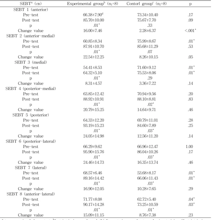
The dynamic balance ability of the FTST and FST groups before and after intervention were also compared (Table 4), using SEBT. The FTST group showed sig- nificant differences in balance before and after inter-
ventions in all eight directions (p<.05), while the FST group showed significant differences in balance before and after interventions in only five directions (p<.05).
The two groups showed significant differences in me- dial, lateral, and anterior-lateral directions (p<.05).
SEBTa (㎝) Experimental groupb (n1=8) Contorl groupc (n2=8) p SEBT 1 (anterior)
Pre-test 66.38±7.90d 73.34±10.40 .17
Post-test 85.70±10.00 75.67±7.70 .09
p .01* .33
Change value 16.00±7.46 2.28±6.37 <.001*
SEBT 2 (anterior-medial)
Pre-test 60.85±8.34 75.99±8.67 .01*
Post-test 87.91±10.70 85.68±11.29 .53
p .01* .07
Change value 22.54±12.25 8.26±10.15 .05
SEBT 3 (medial)
Pre-test 54.41±8.53 71.60±9.12 .01*
Post-test 64.32±5.10 75.53±8.06 .01*
p .01* .29
Change value 8.31±4.57 3.36±7.22 .14
SEBT 4 (posterior-medial)
Pre-test 63.85±12.42 70.94±9.56 .20
Post-test 88.92±10.91 88.10±8.81 .83
p .01* .02*
Change value 20.79±15.25 14.64±9.71 .46
SEBT 5 (posterior)
Pre-test 64.33±12.20 69.79±11.01 .28
Post-test 93.19±15.23 84.60±7.89 .25
p .01* .03*
Change value 24.05±14.98 12.56±11.20 .14
SEBT 6 (posterior-lateral)
Pre-test 66.29±9.62 66.96±12.47 1.00
Post-test 95.90±15.76 86.04±10.26 .17
p .01* .03*
Change value 24.46±14.73 16.35±13.74 .46
SEBT 7 (lateral)
Pre-test 68.57±6.46 53.68±8.17 .01*
Post-test 89.16±14.42 66.06±11.43 .01*
p .01* .03*
Change value 16.90±12.05 10.28±7.65 .29
SEBT 8 (anterior-lateral)
Pre-test 71.77±8.08 62.72±5.40 .04*
Post-test 90.17±14.28 73.25±10.59 .03*
p .01* .01*
Change value 15.09±11.15 8.76±7.38 .23
astar excursion balance test, bfoot intrinsic muscle and tibialis posterior muscle strengthening training, cfoot intrinsic muscle strengthening training, dmean±standard deviation, *p<.05.
Table 4. Comparison of dynamic balance ability outcomes within groups and between groups
Discussion
The purpose of the present study was to examine the effects of exercise to strengthen the tibialis pos- terior muscle, which is an extrinsic muscle, as well as intrinsic foot muscles on foot arch height, foot plantar pressure distribution and dynamic balance among young adults with flexible pes planus.
According to the results, the FTST group, which combined strengthening exercises of the tibialis pos- terior muscle and intrinsic foot muscles, showed de- creased foot arch height differences before and after weight bearing, improved dynamic balance, and sig- nificantly decreased foot plantar pressure in the M1 bone, M24 bones, and MH after intervention. In ad- dition, this group showed significant decreases in MH pressure and significant increases in medial, lat- eral, and anterior-lateral SEBT stretching distances compared to the FST group, which performed only intrinsic foot muscle strengthening exercises (p<.05).
NDTs were conducted to select subjects and meas- ure foot arch heights. Both groups showed sig- nificant decreases in foot arch height differences be- fore and after weight bearing after interventions (p<.05), but the two groups did not show any sig- nificant difference when compared to each other (p>.05). SEBTs were used to evaluate dynamic bal- ance and have been conducted in previous studies as a highly reliable tool for measuring dynamic balance in pes planus patients (Cote et al, 2005; Hyong et al, 2009). According to the results of the present study, the FTST group showed significant increases in stretching distances in all directions, including SEBT-L (foot supination), SEBT-A (forward move- ment of the center of gravity similar to the pro- pulsion stage in gait cycles), and SEBT-AM and SEBT-M directions (moving the center of gravity toward the medial side of the foot; p<.05). The FST group showed significant increases in only five of the eight directions, excluding SEBT-A, SEBT-AM, and SEBT-M (p<.05). Based on these results, rather than strengthen training only the intrinsic foot mus-
cles, training that combines intrinsic muscle strengthening and strengthening of the tibialis poste- rior muscle should be considered to help patients move the center of gravity and during the propulsion stage of gait cycles, which affects gait speed.
Among prior studies, Hyong et al (2009) reported that when extrinsic foot muscle strengthening ex- ercises were implemented with subtalar joint treat- ment, dynamic balance significantly increased, and Panichawit et al (2015) implemented intrinsic and extrinsic muscle strengthening exercises for flexible pes planus patients and reported that foot functions improved. In the present study, the FTST group performed foot intrinsic muscle strengthening ex- ercise along with exercise to strengthen the tibialis posterior muscle, which allowed for longer gaits compared to other supination muscles that are used during the propulsion stage (Neumann, 2011). Thus, strengthening tibialis posterior muscle during sensory receptor activities and neuromuscular functions im- proves dynamic balance due to dynamic support of the foot medial area and static support of the in- trinsic foot muscle. These strengthening exercises are capable of solving balance problems in flexible pes planus patients.
According to Ledoux and Hillstrom (2002), pes planus causes more weight to be applied to the area below the big toe compared to neutral feet, and when compared to pes cavus and neutral feet, pes planus has the most foot plantar pressure in the metatarsal bones and MF region and the least foot plantar pressure in the calcaneus region (Kim, 2013).
During gaits, increased foot plantar pressure is dis- tributed in the medial column (hallux, medial forefoot, medial MF, and medial rear foot), which is medial to the straight line that connects the center of the third metatarsal bone and the calcaneus center compared to normal feet (Sun et al, 2006). The FTST group showed significant decreases in foot plantar pressure in the M1 bone, M24 bones, and MH after the inter- ventions (p<.05), while the FST group showed no significant difference (p>.05). A comparison between
the groups revealed that the FTST group had sig- nificant decreases in MH pressure comparted to the FST group (p<.05), which is attributed to the strengthened tibialis posterior muscle function in the FTST group and posture changes that aligned the rear foot and normal foot arches recovered during weight bearing (Kitaoka et al, 1997; Niki et al, 2001).
This led to reduction in foot plantar pressure in part of the medial column.
The MF did not show a significant difference after interventions for both groups (p>.05). Although the MF was divided into the medial and lateral parts when foot plantar pressure was evaluated in previous studies (Jonely et al, 2011; Tang et al, 2015), the Gaitview equipment used in the present study meas- ured foot plantar pressure in eight zones, including the MF. When posture changed to recover a normal foot arch through exercise interventions, even if the pressure in the medial part of the MF decreased, the pressure in the entire MF increased or was main- tained because of increases in the pressure in the lat- eral part of the foot caused by recovering foot arch.
This study has several limitations. First, the SEBT, which was used to evaluate dynamic balance, is closely related to the range of motion of joints and the forces of surrounding muscles because it requires compositive movements of the foot, ankle joint, knee joint, and hip joint (Cote et al, 2005; Won and Lee, 2010). Second, because young adults with flexible pes planus were selected as study subjects, generalizations of the results to diverse age groups does not apply.
Third, pain in the foot during gait cycles or exercise caused by excessively pronated feet was not considered. When the feet are excessively pronated, pain occurs in the anterior tibialis muscle, the tibialis posterior muscle, the sole, and the plantar fascia, and the feet easily become tired causing. The present study did not consider cases for which subjects’
weight was not balanced between the two feet due to foot arches and pain. Therefore, studies should be continuously conducted with subjects from diverse age groups and conditions to improve these limitations.
Conclusion
This study included 16 male and female adults with flexible pes planus divided into an intrinsic foot muscle and tibialis posterior muscle strengthening exercise group and a intrinsic foot muscle strength- ening exercise group to examine the effects of the exercises on foot arch height, foot plantar pressure distribution, and dynamic balance. According to the results, the combined exercise group showed de- creases in arch height differences before and after weight bearing, improvement of dynamic balance, and significant decreases in foot plantar pressure in the M1 bone, M24 bones, and MH after intervention.
This group also showed decreases in MH pressure and significant increases in stretching distances dur- ing medial and lateral SEBTs, but as the center of gravity moving toward the medial side of the foot during the anterior-lateral SEBT, forward movement of the center of gravity was similar to the propulsion stage of the gait cycle and caused foot supination (p<.05). Therefore combining exercise interventions for flexible pes planus, rather than strengthening on- ly the intrinsic foot muscles, to include strengthening exercises for the tibialis posterior muscle is more ef- fective for medial foot plantar pressure decreases and dynamic balance ability improvement.
References
Abdo RV, Iorio LJ. Rheumatoid arthritis of the foot and ankle. J Am Acad Orthop Surg. 1994;2(6):
326-332.
Allen MK, Glasoe WM. Metrecom measurement of navicular drop in subjects with anterior cruciate ligament injury. J Athl Train. 2000;35(4):403-406.
Beattie P, Isaacson K, Riddle DL, et al. Validity of derived measurements of leg-length differences obtained by use of a tape measure. Phys Ther.
1990;70(3):150-157.
Brown GP, Donatelli R, Catlin PA, et al. The effect
of two types of foot orthoses on rearfoot mechanics. J Orthop Sports Phys Ther. 1995;
21(5):258-267.
Chang JS, Park JW, Kim CS. The changes of plan- tar foot pressure by external loads during walk- ing in flatfoot. J Korean Soc Phys Med.
2010;5(4):543-549.
Cote KP, Brunet II ME, Gansneder BM, et al. Effects of pronated and supinated foot postures on stat- ic and dynamic postural stability. J Athl Train.
2005;40(1):41-46.
Franco AH. Pes cavus and pes planus. Analyses and treatment. Phys Ther. 1987;67(5):688-694.
Freiberger E, Menz HB, Abu-Omar K, et al.
Preventing falls in physically active commun- ity-dwelling older people: A comparison of two intervention techniques. Gerontology. 2007;53(5):
298-305.
Gribble PA, Hertel J, Denegar CR, et al. The effects of fatigue and chronic ankle instability on dy- namic postural control. J Athl Train. 2004;
39(4):321-329.
Hertel J. Functional anatomy, pathomechanics, and pathophysiology of lateral ankle instability. J Athl Train. 2002;37(4):364-375.
Hillstrom HJ, Song J, Kraszewski AP, et al. Foot type biomechanics part 1: Structure and function of the asymptomatic foot. Gait Posture. 2013;37(3):
445-451. https://doi.org/10.1016/j.gaitpost.2012.09.007 Huang CK, Kitaoka HB, An KN, et al. Biomechanical evaluation of longitudinal arch stability. Foot Ankle. 1993;14(6):353-357.
Hyong IH, Kim HS, Lee GC. The effect of muscle activities and dynamic balance ability with mo- bilization and active exercise on pronation foot.
Journal of Sport and Leisure Studies. 2009;
37(2):1023-1032.
Jam B. Evaluation and retraining of the intrinsic foot muscles for pain syndromes related to abnormal control of pronation. Advanced Physical Therapy Education Institute. 2006.
Johanson MA, Donatelli R, Wooden MJ, et al. Effects
of three different posting methods on controlling abnormal subtalar pronation. Phys Ther. 1994;
74(2):149-158.
Jonely H, Brismée JM, Sizer PS Jr, et al.
Relationships between clinical measures of static foot posture and plantar pressure during static standing and walking. Clin Biomech (Bristol, Avon). 2011;26(8):873-879. https://doi.org/10.1016/
j.clinbiomech.2011.04.008
Jung DY, Kim MH, Koh EK, et al. A comparison in the muscle activity of the abductor hallucis and the medial longitudinal arch angle during toe curl and short foot exercises. Phys Ther Sport. 2011;
12(1):30-35. https://doi.org/10.1016/j.ptsp.2010.08.001 Kamiya T, Uchiyama E, Watanabe K, et al. Dynamic
effect of the tibialis posterior muscle on the arch of the foot during cyclic axial loading.
Clin Biomech (Bristol, Avon). 2012;27(9):962-966.
https://doi.org/10.1016/j.clinbiomech.2012.06.006 Kelly LA, Cresswell AG, Racinais S, et al. Intrinsic
foot muscles have the capacity to control de- formation of the longitudinal arch. J R Soc Interface. 2014;11(93):20131188. https://doi.org/
10.1098/rsif.2013.1188
Kim G. Plantar pressure and gait ability analysis for foot deformities with arch support. Jeonnam, Dongshin University, Doctoral Dissertation. 2013.
Kim YT, Lee JS. Normal pressures and reliability of the Gaitview® system in healthy adults. Prosthet Orthot Int. 2012;36(2):159-164. https://doi.org/
10.1177/0309364611433444
Kohls-Gatzoulis J, Angel JC, Singh D, et al. Tibialis posterior dysfunction: A common and treatable cause of adult acquired flatfoot. BMJ. 2004;
329(7478):1328-1333.
Kuhn DR, Shibley NJ, Austin WM, et al.
Radiographic evaluation of weight-bearing or- thotics and their effect on flexible pes planus. J Manipulative Physiol Ther. 1999;22(4):221-226.
Kulig K, Burnfield JM, Requejo SM, et al. Selective activation of tibialis posterior: Evaluation by magnetic resonance imaging. Med Sci Sports
Exerc. 2004;36(5):862-867.
Ledoux WR, Hillstrom HJ. The distributed plantar vertical force of neutrally aligned and pes planus feet. Gait Posture. 2002;15(1):1-9.
Leung AK, Mak AF, Evans JH. Biomedical gait evaluation of the immediate effect of orthotic treatment for flexible flat foot. Prosthet Orthot Int. 1998;22(1):25-34.
Liebenson C. Self-help advice for the clinician:
Sensory-motor training. J Bodyw Mov Ther.
2001;5(1):21-27.
Lynn SK, Padilla RA, Tsang KK. Differences in static- and dynamic-balance task performance after 4 weeks of intrinsic-foot-muscle training:
The shortfoot exercise versus the towel-curl exercise. J Sport Rehabil. 2012;21(4):327-333.
Murley GS, Menz HB, Landorf KB. Foot posture in- fluences the electromyographic activity of se- lected lower limb muscles during gait. J Foot Ankle Res. 2009;2:35. https://doi.org/10.1186/
1757-1146-2-35
Nigg BM, Khan A, Fisher V, et al. Effect of shoe insert construction on foot and leg movement.
Med Sci Sports Exerc. 1998;30(4):550-555.
Neumann DA. Kinesiology of the Musculoskeletal System: Foundation for Rehabilitation. 2nd ed.
St Louis, Mosby, 2011:628-638.
Pandey S, Pal CP, Kumar D et al. Flatfoot in Indian population. J Orthop Surg (Hong Kong). 2013;
21(1):32-36.
Panichawit C, Bovonsunthonchai S, Vachalathiti R, et al.
Effects of foot muscles training on plantar pressure distribution during gait, foot muscle strength, and foot function in persons with flexible flatfoot. J Med Assoc Thai. 2015;98 Suppl 5:S12-S17.
Picciano AM, Rowlands MS, Worrell T. Reliability of open and closed kinetic chain subtalar joint neutral positions and navicular drop test. J Orthop Sports Phys Ther. 1993;18(4):553-558.
Sun SK, Jung DC, Ko KJ, et al. A comparison of physical fitness in people with normal foot and pes planus. Korea Sport Research. 2006;17(6):687-694.
Shrader JA, Popovich JM Jr, Gracey GC, et al.
Navicular drop measurement in people with rheumatoid arthritis: Interrater and intrarater reliability. Phys Ther. 2005;85(7):656-664.
Shumway-Cook A, Horak FB. Assessing the influence of sensory interaction on balance. Suggestion from the field. Phys Ther. 1986;66(10):1548-1550.
Snyder KR, Earl JE, O’Connor KM, et al. Resistance training is accompanied by increases in hip strength and changes in lower extremity bio- mechanics during running. Clin Biomech (Bristol, Avon). 2009;24(1):26-34. https://doi.org/10.1016/
j.clinbiomech.2008.09.009
Tang SF, Chen CH, Wu CK, et al. The effects of total contact insole with forefoot medial post- ing on rearfoot movement and foot pressure distributions in patients with flexible flatfoot.
Clin Neurol Neurosurg. 2015;129 Suppl 1:S8-S11.
https://doi.org/10.1016/S0303-8467(15)30004-4 Tsai LC, Yu B, Mercer VS, et al. Comparison of
different structural foot types for measures of standing postural control. J Orthop Sports Phys Ther. 2006;36(12):942-953.
Vicenzino B, Griffiths SR, Griffiths LA, et al. Effect of antipronation tape and temporary orthotic on vertical navicular height before and after exercise. J Orthop Sports Phys Ther. 2000;
30(6):333-339.
Won KH, Lee MG. Effects of a strength exercise training for foot intrinsic muscle on height of medial longitudinal arch and balance in females aged 20’s. Exercise Science. 2010;19(4):351-360.
This article was received October 7, 2016, was reviewed October 7, 2016, and was accepted November 10, 2016.