INTRODUCTION
The prognosis of advanced gastric cancer (AGC) depends on a variety of different factors, including tumor size, depth of inva- sion, nodal metastases, and pathological type. Among these fac- tors, depth of tumor invasion and nodal metastases are regarded as crucial for treatment planning, as well as determining patient prognosis (1-3). Many investigators have reported that gross classification of Borrmann type may also be a valuable clinico-
pathological factor. Li et al. (4) reported that the prognoses of patients with Borrmann type IV AGCs (infiltrative type) are sig- nificantly worse than those of patients with Borrmann type III AGCs (ulceroinfiltrative type), the 5-year survival rates of Bor- rmann types III and IV were 55.2 and 31.8%, respectively. Fur- thermore, presence of Borrmann type IV AGC is accepted as an independent prognostic factor of decreased patient survival (4- 6). The vast majority of gastric cancers are characterized endo- scopically prior to treatment; however, due to the lack of grossly
J Korean Soc Radiol 2013;68(2):117-124
Received September 19, 2012; Accepted November 26, 2012 Corresponding author: Jeong-Sik Yu, MD
Department of Radiology, Yonsei University College of Medicine, Gangnam Severance Hospital, 211 Eonju-ro, Gangnam-gu, Seoul 135-720, Korea.
Tel. 82-2-2019-3510 Fax. 82-2-3462-5472 E-mail: [email protected]
Copyrights © 2013 The Korean Society of Radiology
Purpose: To characterize Borrmann type IV from Borrmann type III advanced gas- tric cancer (AGC) by two-phased multi-detector row computed tomography (MDCT) using the water filling method.
Materials and Methods: A total of 143 patients (pathologically confirmed Bor- rmann type III and IV - 100 and 43 patients), who underwent preoperative MDCT, were enrolled. Two radiologists, retrospectively and independently, determined tu- mor enhancement pattern using a 5-grade scale without clinical information. A weighted kappa test was applied for interobserver variability. The score of tumor enhancement pattern correlated with Borrmann type as determined by Spearman’s correlation coefficient. The accuracy of differentiation of Borrmann type using MDCT was determined by receiver operating characteristic curves.
Results: Interobserver agreement (weighted kappa = 0.683) was substantial. The tu- mor enhancement pattern score showed a significant correlation with Borrmann type (reviewer 1, r = 0.591, p < 0.001; reviewer 2, r = 0.616, p < 0.001). The accuracy for differentiation of Borrmann type on MDCT was 0.86 (p < 0.001) in both review- ers. The sensitivity and specificity of the diagnosis of Borrmann type IV were 79%
and 82% in reviewer 1, and 88% and 78% in reviewer 2, respectively.
Conclusion: Dual-phased MDCT using the water filling method can differentiate between Borrmann type IV and III AGC with high accuracy.
Index terms Borrmann Type IV Gastric Cancer Computed Tomography
Advanced Gastric Cancer: Differentiation of Borrmann Type IV versus Borrmann Type III by Two-Phased Dynamic Multi-Detector Row CT with Use of the Water Filling Method
1진행성 위암: 물을 경구조영제로 사용한 역동적 CT 검사에서 Borrmann 제4형과 제3형 간의 감별1
Dae Jung Kim, MD
1,2, Jeong-Sik Yu, MD
1, Sang Min Lee, MD
1,3, Hae Youn Kang, MD
4, Joo Hee Kim, MD
1, Jae-Joon Chung, MD
1, Ki Whang Kim, MD
11Department of Radiology, Yonsei University College of Medicine, Gangnam Severance Hospital, Seoul, Korea
2Department of Radiology, CHA Bundang Medical Center, CHA University, Seongnam, Korea
3Department of Radiology, University of Ulsan College of Medicine, Asan Medical Center, Seoul, Korea
4Department of Pathology, CHA Bundang Medical Center, CHA University, Seongnam, Korea
CT Techniques
Two-phased (arterial and portal) contrast enhanced dynamic CT was performed with a 4-MDCT scanner (LightSpeed, GE Healthcare, Milwaukee, WI, USA) and 16-MDCT scanner (So- matom Sensation 16, Siemens Medical Solutions, Forchheim, Germany). Patients fasted for at least 6 hours before examination and then ingested between 600 to 1000 mL of water to distend the stomach immediately prior to CT examination. Because of the middle or distal gastric locations of the primary lesions, most patients were placed in prone position on the scanning table.
However, patients with known primary lesions in the fundal por- tion of the stomach were placed in supine position. All patients received 150 mL of IV nonionic contrast material (iopromide, Ultravist 300, Bayer Health Care, Berlin, Germany) by means of a power injector (Envision CT, Medrad, Indianola, PA, USA) at a rate of 3 mL/s. Scans were acquired in a craniocaudal direction with the following parameters: detector collimation, 16 × 0.75 mm; table feed, 12 mm per rotation; section width, 5 mm; re- construction increment, 5 mm with 5-mm-thick sections; pitch, 1.2; tube current, 120 kVp; and 160 mAs. Acquisition of arterial phase scans was initiated 15 seconds after enhancement of the thoracic aorta, until 100 Hounsfield unit was reached as mea- sured with a bolus-tracking technique after injection of the con- trast material. Portal venous phase scans were acquired 70 sec- onds after the start of the contrast injection.
Image Analysis
Two radiologists with 6 and 7 years of experience in abdomi- nal CT interpretation reviewed all patient CT images without any clinical information. Before the review, Borrmann type IV lesions were presumed to exhibit layered patterns of contrast en- hancement of the diseased gastric wall with time-dependant gradual thickening of an inner layer of contrast enhancement.
Conversely, Borrmann type III lesions were assumed to exhibit rather inhomogeneous contrast enhancement throughout the diseased gastric wall with little difference between the arterial and portal phases. The radiologists retrospectively and indepen- dently scored tumor enhancement patterns of two-phased dy- namic CT images on a picture archiving and communication system monitors using a five-grade scale. The presence or size of ulcerations was not considered as a determinant of the score for each lesion. The five-grade scale was as follows: Grade 1 - defi- distinguishable mucosal lesion, lesion detection and determin-
ing the extent of Borrmann type IV lesions have been challeng- ing for endoscopists (7, 8).
Development of multi-detector row computed tomography (MDCT) has resulted in the ability to obtain improved abdomi- nal imaging resolution with decreased scan time. In addition, the multiplanar capability of MDCT allows for very high quality stomach imaging. Currently, two-phased (arterial and portal phases) dynamic contrast-enhanced MDCT is generally used for the staging of gastric cancers (9, 10). Takao et al. (11) report- ed that 28% of AGCs on triphasic spiral CT demonstrate gradu- al enhancement, and that 73% of these tumors consist of scir- rhous carcinoma. Thus, we hypothesized that there may be different enhancement patterns to characterize Borrmann IV AGCs showing the poorest prognosis from other types of AGCs, including Borrmann III lesions with literally-similar infiltrative pattern of tumor growing on different dynamic phases of con- trast enhanced CT. The purpose of our study was to investigate the capability of two-phased dynamic MDCT, using the water filling method to discern between Borrmann type IV and type III AGCs by evaluating the enhancement patterns.
MATERIALS AND METHODS
Patient Populations
We obtained approval for this study from the institutional re- view board at our hospital, and the requirement for informed consent from individual patients was waived. After reviewing electronically stored clinical records from January 2002 to De- cember 2005, we identified a total of 225 patients who had pathologically proven Borrmann type III (n = 160) and type IV (n = 65) AGCs. All of the pre-operative CT images were initially reviewed by a study coordinator with 10 years of experience with abdominal imaging, and 82 patients including the majority of referred patients from other hospital were excluded because they were not examined by a dual phase CT or water-filling method of gastric lumen by the standard pre-operative protocol of gastric cancer in our institution. Finally, a total of 143 patients [Borrmann type IV, n = 43 (M : F = 25 : 18, 32-75 years old, mean = 56 years old); Borrmann type III, n = 100 (M : F = 65 : 35, 21-87 years old, mean = 54 years old)] were selected for the retrospective analysis of imaging features.
rmann types was substantial (weighted kappa = 0.683) between the two reviewers for each lesion. The grade of tumor enhance- ment pattern showed a significant correlation with Borrmann types (reviewer 1, r = 0.591, p < 0.001; reviewer 2, r = 0.616, p <
0.001), which suggested Borrmann type IV lesions tend to ex- hibit more uniformly layered enhancement patterns, while Bor- rmann type III lesions exhibit more inhomogeneous enhance- ment patterns.
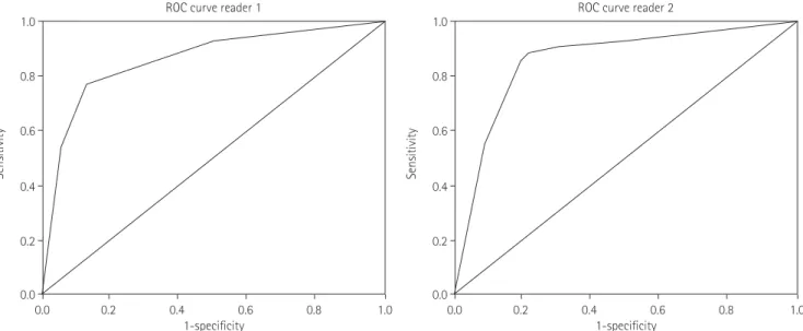
The area under the ROC curve (Az) for differentiation of Bor- rmann type by two-phased dynamic MDCT was 0.86 (p < 0.001) in both reviewers. Using an optimal threshold (Grades 3-5) for the diagnosis of Borrmann type IV distinguished from Bor- rmann type III lesions, the sensitivity and specificity were 79%
and 82% in reviewer 1 and 88% and 78% in reviewer 2, respec- tively (Figs. 1-3, Table 1).
DISCUSSION
In Western countries, more than 80% of patients with gastric cancer have AGC, and the prognosis of gastric cancer is still un- satisfactory, even after radical gastrectomy (15). Morphological classification of AGC using the Borrmann classification system (types I through IV) was described in 1926, and since then, it has been widely used (16, 17). Borrmann type IV has shown an independent poor prognostic factor, and An et al. (5) reported that the 5-year survival rate of Borrmann type IV patients is sig- nificantly lower than that of patients with other Borrmann types nite heterogeneous enhancement of the entire tumor lesion on
arterial and portal phase images; Grade 2 - probably heteroge- neous enhancement of the entire tumor lesion on arterial and portal phase images; Grade 3 - undetermined enhancement pat- tern of the entire tumor on arterial and portal phase images;
Grade 4 - probably homogeneous enhancement of the entire tu- mor lesion with a gradually layered pattern of contrast enhance- ment on arterial and portal phase images; and Grade 5 - definite homogeneous enhancement of the entire tumor lesion with a gradually layered pattern of contrast enhancement on arterial and portal phase images.
Data Analysis
SPSS software (version 15.0; Statistical Package for the Social Science, Chicago, IL, USA) was used for statistical analyses. A weighted kappa test was applied to measure interobserver vari- ability. The analysis of the correlation between the score of tumor enhancement pattern and the macroscopic Borrmann type was determined using Spearman’s correlation coefficient. For evalua- tion of the accuracy of differentiation of Borrmann type IV using MDCT in each reviewer, receiver operating characteristic (ROC) analysis was used (12-14). In all statistical analyses, differences were considered significant when the p value was less than 0.05.
RESULTS
Interobserver agreement for the CT pattern scores of Bor-
Fig. 1. ROC curve for differentiation of Borrmann type using MDCT. The Az was 0.85 (p < 0.001) in both reviewers.
Note.-ROC = receiver operating characteristic
0.0 0.0
0.2 0.2
0.4 0.4
0.6 0.6
0.8 0.8
1.0 1.0
Sensitivity Sensitivity
0.0 0.2 0.4 0.6 0.8 1.0 0.0 0.2 0.4 0.6 0.8 1.0
1-specificity 1-specificity
ROC curve reader 1 ROC curve reader 2
pattern of normal gastric wall and early gastric cancer are mostly visualized in the arterial dominant phase, and the depth of tu- mor invasion, in the case of AGC, is well verified in the equilib- rium phase (11, 21). On two-phased dynamic MDCT using the water filling method, the arterial phase has a role in detection and differentiation of early gastric cancer and AGC. Likewise, the portal phase has a role in evaluation of depth of invasion (T staging). In this study, the arterial phase almost always revealed prominent enhancement of the luminal portion of AGC, while the portal phase identified depth of tumor invasion through the deeper portion of the stomach wall; thereby, allowing the deter- mination of tumor extension by the degree and pattern of en- hancement.
Borrmann type IV AGC tends to be pathologically defined as an infiltration into the serosal layer without any distinct eleva- tion or crater, resulting in a rigid and thick gastric wall with gi- (5-year survival rate on Borrmann type IV versus other Bor-
rmann types, 27.6% versus 61.2%, respectively). At the time of diagnosis, Borrmann type IV tend to show more advanced T- stages of gastric cancer, compared to the other Borrmann types and the rate of T4 lesions are 11.9%, 6.3%, 16.4% and 30.7% in Borrmann types I, II, III and IV, respectively (6). Meanwhile, the overall incidence of Borrmann type IV is reported to represent 11-13% of all AGCs (18, 19).
Dynamic MDCT with the water filling method has several merits, such as clear depiction of the gastric wall, prominent tu- mor enhancement, inexpensive material, good toleration, and lack of overshooting artifacts by air in the lumen. Shimizu et al.
(20) reported that the detection rate of AGC is high (96.2%) when using MDCT with the water filling method and that the accuracy of the depth of invasion of gastric cancer is also high (85%). On dynamic MDCT scan with water filling, the layered Fig. 2. A 76-year-old woman with advanced gastric cancer (Borrmann type III).
A. Arterial phase of contrast enhanced MDCT scan shows inhomogeneous enhancement in mucosa and submucosa areas of the thickened pos- terior wall of the stomach (arrows).
B. Portal phase of contrast enhanced MDCT scan shows inhomogeneous enhancement in the entire posterior wall of the stomach (arrows).
C. Photomicrograph of the specimen shows mucosal ulceration (*) with tumor cell infiltration into the muscular layer (arrows) (hematoxylin and eosin stain, × 10).
D. Schematic illustration of Borrmann type III represents a protruded or elevated lesion with ulceration and diffuse margins.
Note.-MDCT = multi-detector row computed tomography C
A
D
B
Tumor infiltration of stomach wall
Mucosa
Submucosa
Muscularis
Serosa
layers, and desmoplastic infiltrations are rather prominent (Fig.
3). Monzawa et al. (24) reported that delayed enhancement is also frequently demonstrated in tumors with abundant fibrous tissue stoma. Further, Takao et al. (11) reported that 28% of AGCs exhibit gradual enhancement on triphasic spiral CT scan, and that 73% of these tumors have pathologically demonstrated marked fibrous tissue stroma (scirrhoous carcinoma, Borrmann type IV). Based on these previous reports, we hypothesized that Borrmann type IV tumors would show early enhancement of ant mucosal folds. In the early stages of tumor growth in Bor-
rmann type IV AGC, tumor cells individually invade the lamina propria without changing the mucosal surface, grow into the submucosal plane, and invade the whole stomach wall without distinct borders. Thus, Borrmann type IV lesions are not usually detected early and are generally associated with a poor progno- sis (22, 23). Especially during the spread of a tumor into the sub- mucosa and proper muscle layers of the stomach wall, tumor burdens are poorly visualized in the submucosa and muscular Fig. 3. A 55-year-old woman with advanced gastric cancer (Borrmann type IV).
A. Arterial phase of contrast enhanced MDCT scan shows homogeneous enhancement in the inner layer (arrows).
B. Portal phase of contrast enhanced MDCT scan shows homogeneous enhancement in the entire thickened stomach wall (arrows).
C. Endoscopy image demonstrates focal ulcer (arrows) at the antrum of the stomach, suggested by an endoscopist as early gastric cancer.
D. Photomicrograph of the specimen shows maintenance of stomach wall structures with minimal tumor cell infiltration, desmoplastic reaction and reactive lymphoid follicles (hematoxylin and eosin stain, × 10).
E. Schematic illustration of Borrmann type IV represents a diffuse, rather flat lesion with limited ulceration.
Note.-MDCT = multi-detector row computed tomography D
A
E
B C
Table 1. CT Scores of Each Reviewer, According to the Pathologic Results of Borrmann Type
Score* Reviewer 1 Reviewer 2
Borrmann Type III Borrmann Type IV Borrmann Type III Borrmann Type IV
1 69 4 49 3
2 9 1 33 6
3 2 1 5 1
4 10 12 7 10
5 10 25 6 23
100 43 100 43
Note.-*Scores are as follows: Grade 1 – definite heterogeneous enhancement of the entire tumor lesion on arterial and portal phase images; Grade 2 – probably heterogeneous enhancement of the entire tumor lesion on arterial and portal phase images; Grade 3 – undetermined enhancement pattern of the entire tumor on arterial and portal phase images; Grade 4 – probably homogeneous enhancement of the entire tumor lesion with a gradually layered pattern of contrast enhancement on arterial and portal phase images; Grade 5 – definite homogeneous enhancement of the entire tumor lesion with a gradually
Tumor infiltration of stomach wall
Mucosa
Submucosa
Muscularis
Serosa
image analysis. Specifically, if prediction of tumor gross appear- ance had been evaluated, the reviewers would have had a pre- conception during subsequent analysis of tumor enhancement patterns. To minimize such bias, reviewers analyzed only tumor enhancement patterns on images and were not aware of any ad- ditional information. Lastly, for endoscopists, the most impor- tant clinical issue of Borrmann type IV may be lesion delinea- tion rather than differentiation from Borrmann type III lesions;
and, the impact of our present imaging study may be limited to MDCT characterization of Borrmann type IV lesions. However, our results could be helpful for the endoscopically suspicious cases of Borrmann type IV with negative endoscopic biopsy re- sults due to little tumor burden with fibrosis at thickened gastric wall (Fig. 3).
In conclusion, two different tumor enhancement patterns of diffusely heterogeneous versus homogeneous with gradual thick- ening of enhancing portion (layered pattern) were considerably matched with Borrmann type III versus IV, respectively, on du- al-phased dynamic MDCT using the water filling method. Based on the results of this study, with considering the exception of SRCC lesions, MDCT characteristics may offer useful informa- tion for diagnosing Borrmann type IV in AGC, which could be helpful for detection of inexpectant Borrmann type IV and sus- picious Borrmann type IV with negative biopsy by endoscopy.
REFERENCES
1. Adachi Y, Oshiro T, Mori M, Maehara Y, Sugimachi K. Tu- mor size as a simple prognostic indicator for gastric carci- noma. Ann Surg Oncol 1997;4:137-140
2. Yokota T, Ishiyama S, Saito T, Teshima S, Narushima Y, Mu- rata K, et al. Lymph node metastasis as a significant prog- nostic factor in gastric cancer: a multiple logistic regres- sion analysis. Scand J Gastroenterol 2004;39:380-384 3. Michelassi F, Takanishi DM Jr, Pantalone D, Hart J, Chap-
pell R, Block GE. Analysis of clinicopathologic prognostic features in patients with gastric adenocarcinoma. Surgery 1994;116:804-809; discussion 809-810
4. Li C, Oh SJ, Kim S, Hyung WJ, Yan M, Zhu ZG, et al. Macro- scopic Borrmann type as a simple prognostic indicator in patients with advanced gastric cancer. Oncology 2009;77:
197-204 the hypervascular superficial layer, including intact mucosal lin-
ing and subsequent enhancement of relatively hypovascular deeper layers containing abundant fibrosis with scanty tumor cells, resulting in stratification of the enhancement pattern dur- ing dynamic imaging. On the other hand, Borrmann type III le- sions should have a larger cellular component with less fibrotic tumor stroma, and thus, were expected to exhibit a rather inho- mogeneous contrast enhancement of the entire tumor burden depending on the degree of tumor cell infiltration across the gastric wall. In this study, Borrmann type III and IV tumors were significantly correlated with tumor enhancement pattern (diffusely heterogeneous versus homogeneous with gradually layering pattern), and were readily differentiated by enhance- ment pattern on a two-phased dynamic CT. Based on our re- sults, the accuracy of correct diagnosis of Borrmann type IV by enhancement pattern on two-phased dynamic MDCT was sig- nificant (accuracy of 85%) (Fig. 1).
Our study had several limitations. First, we did not evaluate the effect of tumor enhancement by tumor histologic type or tu- mor grading. Borrmann type IV has a high proportion of poorly differentiated adenocarcinoma compared to Borrmann type III (6); however, Yin et al. (25) reported that an enhanced ratio on dual-phased contrast enhanced MDCT is not correlated with histo-differentiation of gastric cancer. Thus, we suggest that tu- mor grade has a minimal effect on tumor enhancement pattern on dynamic MDCT. Second, tumor histologic type, especially mucinous carcinoma and signet ring cell carcinoma, may have affected tumor enhancement pattern. Park et al. (26) reported that mucinous carcinoma has a layering pattern (62%) of en- hancement, while Lee et al. (27) reported that signet ring cell car- cinoma (SRCC) exhibits a high degree of contrast enhancement (37%). Actually we had only two cases of mucinous carcinomas (Borrmann type III, n = 1; type IV, n = 1) with corresponding enhancement patterns of our results; however, a comparison of SRCC lesions (Borrmann type III, n = 26; type IV, n = 11) showed limited specificity (sensitivity 81% and 90%, specificity 43% and 58% for reviewer 1 and 2, respectively) in the diagnosis of type IV lesions by the gradual layering pattern of mural enhancement on the dynamic CT. Considering the exceptional features of SRCC lesions, further study would be needed according to the histologic types of lesions. In addition, we did not evaluate the morphologic appearance of Borrmann types on MDCT during
rmann type 4 gastric cancer. J Korean Surg Soc 2003;64:
127-133
19. Kitamura K, Beppu R, Anai H, Ikejiri K, Yakabe S, Sugimachi K, et al. Clinicopathologic study of patients with Borrmann type IV gastric carcinoma. J Surg Oncol 1995;58:112–117 20. Shimizu K, Ito K, Matsunaga N, Shimizu A, Kawakami Y.
Diagnosis of gastric cancer with MDCT using the water- filling method and multiplanar reconstruction: CT-histo- logic correlation. AJR Am J Roentgenol 2005;185:1152- 1158
21. Mani NB, Suri S, Gupta S, Wig JD. Two-phase dynamic con- trast-enhanced computed tomography with water-filling method for staging of gastric carcinoma. Clin Imaging 2001;25:38-43
22. Maehara Y, Moriguchi S, Orita H, Kakeji Y, Haraguchi M, Korenaga D, et al. Lower survival rate for patients with carcinoma of the stomach of Borrmann type IV after gas- tric resection. Surg Gynecol Obstet 1992;175:13-16 23. Lauren P. The two histological main types of gastric carci-
noma: diffuse and so-called intestinal-type carcinoma. An attempt at a histo-clinical classification. Acta Pathol Mi- crobiol Scand 1965;64:31-49
24. Monzawa S, Omata K, Nakazima H, Yokosuka N, Ito A, Araki T. [Advanced gastric cancer: the findings of delayed phase dynamic CT and radiologic-histopathologic correla- tion]. Nihon Igaku Hoshasen Gakkai Zasshi 2000;60:508- 513
25. Yin XD, Zhao JH, Zhang L, Wang LP, Lu LQ, Wang LW, et al.
Multi-slice CT contrast-enhanced presentations of ad- vanced gastric cancer: associations with histo-differentia- tion and expression of p53 and P-glycoprotein. Chin Med J (Engl) 2008;121:2487-2491
26. Park MS, Yu JS, Kim MJ, Yoon SW, Kim SH, Noh TW, et al.
Mucinous versus nonmucinous gastric carcinoma: differ- entiation with helical CT. Radiology 2002;223:540-546 27. Lee JH, Park MS, Kim KW, Yu JS, Kim MJ, Yang SW, et al.
Advanced gastric carcinoma with signet ring cell carcino- ma versus non-signet ring cell carcinoma: differentiation with multidetector CT. J Comput Assist Tomogr 2006;30:
880-884 5. An JY, Kang TH, Choi MG, Noh JH, Sohn TS, Kim S. Bor-
rmann type IV: an independent prognostic factor for sur- vival in gastric cancer. J Gastrointest Surg 2008;12:1364- 1369
6. Kim DY, Kim HR, Kim YJ, Kim S. Clinicopathological fea- tures of patients with Borrmann type IV gastric carcinoma.
ANZ J Surg 2002;72:739-742
7. Levine MS, Kong V, Rubesin SE, Laufer I, Herlinger H. Scir- rhous carcinoma of the stomach: radiologic and endo- scopic diagnosis. Radiology 1990;175:151-154
8. Park MS, Ha HK, Choi BS, Kim KW, Myung SJ, Kim AY, et al. Scirrhous gastric carcinoma: endoscopy versus upper gastrointestinal radiography. Radiology 2004;231:421-426 9. Horton KM, Fishman EK. Current role of CT in imaging of
the stomach. Radiographics 2003;23:75-87
10. Kim AY, Kim HJ, Ha HK. Gastric cancer by multidetector row CT: preoperative staging. Abdom Imaging 2005;30:
465-472
11. Takao M, Fukuda T, Iwanaga S, Hayashi K, Kusano H, Oku- daira S. Gastric cancer: evaluation of triphasic spiral CT and radiologic-pathologic correlation. J Comput Assist To- mogr 1998;22:288-294
12. Hanley JA, McNeil BJ. A method of comparing the areas under receiver operating characteristic curves derived from the same cases. Radiology 1983;148:839-843 13. Swets JA. Measuring the accuracy of diagnostic systems.
Science 1988;240:1285-1293
14. Greiner M, Pfeiffer D, Smith RD. Principles and practical application of the receiver-operating characteristic analy- sis for diagnostic tests. Prev Vet Med 2000;45:23-41 15. Roukos DH. Current status and future perspectives in gas-
tric cancer management. Cancer Treat Rev 2000;26:243- 255
16. Borrmann R. Geschwulste Des Magens und Des Duode- nums. In Henke F, Lubarsch O. Handbuch der speziellen pathologichen anatomie und histologic Vol. IV/I. Berlin:
Springer, 1986:812-1054
17. Borchard F. Classification of gastric carcinoma. Hepato- gastroenterology 1990;37:223-232
18. Kwon SJ, Lee GJ. Clinicopathologic characteristics of Bor-
진행성 위암: 물을 경구조영제로 사용한 역동적 CT 검사에서 Borrmann 제4형과 제3형 간의 감별1
김대중
1,2· 유정식
1· 이상민
1,3· 강해윤
4· 김주희
1· 정재준
1· 김기황
1목적: 물을 경구조영제로 사용한 역동적 CT 검사가 진행성 위암 Borrmann 제4형과 제3형의 감별의 유용성을 알아보기 위해 연구를 시행하였다.
대상과 방법: 진행성 위암 환자 143명(조직학적 Borrmann 제3형과 제4형 - 100명, 43명)이 후향적 연구에 포함됐으며 모두 술전 CT 검사를 시행하였다. 영상의학과 의사 두 명이 임상 정보 없이 독립적으로 병변의 조영 양상을 다섯 단계로 나누어 평가하였다. 측정자들의 통계적 일치는 가중 kappa를 사용하였고 병변의 조영 양상 단계와 Borrmann 분류의 통 계적 관계분석은 Spearman 상관 분석을 시행하였다. 수신기작동특성곡선으로 CT를 이용한 Borrmann 제4형과 제3형 의 감별의 정확도를 알아보고자 하였다.
결과: 측정자 간의 일치도는 가중 kappa값이 0.683으로 통계적으로 충분하였다. 병변의 조영 양상의 단계와 Borrmann 분 류는 통계학적으로 유의있는 관련성을 보였다(관찰자 1, r = 0.591, p < 0.001; 관찰자 2, r = 0.616, p < 0.001). 역동적
CT 검사의 Borrmann 제4형과 제3형의 감별의 정확도는 모두 0.86(p < 0.001)의 결과를 보였으며 적정한 기준점으로
Borrmann 제4형의 진단의 민감도와 특이도는 관찰자 1에서 79%와 82%, 관찰자 2에서 88%와 78%의 결과를 보였다.
결론: 물을 경구조영제로 사용한 역동적 CT 검사는 진행성 위암의 Borrmann 제4형과 제3형 간의 감별에 높은 정확성 을 보인다.
1연세대학교 의과대학 강남세브란스병원 영상의학과, 2CHA의과학대학교 분당차병원 영상의학과,
3울산대학교 의과대학 서울아산병원 영상의학과, 4CHA의과학대학교 분당차병원 병리과