The Effects of Rituximab on Lipids, Arterial Stiffness, and Carotid Intima-Media Thickness in Rheumatoid Arthritis
The aim of the study was to examine lipid profiles, arterial stiffness (AS), and carotid intima-media thickness (cIMT), in 55 women with RA without overt cardiovascular disease (CVD) treated with rituximab (RTX).The following parameters were recorded before and 24 weeks after RTX therapy (2 infusions of 500 or 1,000 mg RTX intravenously, fortnightly):
plasma total cholesterol (TC), high-density lipoprotein cholesterol (HDL-C), low-density lipoprotein cholesterol (LDL-C), triglycerides, DAS 28-ESR, serum C-reactive protein (CRP), RF IgM, AS (SI - stiffness index, RI – reflection index) by digital volume pulse contour analysis (Micro Medical, UK), and common cIMT by high-resolution B-mode carotid ultrasound. Based on the European League Against Rheumatism (EULAR) criteria, patients were divided into two groups: 1) moderate/good response to RTX therapy after 24 weeks (41 patients, 75%), 2) no response to RTX therapy (14 patients, 25%). Effective RTX therapy resulted in 9% increase in TC, 23% increase in HDL-C and 14% decrease in atherogenic index, 57% decrease in SI and 24% decrease in RI. We observed a 9% decrease of cIMTmax at 24 weeks. The improvement of cardiovascular parameters was accompanied by statistically significant decreases of CRP, ESR, RF IgM, and DAS 28 in group 1
(P < 0.05). There were no significant changes in lipid profile, AS parameters, and cIMT in group 2. Two infusions of RTX in case of moderate/good EULAR effect of therapy exerted favorable effects on lipid profile, AS and cIMT in women with RA without overt CVD.
Keywords: Rheumatoid Arthritis; Lipids; Carotid Atherosclerosis; Arterial Stiffness;
Rituximab Diana S. Novikova,1 Tatiana V. Popkova,2
Galina V. Lukina,3 Elena L. Luchikhina,3 Dmitry E. Karateev,3 Alexander V. Volkov,1 Alexander A. Novikov,4
Elena N. Aleksandrova,4 and Evgeny L. Nasonov5
Departments of 1Instrumental and Ultrasound Diagnostics, 2Systemic Rheumatic Diseases, 3Early Arthritis, 4Immunology and Molecular Biology, V.A.
Nasonova Research Institute of Rheumatology, Moscow; 5V.A. Nasonova Research Institute of Rheumatology, Moscow, Russian Federation Received: 3 November 2015
Accepted: 18 December 2015 Address for Correspondence:
Diana S. Novikova, MD
Department of Instrumental and Ultrasound Diagnostics, V.A.Nasonova Research Institute of Rheumatology, 34a, Kashirskoe shosse, Moscow, 115522, Russian Federation Tel: +7.499-614-44-90; +7.965-165-04-21, Fax: +7.499-614-44-90 E-mail: diananovikova75@yandex.ru
http://dx.doi.org/10.3346/jkms.2016.31.2.202 • J Korean Med Sci 2016; 31: 202-207
INTRODUCTION
Rheumatoid arthritis (RA) confers a high risk of cardiovascular morbidity and mortality due to traditional cardiovascular risk factors, uncontrolled systemic inflammation, endothelial dys- function, enhanced atherosclerosis, and prothrombotic state (1-4). Proinflammatory states in RA may also damage the elas- tic structures of arteries, increase arterial stiffness, and eventu- ally contribute to peripheral overload and heart failure (5). De- teriorated elastic properties of the arterial wall and thickened carotid artery intima-media complex are independent predic- tors of cardiovascular events in the general population (6,7).
Enhanced arterial stiffness and carotid intima-media thickness (cIMT) are both associated with systemic rheumatoid inflam- mation (5,8,9), all of which add to the cardiovascular phenom- enon in RA.
Over the past decade, significant achievements in the treat- ment of RA have resulted in improved prognosis of the disease due to the prevention of irreversible changes in the musculo- skeletal system (10). Drug-induced remission and low activity of RA are also associated with lowered cardiovascular risk (11).
Rituximab, the chimerical monoclonal antibodies to CD20 of
B-lymphocytes, has been successfully used in the treatment of highly active RA, and few studies pointed to the beneficial ef- fects of the biologic therapy on systemic inflammation, endo- thelial function and arterial stiffness of patients with RA (12-14).
Pilot studies of rituximab in RA have also proved that the drug therapy ameliorates lipid profiles and variability of cardiac rhythm in RA (15-17).
The aim of the current study was to examine blood lipid pro- file, systolic and diastolic blood pressure (SBP and DBP), body mass index (BMI), blood glucose level, elastic properties of the arterial wall and cIMT in women with RA without clinical man- ifestations of cardiovascular disease during 6-month rituximab therapy.
MATERIALS AND METHODS Subjected patients
A total of 55 women with definite diagnosis of RA (18) and high disease activity (DAS28-ESR ≥ 5.2) (18) were enrolled in the study. Mean age of the patients was 50.4 years, mean disease duration ≤ 98 months, and DAS28-ESR ≤ 6.2 (Table 1). Most patients were positive for rheumatoid factor (RF) and antibod- Immunology, Allergic Disorders & Rheumatology
ies to cyclic citrullinated peptide (anti-CCP), with Steinbrock- er’s radiological stage ≥ II. Mean score of functional insufficien- cy of joints HAQ (20) was 1.77. Extraarticular features of RA were present in 26 patients (47%), including 12 (22%) with rheuma- toid nodules, 11 (20%) with neuropathy, 4 (7%) with dermato- vasculitis (erosive-ulcerative vasculitis, nail-bed hemorrhage, digital arteritis), 2 (3.5%) with pleuritis/pericarditis, 3 (5.2%) with eye lesions.
Medication
Disease-modifying anti-rheumatic drugs (DMARDs) were ad- ministered in 47 patients (86%): methotrexate-in 26 (55%), lefl- unomide-in 17 (38%), and other DMARDs (hydroxychloroquine, sulfasalazine, chlorobutine) in 4 (7%). Doses of DMARDs were constant during the 6-month study period. Nonsteroidal anti- inflammatory drugs (NSAIDs) were used by 54 patients (98%) and glucocorticoids (GC < 10 mg/day) in 44 patients (80%). In all the patients previous DMARD therapies were not efficient for decreasing DAS28-ESR or they were intolerant to two and more DMARDs. Inhibitors of tumor necrosis factor alpha were inefficient in 38% of them before the start of rituximab therapy.
Rituximab was administered intravenously, fortnightly: 500 mg in 12 patients (22%), 1,000 mg in 43 (78%) on top of DMARDs, NSAIDs and GCs. All patients were tolerant to the biological ther- apy. The clinical efficiency of the therapy was assessed by the EULAR criteria at a 6-month point (21). Based on the criteria, pa- tients were divided into two groups: group 1-41 (75%) with good and satisfactory response to the therapy and group 2-14 (25%) with no response.
Inclusion criteria
Patients fulfillingthe American College of Rheumatology (ACR) classification criteria for RA, 18-60 years ofage, and designated to receive rituximab, after evaluation by a clinicalrheumatolo- gist independent of this study.
Exclusion criteria
Age above 60 years, established coronary artery disease (angina pectoris, previous myocardial infarction), clinical manifesta- tions of chronic heart failure of New York Heart Association (NYHA) functional class II-IV, cerebral stroke, diabetes mellitus (DM), valvular heart defects, 3-4 degree of obesity, gastric and duodenal ulcers, oncological diseases, infectious diseases, thy- roid disease and refusal to participate in the study. Patients on regular drug therapies for concomitant diseases (hypotensive, hypolipidemic, and anti-arrhythmic) were also excluded.
Clinical examination
Patients were examined before and after 6 months following rituximab administration. Descriptive information about tradi- tional cardiovascular risk factors (hypertension, smoking, over- weight, family anamnesis of CVD, menopause, dyslipidemia, and hypodynamia) and aggregate cardiovascular risk by the SCORE (22) were recorded.
Biomarkers
To assess elastic properties of the arterial wall, indicators of digi- tal volumetric pulse were recorded by the Pulse Trace apparatus (Micro Medical, UK). The stiffness index (SI, m/s) and the pulse wave reflection index (RI, %) were analyzed. In cases of inability to estimate the value of arterial stiffness (“stiff arteries”), maxi- mal values for SI (20 m/s) and RI (90%) were recorded.
Duplex scanning of carotid arteries was performed the Volu- son 730 Expert device (Austria) using a transducer probe with a frequency of 7.5 MHz. The cIMT values in mm were recorded at three points from each side: at 10 mm to the carotid bulb on the common carotid artery; 5-10 mm cranially from the bulb; and at a 10 mm distance from the bifurcation on the internal carotid artery. The mean IMT was calculated based on three values from each side. Thickened IMT corresponded to values from 0.9 to 1.2 mm, and values of IMT ≥ 1.2 mm were interpreted as atherosclerotic plaques (23).
Total cholesterol (TC) values were recorded using the fer- mentative photometric test Chod-PAP, and triglycerides (TG) concentrations were identified using the fermentative colori- metric method (GPO-PAP) with glycerol-3-phosphatoxidase (variation coefficient < 5%). The values of high density lipopro- tein cholesterol (HDL-C) were estimated by the fermentative method on the biochemical analyzer “Bayer” (Germany) using
“DiaSys” reagents (Germany). The values of low density lipo- protein cholesterol (LDL-C) were calculated using the Fried- Table 1. Baseline characteristics of women with RA (n = 55)
Parameters No. of patients
Age, yr 50.4 ± 1.7
Disease duration, mo 98 ± 9
DAS 28-ESR 6.2 ± 0.1
HAQ score, points 1.77 ± 0.1
Radiological stage (I/ /II/ III/ IV), % 2/33/38/27
RF+, % 82
Anti-CCP+, % 79
Extraarticular manifestations, % 47
NSAIDs, % 98
GC, % 80
GC dose at the moment of estimation, mg per day 8.0 ± 0.5
DMARDs use, % 86
Methotrexate use, % 55
Dose of methotrexate, mg/wk 12.1 ± 0.38
Leflunomide use, % 38
Dose of leflunomide, mg/day 20
Other DMARDs, % 7
Data are presented as M ± m, unless otherwise noted. RF+, rheumatoid factor posi- tivity; anti-CCP+, positivity for anti-cyclic citrullinated peptide antibodies; NSAIDs, use of non-steroidal anti-inflammatory drugs; DAS28-ESR, disease activity score 28-joints based on erythrocyte sedimentation rate; DMARDs, disease-modifying anti-rheumatic drugs; RA, rheumatoid arthritis; GC, glucocorticoids.
wald formula with concentration of TG lower than 400 mg/dL (LDL-C = TC–[HDL-C +TG/2.19] in mmol/L). To determine the correlation between atherogenic and antiatherogenic lipopro- teins, atherogenicity index (AI) was calculated: (TC–HDL-C)/
HDL-C: the level > 4 reflects the atherogenicity of lipoproteins).
C-reactive protein (CRP) and IgM RF in blood serum were determined by highly sensitive immune nephelometric meth- od using an automatic analyzer BN 100, BEHRING (Germany).
Concentrations of anti-CCP were determined by immune en- zyme analysis with the use of the commercial kit “Axis – Shield Diagnostic Limited” (UK).
Statistical analysis
Statistical analyses were performed by SPSS 15.0 software. Dis- tribution of all parameters was checked by the Kolmogorov-Sm- irnov test. Normally distributed data were presented as M ± m.
Statistical significance of the difference between not normally distributed variables was checked by the Mann-Whitney test.
Matched samples were analyzed by the Wilcoxon Z-criterion.
Significance of the differences between percentages was tested by the chi-squared test or the Fisher’s exact test. Association between continuous variables was analyzed by the Spearman’s rank correlation coefficient. Two-sided P values < 0.05 were set as significant.
Ethics statement
The local ethics committee of the V.A.Nasonova Research Insti- tute of Rheumatology, Moscow, Russia approved the study and the patients gave a signed informed consent on inclusion (IRB No. 19-18-09-2009f).
RESULTS
At 6-month, values of DAS28-ESR and HAQ score, concentra- tions of IgM RF, CRP, and ESR were significantly lower in group 1 compared to group 2. There was only a tendency to a decrease of these indicators in group 2 (Table 2).
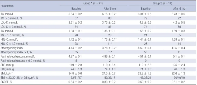
Group 1 demonstrated a significant increase in TC (9%) and HDL-C (23%) without any significant changes in LDL-C and TG, resulting in lowered atherogenicity index (by 14%) (Table 3). No statistically significant differences in fasting blood glu- cose, SBP, DBP, BMI, and the aggregate cardiovascular risk (SCORE) were noted in group 1. No significant changes in indi- cators of the lipid profile, glucose, SBP, DBP, BMI, and SCORE were recorded in group 2 (Table 3).
Rituximab therapy led to the improvement of elastic proper- ties of the arterial walls in group 1 due to the decreased stiffness in major arteries (SI–decreased by 57%) and arterioles (RI–de- creased by 24%) (Table 4). The rate of “very stiff” arteries low- ered 3.5 times in group 1, whereas no significant changes in pa- rameters of arterial stiffness were recorded in group 2 (Table 4).
Table 2. Dynamics of DAS28, HAQ score, RF, ACCP, CRP, and ESR in patients with RA on rituximab therapy
Parameters Group 1 (n = 41) Group 2 (n = 14) Baseline After 6 mo Baseline After 6 mo DAS 28-ESR 6.3 ± 0.12 3.5 ± 0.1* 6.2 ± 0.3 5.6 ± 0.3 HAQ score, points 1.78 ± 0.09 0.80 ± 0.07* 1.80 ± 0.1 1.35 ± 0.16 IgM RF, IU/mL 488 ± 138 100 ± 34* 392 ± 179 243 ± 109
Anti-CCP, U/mL 70 ± 6 67 ± 7 64 ± 12 59 ± 12
CRP, mg/L 46 ± 6 10 ± 3* 28 ± 7 24 ± 10
ESR, mm/hr 53 ± 3 21 ± 3* 60 ± 7 47 ± 6
Data are presented as M ± m. *P < 0.05 before and after rituximab therapy.
Table 3. Dynamics of lipids profile, glucose level, systolic blood pressure, diastolic blood pressure, body mass index, and SCORE in patients with RA on rituximab therapy
Parameters Group 1 (n = 41) Group 2 (n = 14)
Baseline After 6 mo Baseline After 6 mo
TC, mmol/L 5.64 ± 0.2 6.15 ± 0.2* 6.34 ± 0.5 6.73 ± 0.5
TC > 5 mmol/L, % 67 88 79 92
LDL-C, mmol/L 3.61 ± 0.2 3.73 ± 0.2 4.2 ± 0.5 4.2 ± 0.5
LDL-C > 3 mmol/L, % 74 85 74 85
TG, mmol/L 1.33 ± 0.1 1.36 ± 0.1 1.55 ± 0.2 1.59 ± 0.3
TG > 1.7 mmol/L, % 28 28 21 35
HDL-C, mmol/L 1.42 ± 0.1 1.73 ± 0.1* 1.44 ± 0.1 1.59 ± 0.1
HDL-C < 1.2 mmol/L, % 28 11* 28 14
Atherogenicity index 4.14 ± 0.2 3.78 ± 0.2* 4.52 ± 0.4 4.35 ± 0.4
Atherogenicity index > 4, % 55 31* 56 41
Fasting blood glucose, mmol/L 4.87 ± 0.1 4.96 ± 0.1 4.51 ± 0.1 5.1 ± 0.1
Fasting blood glucose > 6.0 mmol/L, % 5 2 0 0
SBP, mmHg 119 ± 2.8 119 ± 2.4 112 ± 2.8 125 ± 2.4
DBP, mmHg 74 ± 1.3 74 ± 1.3 71 ± 1.3 76 ± 1.3
BMI, kg/m2 24.8 ± 0.6 24.5 ± 0.7 23.8 ± 1.3 22.6 ± 1.3
BMI < 20/20-25/ > 25 kg/m2, % 52/31/17 50/33/17 43/36/21 30/40/40
SCORE, % 0.84 ± 0.2 0.83 ± 0.2 0.59 ± 0.2 0.61 ± 0.2
Data are presented as M ± m unless otherwise noted. *P < 0.05 before and after RTX treatment.
Finally, significant changes of cIMT were noted in group 1 at 6-month: mean cIMT decreased by 11% and maximal cIMT - by 9% (Table 4). There was a correlation between decreasing cIMT and IgM RF (r = 0.49, P < 0.001). No significant changes of cIMT were recorded in group 2.
DISCUSSION
Controlling systemic inflammation is currently viewed as an ef- fective strategy for modifying cardiovascular risk factors and preventing vascular events in RA (24). Recent cohort data sug- gest that moderate doses of glucocorticoids (above 7.5 mg/day) increase HDL in patients with RA at high risk of cardiovascular events without increasing the atherogenicity index (25). How- ever, long-term use of these powerful anti-inflammatory agents is associated with numerous side effects, limiting their cardio- vascular benefits (26). Widely used anti-TNF agents seem to be safer for controlling risk of vascular events in RA (27), though some patients fail to respond due to intolerance and secondary inefficacy. In case of anti-TNF failure, switching to other biolog- ic agents is advisable, and rituximab hold promise as a safe and effective alternative agent (28).
Rituximab therapy leads to the depletion of B-lymphocytes, which is facilitated by the system of complement, antibody-de- pendent cytotoxicity, and apoptosis (29). Recent experimental and clinical studies have demonstrated that anti-CD20 therapy modulates the course of atherosclerosis in RA (30). However, there are two different subtypes of B-lymphocytes, which have contrary effects on atherosclerosis: B1-cells are atheroprotec- tive and B2-cells represent proatherogenic family of B-cells (30). Rituximab therapy deplets proatherogenic B2-cells and preserves atheroprotective B1-cells, resulting in inactivation of B-cells and macrophages as well as lowered production of pro- inflammatory cytokines and antibodies to oxidized LDL. The selective inhibition of B-cells may also indirectly affect endo- thelial function and prevent vascular events in RA (29).
The present study demonstrated that rituximab therapy in- creases HDL-C by 22% and ameliorates lipid profile in patients with RA, decreasing the atherogenicity index. Our results are in line with the data of the pilot study by Kerekes et al. (31), who re-
ported an improved lipid profile (lowered TC and raised HDL-C in 5 female patients with RA on rituximab. Our results are also in agreement with the data by Raterman et al. (15), who reported a 9% decrease of the atherogenicity index in 49 patients after 6- month rituximab therapy. In contrast, another small study on the effects of one-year anti-TNF and rituximab therapies in pa- tients with RA, showed anti-CD20 therapy (n = 53) decreased level of atheroprotective IgM antibodies against phosphorylcho- line by 14% (32). Additionally, a one-year study of arterial stiff- ness, lipid profile and systemic inflammation in 33 patients with RA on rituximab, mostly nonrespondents to previous anti-TNF therapies, found no improvement of arterial function and even an increase of LDL-C and the atherogenicity index (33). To sum up, preliminary studies of rituximab in RA have provided contra- dictory data, which highlight multifaceted effects of anti-CD20 therapies on cardiovascular risk factors. The prognostic cardio- vascular significance of the shifts in lipid and vascular parame- ters in response to rituximab remains uncertain.
The results of the current study point to an improvement of elastic properties of the arterial wall in patients with RA without cardiovascular disease responding to 6-month rituximab thera- py. In nonrespondents, we found a tendency toward an aggra- vation of elastic properties of arteries. These findings are sup- ported by the literature data, suggesting that the dampening of systemic inflammation by rituximab is not always accompa- nied by the amelioration of arterial stiffness in RA (33). Overall, the effects of rituximab therapy on the vascular wall should be interpreted in the context of the patients’ age, preexisting car- diovascular disease, and duration and activity of RA. An impor- tant confounding factor, preventing the generalization of the results, is the difference in methodologies for assessing arterial stiffness in different studies.
Previous pilot studies have demonstrated that the depletion of B-cells in RA leads to the ameliorated endothelial function on the background of decreased disease activity (low DAS28) (12,14). A 24-month study of rituximab therapy in 38 patients with RA proved that the biologic therapy may not only improve endothelial function, but also decrease carotid IMT and reduce the progression of atherosclerosis (13). The results of our study prove that rituximab therapy may have positive effects on sub- clinical atherosclerosis as early as at 6-month, which is associ- ated the reduction of IgM RF - an independent predictor of car- diovascular mortality (34).
Overall, rituximab therapy is well tolerated, and there is no evidence of a direct cardiotoxic effect of the biologic agent (35).
One of the largest and longest studies of the safety of rituximab (3,194 patients with RA, who had received up to 17 courses of rituximab over 9.5 years) showed no increased risk of myocar- dial infarction and stroke over the time (33). The development of infusion reactions and vascular events in the course of ritux- imab therapy are rarely reported, and their association with the Table 4. Dynamics of arterial stiffness and carotid intima-media thickness in patients
with RA on rituximab therapy
Parameters Group 1 (n = 41) Group 2 (n = 14) Baseline After 6 mo Baseline After 6 mo
SI, m/s 14.7 ± 0.9 9.9 ± 0.9* 12 ± 3 12 ± 3
RI, % 76.5 ± 3 67 ± 3 68 ± 2 75 ± 2
Stiffarteries, % 52 15* 28 36
IMTmean, mm 0.77 ± 0.03 0.69 ± 0.02* 0.77 ± 0.02 0.73 ± 0.03 IMTmax, mm 0.97 ± 0.03 0.87 ± 0.02* 0.93 ± 0.04 0.92 ± 0.04 Data are presented as M ± m unless otherwise noted. *P < 0.05 before and after rituximab therapy. SI, stiffness index; RI, reflection index; IMT, intima-media thickness.
biologic therapy is debatable (37).
The mechanisms of adverse cardiovascular effects in the course of rituximab therapy in RA remain uncertain. It is possi- ble that platelet activation, overproduction of interleukine-6 and tumor necrosis factor alpha, subsequent coronary spasm, and rupture of atheroscleroticplaques underlie related vascular events. Perhaps the most important driving factor of the ad- verse vascular events is the preexisting overt or silent cardiovas- cular disease, necessitating comprehensive cardiovascular as- sessment prior to the initiation of the therapy. In our study, we enrolled patients without cardiovascular disease, and there were no major and minor side effects over the 6-month period.
In conclusion, rituximab therapy potently suppresses sys- temic inflammation, improves lipid profile and the atheroge- nicity index, decreases carotid IMT, and ameliorates elastic properties of the arterial wall in patients with RA without estab- lished cardiovascular disease. Further prospective studies are warranted to assess the effects of rituximab on cardiovascular risk factors and associated vascular events in RA.
ACKNOWLEDGMENT
The authors are grateful to Dr. Armen Yuri Gasparyan for his critical reading, comments, and editing.
DISCLOSURE
There are no potential conflicts of interest related to the con- tents of the article. No any pharmaceutical agency was involved in the study design, collection, management, analysis, interpre- tation of the data, writing of the manuscript, and decision to submit the manuscript for publication.
AUTHOR CONTRIBUTION
Conception & design of the study: Novikova DS, Popkova TV, Nasonov EL. Data collection: Novikova DS, Popkova TV, Lukina GV, Luchikhina EL, Volkov AV, Novikov AA, Nasonov EL. Statis- tical analysis: Novikova DS, Popkova TV. Interpretation and analysis of the data: Novikova DS, Popkova TV. Writing the draft and paper: Novikova DS, Popkova TV. Critical review and revi- sion: Novikova DS, Popkova TV, Lukina GV, Karateev DE, Volkov AV, Novikov AA, Aleksandrova EN, Nasonov EL. Ap- proval of the final manuscript and submission: all authors.
ORCID
Diana S. Novikova http://orcid.org/0000-0003-0840-1549 Tatiana V. Popkova http://orcid.org/0000-0001-5793-4689 Galina V. Lukina http://orcid.org/0000-0001-7958-5926 Elena L. Luchikhina http://orcid.org/0000-0002-6519-1106
Dmitry E. Karateev http://orcid.org/0000-0002-2352-4080 Alexander V. Volkov http://orcid.org/0000-0003-1784-3699 Alexander A. Novikov http://orcid.org/0000-0002-2738-2956 Elena N. Aleksandrova http://orcid.org/0000-0003-4074-5907 Evgeny L. Nasonov http://orcid.org/0000-0002-7177-2409
REFERENCES
1. Mellana WM, Aronow WS, Palaniswamy C, Khera S. Rheumatoid ar- thritis: cardiovascular manifestations, pathogenesis, and therapy. Curr Pharm Des 2012; 18: 1450-6.
2. Aviña-Zubieta JA, Choi HK, Sadatsafavi M, Etminan M, Esdaile JM, La- caille D. Risk of cardiovascular mortality in patients with rheumatoid arthritis: a meta-analysis of observational studies. Arthritis Rheum 2008;
59: 1690-7.
3. Kerekes G, Szekanecz Z, Dér H, Sándor Z, Lakos G, Muszbek L, Csipö I, Sipka S, Seres I, Paragh G, et al. Endothelial dysfunction and atheroscle- rosis in rheumatoid arthritis: a multiparametric analysis using imaging techniques and laboratory markers of inflammation and autoimmuni- ty. J Rheumatol 2008; 35: 398-406.
4. del Rincón I, Polak JF, O’Leary DH, Battafarano DF, Erikson JM, Restre- po JF, Molina E, Escalante A. Systemic inflammation and cardiovascu- lar risk factors predict rapid progression of atherosclerosis in rheumatoid arthritis. Ann Rheum Dis 2015; 74: 1118-23.
5. Ambrosino P, Tasso M, Lupoli R, Di Minno A, Baldassarre D, Tremoli E, Di Minno MN. Non-invasive assessment of arterial stiffness in patients with rheumatoid arthritis: a systematic review and meta-analysis of lit- erature studies. Ann Med 2015; 47: 457-67.
6. Van Bortel LM, Laurent S, Boutouyrie P, Chowienczyk P, Cruickshank JK, De Backer T, Filipovsky J, Huybrechts S, Mattace-Raso FU, Protoger- ou AD, et al. Expert consensus document on the measurement of aortic stiffness in daily practice using carotid-femoral pulse wave velocity. J Hy- pertens 2012; 30: 445-8.
7. Peters SA, den Ruijter HM, Bots ML, Moons KG. Improvements in risk stratification for the occurrence of cardiovascular disease by imaging subclinical atherosclerosis: a systematic review. Heart 2012; 98: 177-84.
8. Gasparyan AY, Stavropoulos-Kalinoglou A, Mikhailidis DP, Toms TE, Douglas KM, Kitas GD. The rationale for comparative studies of acceler- ated atherosclerosis in rheumatic diseases. Curr Vasc Pharmacol 2010; 8:
437-49.
9. Im CH, Kim NR, Kang JW, Kim JH, Kang JY, Bae GB, Nam EJ, Kang YM.
Inflammatory burden interacts with conventional cardiovascular risk factors for carotid plaque formation in rheumatoid arthritis. Rheuma- tology (Oxford) 2015; 54: 808-15.
10. Emery P. Rheumatoid arthritis in 2014: exciting times for RA research.
Nat Rev Rheumatol 2015; 11: 69-70.
11. Solomon DH, Reed GW, Kremer JM, Curtis JR, Farkouh ME, Harrold LR, Hochberg MC, Tsao P, Greenberg JD. Disease activity in rheuma- toid arthritis and the risk of cardiovascular events. Arthritis Rheumatol 2015; 67: 1449-55.
12. Gonzalez-Juanatey C, Llorca J, Vazquez-Rodriguez TR, Diaz-Varela N, Garcia-Quiroga H, Gonzalez-Gay MA. Short-term improvement of en- dothelial function in rituximab-treated rheumatoid arthritis patients re- fractory to tumor necrosis factor alpha blocker therapy. Arthritis Rheum
2008; 59: 1821-4.
13. Benucci M, Saviola G, Manfredi M, Sarzi-Puttini P, Atzeni F. Factors cor- related with improvement of endothelial dysfunction during rituximab therapy in patients with rheumatoid arthritis. Biologics 2013; 7: 69-75.
14. Hsue PY, Scherzer R, Grunfeld C, Imboden J, Wu Y, Del Puerto G, Nitta E, Shigenaga J, Schnell Heringer A, Ganz P, et al. Depletion of B-cells with rituximab improves endothelial function and reduces inflamma- tion among individuals with rheumatoid arthritis. J Am Heart Assoc 2014; 3: e001267.
15. Raterman HG, Levels H, Voskuyl AE, Lems WF, Dijkmans BA, Nurmo- hamed MT. HDL protein composition alters from proatherogenic into less atherogenic and proinflammatory in rheumatoid arthritis patients responding to rituximab. Ann Rheum Dis 2013; 72: 560-5.
16. Novikova DS, Popkova TV, Gerasimova EV, Novikov AA, Aleksandrova EN, Nasonov EL. Changes in heart rate, heart rate variability and QT interval in women with rheumatoid arthritis during rituximab treat- ment. Rheumatol Sci Pract 2014; 52: 270-6.
17. Provan SA, Berg IJ, Hammer HB, Mathiessen A, Kvien TK, Semb AG.
The impact of newer biological disease modifying antirheumatic drugs on cardiovascular risk factors: a 12-month longitudinal study in rheu- matoid arthritis patients treated with rituximab, abatacept and tociliziu- mab. PLoS One 2015; 10: e0130709.
18. Arnett FC, Edworthy SM, Bloch DA McShane DJ, Fries JF, Cooper NS, Healey LA, Kaplan SR, Liang MH, Luthra HS, et al. The American Rheu- matism Association 1987 revised criteria for the classification of rheu- matoid arthritis. Arthritis Rheum 1988; 31: 315-24.
19. Smolen JS, Breedveld FC, Schiff MH, Kalden JR, Emery P, Eberl G, van Riel PL, Tugwell P. A simplified disease activity index for rheumatoid ar- thritis for use in clinical practice. Rheumatology (Oxford) 2003; 42: 244- 57.
20. The Arthritis, Rheumatism, and Aging Medical Information System.
Aramis: HAQ. Available at http://aramis.stanford.edu/HAQ.html [ac- cessed on 29 October 2015].
21. van Gestel AM, Prevoo ML, van ‘t Hof MA, van Rijswijk MH, van de Putte LB, van Riel PL. Development and validation of the European League Against Rheumatism response criteria for rheumatoid arthritis. Com- parison with the preliminary American College of Rheumatology and the World Health Organization/International League Against Rheuma- tism Criteria. Arthritis Rheum 1996; 39: 34-40.
22. Conroy RM, Pyörälä K, Fitzgerald AP, Sans S, Menotti A, De Backer G, De Bacquer D, Ducimetière P, Jousilahti P, Keil U, et al. Estimation of ten-year risk of fatal cardiovascular disease in Europe: the SCORE proj- ect. Eur Heart J 2003; 24: 987-1003.
23. Touboul PJ, Hennerici MG, Meairs S, Adams H, Amarenco P, Desvarieux M, Ebrahim S, Fatar M, Hernandez Hernandez R, Kownator S, et al. Ma- nnheim intima-media thickness consensus. Cerebrovasc Dis 2004; 18:
346-9.
24. Mason JC, Libby P. Cardiovascular disease in patients with chronic in- flammation: mechanisms underlying premature cardiovascular events in rheumatologic conditions. Eur Heart J 2015; 36: 482-489c.
25. Schroeder LL, Tang X, Wasko MC, Bili A. Glucocorticoid use is associat- ed with increase in HDL and no change in other lipids in rheumatoid arthritis patients. Rheumatol Int 2015; 35: 1059-67.
26. Avouac J, Allanore Y. Cardiovascular risk in rheumatoid arthritis: effects of anti-TNF drugs. Expert Opin Pharmacother 2008; 9: 1121-8.
27. Rasch LA, Bultink IE, van Tuyl LH, Lems WF. Glucocorticoid safety for treating rheumatoid arthritis. Expert Opin Drug Saf 2015; 14: 839-44.
28. Chatzidionysiou K, van Vollenhoven RF. Rituximab versus anti-TNF in patients who previously failed one TNF inhibitor in an observational co- hort. Scand J Rheumatol 2013; 42: 190-5.
29. Novikova DS, Popkova TV, Nasonov EL. The effect of anti-B-cell therapy on the development of atherosclerosis in patients with rheumatoid ar- thritis. Curr Pharm Des 2012; 18: 1512-8.
30. Tsiantoulas D, Sage AP, Mallat Z, Binder CJ. Targeting B cells in athero- sclerosis: closing the gap from bench to bedside. Arterioscler Thromb Vasc Biol 2015; 35: 296-302.
31. Kerekes G, Soltész P, Dér H, Veres K, Szabó Z, Végvári A, Szegedi G, Sho- enfeld Y, Szekanecz Z. Effects of rituximab treatment on endothelial dys- function, carotid atherosclerosis, and lipid profile in rheumatoid arthri- tis. Clin Rheumatol 2009; 28: 705-10.
32. Ajeganova S, Fiskesund R, de Faire U, Hafström I, Frostegård J. Effect of biological therapy on levels of atheroprotective antibodies against phos- phorylcholine and apolipoproteins in rheumatoid arthritis - a one year study. Clin Exp Rheumatol 2011; 29: 942-50.
33. Mathieu S, Pereira B, Dubost JJ, Lusson JR, Soubrier M. No significant change in arterial stiffness in RA after 6 months and 1 year of rituximab treatment. Rheumatology (Oxford) 2012; 51: 1107-11.
34. Tomasson G, Aspelund T, Jonsson T, Valdimarsson H, Felson DT, Gud- nason V. Effect of rheumatoid factor on mortality and coronary heart disease. Ann Rheum Dis 2010; 69: 1649-54.
35. Gasparyan AY, Ayvazyan L, Cocco G, Kitas GD. Adverse cardiovascular effects of antirheumatic drugs: implications for clinical practice and re- search. Curr Pharm Des 2012; 18: 1543-55.
36. van Vollenhoven RF, Emery P, Bingham CO 3rd, Keystone EC, Fleisch- mann RM, Furst DE, Tyson N, Collinson N, Lehane PB. Long-term safe- ty of rituximab in rheumatoid arthritis: 9.5-year follow-up of the global clinical trial programme with a focus on adverse events of interest in RA patients. Ann Rheum Dis 2013; 72: 1496-502.
37. van Sijl AM, van der Weele W, Nurmohamed MT. Myocardial infarction after rituximab treatment for rheumatoid arthritis: Is there a link? Curr Pharm Des 2014; 20: 496-9.