ⓒ 2019 Korean Association of Physical Anthropologists
This is an Open Access article distributed under the terms of the Creative Commons Attribution Non-Commercial License(http://creativecommons.org/ licenses/by-nc/3.0) which permits unrestricted non-commercial use, distribution, and reproduction in any medium, provided the original work is properly cited.
ISSN 2671-566X (Online)·ISSN 2671-5651 (Print) Anat Biol Anthropol Vol. 32, No. 3(2019) pp.93~99
https://doi.org/10.11637/aba.2019.32.3.93
교정용 미니스크류 식립을 위한 입천장뼈 두께의 골격 분석
유선경
1, 조용화
1, 임진웅
1, 김흥중
11조선대학교 치과대학 구강해부학교실
Osteometric Analysis of Palatal Bone Thickness for Orthodontic Miniscrew Placement
Sun-Kyoung Yu
1, Yonghwa Cho
1, Jin Woong Lim
1, Heung-Joong Kim
11Department of Oral Anatomy, College of Dentistry, Chosun University
Abstract : Orthodontic miniscrews have been widely used in various areas, because they are stable, easy to use, and inexpensive. Therefore, the aims of this study are to measure the palatal bone thickness, to analysis the correlation between the size of the alveolar arch and palatal bone thickness, and to discuss the skeletal structure of the hard palate for miniscrew placement. Twenty-four dry skulls in Koreans were used. The three different horizontal reference lines were established at first premolar and second premolar, between second premolar and first molar, and first molar. And then, a total of 12 points were set up in relation to each horizontal reference line by drawing a vertical reference line perpendicular to central incisor, between central incisor and lateral incisor, lateral incisor, between lateral incisor and canine. At each point, the palatal bone thickness, the width and length of the alveolar arch, and the width and length of the incisive foramen were measured directly with the bone caliper and the digital caliper. The correlation between the width and the length of alveolar arch and the palatal bone thickness was analyzed. The mean of palatal bone thickness based on the horizontal reference line was 11.4±3.2mm in between first premolar and second premolar, 7.4±2.4mm in between second premolar and first molar, and 5.2±1.5mm in first molar, decreased posteriorly with statistically significant difference. The position between first premolar and second premolar showed a constant thickness, and thickened laterally from the median palatal suture due to the alveolar process, but no statistically significant difference. At the position between second premolar and first molar and the position in first molar, it were also constant, then became significantly thicker toward point between lateral incisor and canine due to the alveolar process and the palatal spine. The width of the alveolar arch was correlated with the length of the alveolar arch and the palatal bone thickness of between first premolar Original Article
*이 논문은 2016학년도 조선대학교 학술연구비의 지원을 받아 연구되었음.
저자 (들)는 ‘의학논문 출판윤리 가이드라인’을 준수합니다.
저자 (들)는 이 연구와 관련하여 이해관계가 없음을 밝힙니다.
Received: August 29, 2019; Revised: September 18, 2019; Accepted: Sep- tember 20, 2019
Correspondence to: 김흥중(조선대학교 치과대학 구강해부학교실) E-mail: [email protected]
서 론
위턱뼈의 입천장돌기는 위턱뼈몸통 아래에서 두껍고 수 평의 판모양으로 좌우에서 만나 정중입천장봉합(median palatine suture)을 만들며 단단입천장의 앞쪽 3/4 부분을 형 성하고, 입안과 코안의 경계를 이룬다. 이러한 입천장은 정 중입천장봉합 부위 앞쪽의 앞니구멍을 제외하고 특별한 해 부학적 구조물이 없으며, 충분한 두께와 치밀한 뼈 구조 및 치밀결합조직으로 인하여 교정용 미니스크류 식립을 위해 자주 이용된다[1-3]. 이때 입천장뼈의 두께를 최대한 활용하 여 코안으로 천공없이 적절한 초기 고정과 안정적인 유지력 을 얻기 위하여, 미니스크류의 식립 위치와 길이를 결정하기 위해 입천장뼈 두께와 밀도, 겉질뼈의 두께 및 연조직의 두 께 등에 관한 많은 연구가 진행되어 왔다[1-8].
교정용 미니스크류 식립을 위해서는 최소한 4~5mm 뼈 두께가 필요한데, 이때 입천장에서 가장 먼저 추천되는 부위 는 정중입천장봉합 부위로, 충분한 뼈 두께와 치밀한 골밀도 및 약 1mm의 균일한 입천장 점막 두께로 염증의 합병증 발 생 가능성이 적고 최대한의 유지를 얻을 수 있기 때문이다 [1,4,5,7,8]. 그 다음으로 앞니구멍에서 뒤로 3mm, 가쪽으로 5mm 부위의 첫째작은어금니와 둘째작은어금니 위치가 일 정한 뼈 두께와 앞니구멍을 통해 나오는 신경과 혈관의 손 상을 피할 수 있어서 추천된다[2,7-12]. 그러나 이러한 연구 에서 저자들은 사람 개인에 따라 입천장뼈의 두께가 계측 지점 모두에서 큰 차이가 있기 때문에 수술 전 치료 계획을 세울 때 전산화단층촬영(cone beam computed tomography) 을 통한 분석을 권장하고 있다[8,11,13].
입천장뼈의 두께는 남자에서 여자보다 유의적으로 두꺼우 며[6-8], 이틀활(alveolar arch)의 형태[11], 얼굴 형태[14] 및 부정교합의 유형[15]에 따라서도 입천장뼈의 두께가 차이가 나타난다. 또한 계측 방법의 차이도 원인이 될 수 있다[16].
전산화단층촬영의 경우, 화소(pixel) 크기와 슬라이스 두께에 따라 영상의 해상도에 큰 영향을 미치며, 환자의 잘못된 머리 위치와 흔들거림과 같은 측정 오차 역시 영향을 미쳐 영상의 분석을 어렵게 한다[8]. 가쪽머리계측X선사진(cephalogram) 의 경우 약 110%의 확대율을 보이며, 양쪽 바깥귀길의 위치
차이로 인하여 입천장뼈의 흐림(blurring)이 발생할 수 있다 [11]. 이처럼 이전 연구에서는 앞니구멍을 기준으로 수평 수 직 방향으로 일정 간격으로 영상의 단면을 얻어 하나의 지 점을 계측함으로써, 이틀활의 크기 및 입천장가시(palatal spine)와 입천장융기(palatal torus)와 같이 입체적으로 나타 나는 정상 골격 구조물이 고려되지 못하였다[7-9.13,16].
따라서 본 연구에서는 이틀활의 크기를 고려하고 진료 시 쉽게 위치 관계를 확인할 수 있도록 치아를 기준으로 하여, 한국인 마른 머리뼈에서 교정용 미니스크류 식립을 위해 추 천되는 부위의 입천장뼈의 두께와 앞니구멍의 크기를 직접 계측하고 이틀활의 크기와 입천장뼈 두께와의 상관관계를 분석하였다. 이렇게 얻어진 결과를 바탕으로 단단입천장의 골격 구조물을 비교·평가하여 미니스크류 식립 시 유용한 해부학적 자료를 제공하고자 한다.
재료 및 방법
1. 재료
본 연구는 조선대학교 의과대학 해부학교실에 교육 용 목적으로 기증되어 실습용으로 사용되는 마른 머 리뼈 32개를 사용하였으며, 사망 시 연령과 성별은 알 수 없었다. 교정용 미니스크류 식립을 위한 단단입천 장의 골격 구조물의 분석을 위하여 머리뼈의 손상 또 는 파절이 심한 경우, 3개 이상의 치아가 좌우 양쪽에 서 연속적으로 상실되어 치아의 위치 관계를 알 수 없 는 경우, 완전 무치악 및 이틀돌기(alveolar process) 가 심하게 손상되어 이틀활을 분석하기 어려운 8개의 머리뼈를 제외하고, 총 24개의 머리뼈가 분석에 사용되었다.
2. 측정
치아 위치에 따른 입천장뼈의 두께를 계측하기 위하여 총 12개의 지점을 설정하였다. 먼저 수평기준선으로 양쪽 첫 째작은어금니와 둘째작은어금니 사이를 연결한 첫 번째 기 준선(P1-P2), 둘째작은어금니와 첫째큰어금니 사이를 연결 and second premolar, but not with the length of the alveolar arch and the palatal bone thickness. These results can provide useful anatomical data on palatal bone thickness including skeletal structures of hard palate for orthodontic miniscrew placement.
Keywords : Orthodontic miniscrew, Palatal bone thickness, Alveolar arch, Incisive foramen, Palatal spine
한 두 번째 기준선(P2-M1), 첫째어금니의 중심오목(central fossa) 또는 뿌리사이중격(interradicular septum)을 연결한 세 번째 기준선(M1)을 설정하였다. 이어 수직기준선으로 안쪽앞니(CI), 안쪽앞니와 가쪽앞니 사이(CI-LI), 가쪽앞니 (LI), 가쪽앞니와 송곳니 사이(LI-C)의 수직으로 연장선을 그어 각 수평기준선과 만나는 4개의 지점을 표시하였다. 이 때 치아가 있는 경우 치아머리의 중앙 또는 접촉점을 치아 가 탈락한 경우 치아확(dental alveoli)의 중앙 또는 확사이 중격(interalveolar septum)을 기준으로 하였다(Fig. 1). 각각 의 지점에서 40mm Curved Castroviejo Bone Caliper(CL- C40C; Hu-Friedy Mfg. Co., Inc., Chicago, IL, USA)를 이용 하여 입천장뼈의 두께를 좌우 양쪽 모두에서 0.5mm 수준까 지 측정하였다.
이틀활의 크기를 분석하기 위하여 세 번째 수평 기준선의 좌우 너비와 양쪽 안쪽앞니 사이에서 세 번째 기준선까지의 앞뒤 길이를 계측하였다. 또한 앞니구멍의 앞뒤 길이와 좌 우 너비를 계측하였다(Fig. 1). 측정은 Digital Caliper(CD- 15CP, Mitutoyo Co., Japan)를 사용하여 0.1mm 수준까지 측정하였다. 해부학적 지식을 갖춘 두 명의 계측자가 동일한 기준에 따라 계측을 시행하였다.
3. 통계분석
모든 통계분석은 IBM SPSS Statistics(version 23.0, IBM Corporation, Somers, NY, USA)을 이용하여 시행하였다. 먼 저 입천장을 분석하기 위한 28개 항목에서 계측자 간의 신
뢰도(reliability)를 분석한 결과, Cronbach a는 .981로 매우 높게 나타나 계측값의 신뢰성을 얻었다. 입천장뼈 두께의 좌 우 차이를 확인하기 위하여 두 대응표본 t 검정(two-depen- dent samples t test)을 시행한 결과 12개 지점 모두에서 유의 수준 0.05에서 유의적 차이가 나타나지 않아, 좌우를 구분하 지 않고 하나의 집단으로 평가하여 분석하였다.
입천장뼈 두께의 각 치아 위치에서 지점 간의 차이와 각 지점에서 치아 위치에 따른 차이를 일원배치분산분석(one- way ANOVA)을 이용하여 통계적 유의성을 검사한 후, Scheeffe의 기준에 따라 사후검정을 시행하였다. 또한 이틀 활의 너비, 길이 및 입천장뼈 두께와의 상관관계를 분석하기 위하여 Pearson의 적률상관계수(correlation coefficient)를 구하여 통계적 유의성을 검증하였다. 모든 계측값은 평균과 표준편차로 나타내었으며, 유의수준 0.05에서 이루어졌다.
이후 통계 분석을 하여 얻어진 결과를 바탕으로 단단입천장 의 골격 구조물을 평가하여 고찰하였다.
결 과
1. 단단입천장의 뼈 두께
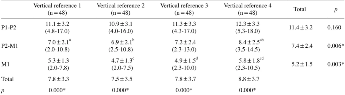
수평기준선에서 입천장뼈의 평균 두께는 P1-P2에서 11.4±3.2mm, P2-M1에서 7.4±2.4mm, M1에서 5.2±1.5 mm로 뒤쪽으로 갈수록 두께가 유의적으로 감소하였다. 각 치아 위치에서 살펴보면, P1-P2의 입천장뼈 두께는 정중입
A B
Fig. 1. Photographs showing the parameters of the palatal bone thickness, the width and length of the alveolar arch, and the width and length of the incisive foramen. A represents a total of 12 points(red dots) that are measurement points crossing between horizontal reference line(solid lines) and vertical reference line(dotted lines) at each tooth position. B represents the width and length of the alveolar arch(white dotted double arrows) and the width and length of the incisive foramen(red solid double arrows). CL, central incisor; LI, lateral incisor; C, canine; P1, first premolar; P2, second premolar; M1, first molar.
천장봉합에서 가쪽으로 일정한 두께를 보이다가 LI-C 지점 에서 두꺼워졌으나 통계적으로 유의한 차이를 보이지는 않 았다. 반면 뒤쪽의 치아 위치와 비교하여 두께도 크고, 최소 값과 최대값의 범위 및 분포의 편차가 컸다. P2-M1와 M1 위치에서는 역시 가쪽으로 일정한 두께를 보이다가, LI-C 지 점에서 두께가 유의적으로 두꺼워졌다. 수직기준선에서 입 천장뼈의 정중입천장봉합에서 가쪽으로의 평균 두께는 CI 에서부터 순서대로 7.8±3.3, 7.5±3.5, 7.8±3.7, 8.8±3.7 mm였으며, 각각의 수직기준선을 기준으로 치아 위치에 따 른 두께가 매우 유의적 차이를 보였다(Table 1).
2. 이틀활의 너비와 길이 및 단단입천장의 뼈 두께와의 상관관계
M1 기준에서 살펴 본 이틀활의 평균 너비는 49.1±2.8 mm이고 평균 길이는 23.3±2.3mm였다. 이틀활 너비와 길 이, 각 치아 위치에서 입천장뼈 두께와의 상관계수를 살펴 보면 이틀활의 너비는 이틀활의 길이 및 P1-P2의 두께와는 상관이 있는 것으로 나타났으나, 이틀활의 길이와 입천장 뼈 두께와는 상관이 없는 것으로 검증되었다(Table 2). 즉 이
틀활의 너비가 커지면 길이도 커져 큰 이틀활이 되고 이때 P1-P2의 두께도 크다고 할 수 있는데, 반면 이틀활의 길이가 길어져도 입천장 두께와는 상관이 없다고 할 수 있다. 또한 각 치아 위치에서 수평기준선에서 입천장뼈 두께는 P1-P2와 P2-M1, P2-M1와 M1 간은 높은 상관을 나타내었고, P1-P2 와 M1 역시 상관이 있는 것으로 나타났다(Table 2). 즉 입 천장뼈의 앞부위의 두께가 두꺼우면 뒤부위의 뼈 두께 역시 두껍다고 할 수 있다.
3. 앞니구멍의 크기
앞니구멍의 평균 너비는 4.2±0.9mm이고 평균 길이는 7.8±1.8mm로, 수직으로 긴 타원(oval) 형태로 나타났다 (Table 3).
Table 1. Palatal bone thickness according to tooth position at a total of 12 measurement points Vertical reference 1
(n=48) Vertical reference 2
(n=48) Vertical reference 3
(n=48) Vertical reference 4
(n=48) Total p
P1-P2 11.1±3.2
(4.8-17.0) 10.9±3.1
(4.0-16.0) 11.3±3.3
(4.3-17.0) 12.3±3.3
(5.3-18.0) 11.4±3.2 0.160
P2-M1 7.0±2.1a
(2.0-10.8) 6.9±2.1b
(2.5-10.8) 7.2±2.4
(2.3-13.0) 8.4±2.5ab
(3.5-14.5) 7.4±2.4 0.006*
M1 5.3±1.3
(2.0-7.8) 4.7±1.3c
(2.0-7.5) 4.9±1.5d
(2.3-10.0) 5.8±1.8cd
(2.3-10.5) 5.2±1.5 0.003*
Total 7.8±3.3 7.5±3.5 7.8±3.7 8.8±3.7
p 0.000* 0.000* 0.000* 0.000*
The data(in mm) are mean±SD(minimum – maximum).
Vertical reference 1, vertical extension line of central incisor; Vertical reference 2, vertical extension line of between central incisor and lateral incisor; Vertical reference 3, vertical extension line of lateral incisor; Vertical reference 4, vertical extension line of between lateral incisor and canine.
P1, first premolar; P2, second premolar; M1, first molar.
abcdIdentical superscripted letters indicate statistically significant differences among the measurement points at each tooth position.
*p-values represent the difference among the measurement points at each tooth position and among the tooth positions at each measurement point using one- way ANOVA(p<.05).
Table 2. Correlation coefficient between the width and the length of alveolar arch and the palatal bone thickness according to tooth position Alveolar arch width
(n=24) Alveolar arch length
(n=24) P1-P2(n=48) P2-M1(n=48) M1(n=48)
Alveolar arch width 1 0.46* 0.42* 0.05 0.02
Alveolar arch length 1 0.08 -0.02 -0.08
P1-P2 1 0.76** 0.48**
P2-M1 1 0.74**
*p<.05, **p<.01.
Table 3. Width and length of incisive foramen(mm)
Minimum Maximum Mean±SD(n=24) Incisive foramen width 2.4 5.7 4.2±0.9 Incisive foramen length 5.1 11.4 7.8±1.8
고 찰
교정치료에서 뼈대 고정원으로 미니스크류는 시술이 간 편하고 비용이 저렴하여 여러 부위에 다양하게 적용된다. 주 요 식립 부위로는 이틀돌기, 단단입천장, 광대아래돌기 및 위턱뼈융기가 주로 이용되는데, 그 중 입천장이 충분한 뼈 두께, 얇은 각화 연조직 및 환자의 선호로 인하여 가장 널 리 이용된다[5,16,17]. 이때 미니스크류의 적절한 식립 위 치와 길이를 결정하기 위하여 치아 이동량과 고정원으로서 생역학 이외에도 해부학적 구조물에 대한 평가가 중요하다 [2,7,8]. 따라서 본 연구에서는 입천장뼈와 앞니구멍을 직접 계측하고 이틀활의 크기와 입천장뼈 두께와의 상관관계를 분석하여, 단단입천장의 골격 구조물을 평가하여 고찰하고 자 하였다.
전산화단층촬영을 이용하여 앞니구멍을 기준으로 입천장 뼈의 두께를 분석한 이전 연구 결과, 앞니구멍으로부터 뒤쪽 및 가쪽으로 갈수록 유의적으로 감소하기 때문에 앞니구멍 을 주의한 앞쪽 입천장 부위가 안정적인 식립 위치로 추천 되었다[2,8,16]. 본 연구의 치아 위치를 기준으로 한 결과에 서도 입천장뼈의 두께는 유사한 두께로 나타났으며, P1-P2 에서 M1의 뒤쪽으로 갈수록 두께 역시 유의적으로 감소하 였다. 그러나 각 치아 위치에서 수평방향의 가쪽으로 일정 한 두께를 보이다 LI-C의 수직 연장선과 만나는 지점에서는 유의적으로 두께가 증가하여, Chhatwani 등[7]의 연구 결과 와 유사한 경향을 보였다. P1과 P2 위치는 충분한 뼈 두께를 가졌으나, 특히 가쪽으로의 두께 증가는 단단입천장이 위턱 뼈의 이틀돌기로 이어지기 때문으로 치아의 뿌리와 만날 수 있어 정중입천장봉합에서 가쪽으로 먼 지점에서의 식립은 피하여야 할 것으로 사료된다. 또한 뼈 밀도를 조사한 연구 에서도 C에서 P1 부위는 뼈가 두껍고 밀도가 높으므로 식립 시 높은 회전력(torque)을 필요로 하여 미니스크류가 파절될 수 있으므로 주의할 필요가 있다고 하였다[6].
M1 위치에서 입천장뼈의 두께는 CI에서 가쪽으로 감소하 였다가 LI-C의 수직 연장선과 만나는 지점에서는 유의적으 로 두께가 증가하였으며, King 등[9]의 연구 결과와 경향은 유사하지만 두께는 더 두껍게 나타났다. 이는 중앙에서 가까 운 CI 지점에서 두꺼운 계측값을 나타내는 경우 입천장융기 가 관찰되어 평균 두께를 증가시켰을 것으로 생각된다. 또한 LI-C 지점에서의 유의적 두께 증가는 입천장가시의 골격 구 조물로 사료된다. Yu 등[18]의 연구 결과에서 입천장가시는 둘째큰어금니(M2)에서 시작하여 M1에서 끝나는 약 1cm의 뼈 융기 구조물로 대략 85%에서 나타나며, 입천장가시의 양 쪽으로 안쪽과 가쪽 고랑을 형성하여 입천장에 주요 신경과 혈관을 공급하는 큰입천장신경과 동맥이 주행한다고 보고
하였다. 따라서 전산화단층촬영 영상에서 한 단면을 잡아 계 측하여 해당 부위에서 두께가 두껍게 나왔다면, 정상 구조물 인 입천장가시 및 그 주위 신경과 혈관의 주행을 주의하여 야 한다.
본 연구에서 마른 머리뼈의 직접 계측으로 인하여 정중입 천장봉합의 중심선에서의 두께는 계측하지 못하였지만, 각 치아 위치에서 봉합 주위 CI의 수직 연장선과 만나는 지점 에서의 두께 역시 뒤쪽으로 갈수록 감소하였고 전체적으로 다른 지점과 비교하여 두꺼웠다. 이는 이전 연구에서 보고 된 것처럼 코안쪽으로 코중격을 이루기 위하여 코능선(nasal crest)이 봉합의 위쪽으로 솟아 있기 때문이다[8]. 또한 입 천장 점막(palatal mucosa)을 조직학적으로 분석한 연구 결 과에 따르면 입천장쪽 잇몸과 정중입천장봉합 부위에서는 치밀결합조직으로 구성된 고유판으로 주로 이루어졌으며, 성긴 지방조직으로 구성된 점막밑층은 상대적으로 적었다 [19]. 따라서 정중입천장봉합 부위는 이전 연구에서처럼 미 니스크류를 식립하기에 적합한 위치이다[1,4,5,7,8]. 하지만 큰어금니 부위에서 봉합 가까운 곳에 샘조직이 집중되어 있 고[19], 본 연구에서 최소값이 2mm로 매우 작게 나타나므 로 수술 전 평가가 중요하다.
본 연구에서 이틀활의 크기와 입천장뼈 두께와의 상관관 계를 분석한 결과, 이틀활의 너비가 커지면 길이도 커져 큰 이틀활이 되고 이때 P1-P2의 두께도 두꺼웠지만, 반면 이틀 활의 길이는 입천장 두께와는 상관이 없었다. 또한 각 치아 위치에서 입천장뼈 두께는 앞부위가 두꺼우면 뒤쪽 부위 역 시 뼈 두께가 두껍다고 할 수 있었다. 이러한 결과는 이틀활 이 더 큰 남자에게서 이전 연구 결과에서처럼 입천장뼈가 더 두껍게 나타나는 것을 확인시켜 줄 수 있다[6-8]. 더불어 다른 연구에서 깊은 입천장, V 형태의 이틀활, 긴 얼굴형 및 수직 골격성 개방교합의 1급 부정교합에서 더 얇은 입천장 뼈의 두께를 보였다[11,14,15]. 따라서 이후 연구에서 이틀 활의 크기와 형태에 따른 입천장의 전체 뼈와 겉질뼈의 두 께를 비교하여 분석할 필요가 있다.
앞니구멍은 앞니관(incisive canal)이 입천장에 열리는 구 조물로, 몽골인에서 전산화단층촬영의 결과 평균 너비 3.5 mm 평균 길이 2.8mm의 수평으로 긴 타원(oval) 형태이며 남자에게서 유의적으로 크게 나타났다[20]. 반면 본 연구에 서는 평균 너비 4.2mm 평균 길이 7.8mm로 수직으로 긴 타원 형태로 나타났는데, 이러한 차이는 인종과 계측 지점 의 차이 및 본 연구의 적은 표본 수를 고려해 볼 수 있다. 이 러한 앞니구멍은 실제 환자의 입안에서 앞니유두(incisive papilla)와 위치적으로 가깝지만 실제 관은 더 위쪽 뒤쪽에 위치하여, 앞니관은 P1-P2에서 60% P2에서 13% 정도로 위 치하게 된다[2,11]. 또한 정중시상단면에서 관의 주행 경로
분류에서도 앞니관이 앞니구멍에서 비스듬하게 뒤위쪽으 로 열리는 경우가 대략 40% 정도로 나타났다[21]. 따라서 이 지점에서 입천장뼈의 두께는 8.6mm이지만 앞니관의 직 경은 2.5mm로 실제 미니스크류 식립 시 얻을 수 있는 뼈의 두께는 줄어들게 된다[22]. 즉 입천장 앞쪽 부위가 상대적으 로 안전한 부위이지만 앞니관의 비스듬한 주행과 수직으로 긴 앞니구멍의 열림으로 인하여 주의가 요구된다.
본 연구에서 적은 표본 수 및 마른 머리뼈의 직접 계측 시 기구 접근의 어려움으로 계측 시 오차가 발생하였다. 그러나 진료 시 쉽게 위치 관계를 확인할 수 있도록 치아를 기준으 로 하여 입천장뼈 두께를 계측하고 이틀활의 크기와의 상관 관계를 분석하여, 입천장가시와 입천장융기와 같이 입체적 으로 나타나는 정상 구조물을 평가하여 고찰하였다. 이러한 결과는 교정용 미니스크류 식립 시 입천장뼈 두께를 포함하 여 단단입천장의 골격 구조물에 대한 유용한 해부학적 자료 를 제공할 수 있다.
REFERENCES
1. Wehrbein H, Merz BR, Diedrich P. Palatal bone support for orthodontic implant anchorage - a clinical and radiological study. Eur J Orthod. 1999;21:65-70.
2. Baumgaertel S. Quantitative investigation of palatal bone depth and cortical bone thickness for mini-implant placement in adults. Am J Orthod Dentofacial Orthop. 2009;136:104-8.
3. Lai RF, Zou H, Kong WD, Lin W. Applied anatomic site study of palatal anchorage implants using cone beam com- puted tomography. Int J Oral Sci. 2010;2:98-104.
4. Kim HJ, Yun HS, Park HD, Kim DH, Park YC. Soft-tissue and cortical-bone thickness at orthodontic implant sites. Am J Orthod Dentofacial Orthop. 2006;130:177-82.
5. Park JT, Jeong RR, Kim KT, Kim SB, Hu KS, Kim HJ, et al.
Maxillary soft tissue and cortical bone thickness for mini-im- plant placement. Korean J Phys Anthropol. 2008;21:215-24.
6. Yadav S, Sachs E, Vishwanath M, Knecht K, Upadhyay M, Nanda R, et al. Gender and growth variation in palatal bone thickness and density for mini-implant placement. Prog Or- thod. 2018;19:43.
7. Chhatwani S, Rose-Zierau V, Haddad B, Almuzian M, Kirschneck C, Danesh G. Three-dimensional quantitative assessment of palatal bone height for insertion of orthodon- tic implants - a retrospective CBCT study. Head Face Med.
2019;15:9.
8. Kang S, Lee SJ, Ahn SJ, Heo MS, Kim TW. Bone thickness of the palate for orthodontic mini-implant anchorage in adults. Am J Orthod Dentofacial Orthop. 2007;131(4 Sup-
pl):S74-81.
9. King KS, Lam EW, Faulkner MG, Heo G, Major PW. Verti- cal bone volume in the paramedian palate of adolescents: a computed tomography study. Am J Orthod Dentofacial Or- thop. 2007;132:783-8.
10. Bernhart T, Vollgruber A, Gahleitner A, Dörtbudak O, Haas R. Alternative to the median region of the palate for place- ment of an orthodontic implant. Clin Oral Implants Res.
2000;11:595-601.
11. Kim YJ, Lim SH, Gang SN. Comparison of cephalometric measurements and cone-beam computed tomography-based measurements of palatal bone thickness. Am J Orthod Den- tofacial Orthop. 2014;145:165-72.
12. AlSamak S, Gkantidis N, Bitsanis E, Christou P. Assessment of potential orthodontic mini-implant insertion sites based on anatomical hard tissue parameters: a systematic review. Int J Oral Maxillofac Implants. 2012;27:875-87.
13. Wang M, Sun Y, Yu Y, Ding X. Evaluation of palatal Bone thickness for insertion of orthodontic mini-implants in adults and adolescents. J Craniofac Surg. 2017;28:1468-71.
14. Wang Y, Qiu Y, Liu H, He J, Fan X. Quantitative evaluation of palatal bone thickness for the placement of orthodontic miniscrews in adults with different facial types. Saudi Med J.
2017;38:1051-7.
15. Suteerapongpun P, Wattanachai T, Janhom A, Tripuwabhrut P, Jotikasthira D. Quantitative evaluation of palatal bone thick- ness in patients with normal and open vertical skeletal con- figurations using cone-beam computed tomography. Imaging Sci Dent. 2018;48:51-7.
16. Ryu JH, Park JH, Vu Thi Thu T, Bayome M, Kim Y, Kook YA. Palatal bone thickness compared with cone-beam com- puted tomography in adolescents and adults for mini-implant placement. Am J Orthod Dentofacial Orthop. 2012;142:207- 17. Gündüz E, Schneider-Del Savio TT, Kucher G, Schnei-12.
der B, Bantleon HP. Acceptance rate of palatal implants:
a questionnaire study. Am J Orthod Dentofacial Orthop.
2004;126:623-6.
18. Yu SK, Lee MH, Park BS, Jeon YH, Chung YY, Kim HJ.
Topographical relationship of the greater palatine artery and the palatal spine. Significance for periodontal surgery. J Clin Periodontol. 2014;41:908-13.
19. Yu SK, Lim JW, Cho YH, Kim HJ. Anatomical assessment of the palatal mucosa for connective tissue grafts. Oral Biol Res. 2018;42:156-62.
20. Al-Amery SM, Nambiar P, Jamaludin M, John J, Ngeow WC. Cone beam computed tomography assessment of the maxillary incisive canal and foramen: considerations of an- atomical variations when placing immediate implants. PLoS One. 2015;10:e0117251.
21. Song WC, Jo DI, Lee JY, Kim JN, Hur MS, Hu KS, et al. Microanatomy of the incisive canal using three-di- mensional reconstruction of microCT images: an ex vivo study. Oral Surg Oral Med Oral Pathol Oral Radiol Endod.
2009;108:583-90.
22. Henriksen B, Bavitz B, Kelly B, Harn SD. Evaluation of bone thickness in the anterior hard palate relative to midsag- ittal orthodontic implants. Int J Oral Maxillofac Implants.
2003;18:578-81.
간추림 : 교정치료에서 뼈대 고정원으로 미니스크류는 시술이 간편하고 비용이 저렴하여 여러 부위에 다양하게 적용되
는데, 그 중 입천장이 충분한 뼈 두께, 얇은 각화 연조직 및 환자의 선호로 인하여 가장 널리 이용된다. 따라서 본 연구에 서는 교정용 미니스크류 식립을 위해 추천되는 부위의 입천장 뼈 두께를 계측하고 이틀활의 크기와의 상관관계를 분석
하여, 입천장가시와 입천장융기와 같이 입체적으로 나타나는 정상 구조물을 평가하여 고찰하고자 한다. 한국인 마른 머
리뼈 24개를 사용하였다. 치아 위치에 따른 입천장 뼈의 두께를 계측하기 위하여 첫째작은어금니와 둘째작은어금니 사
이, 둘째작은어금니와 첫째큰어금니 사이, 첫째어금니에서 3개 수평기준선을 설정하고, 이어 수직기준선으로 안쪽앞니, 안쪽앞니와 가쪽앞니 사이, 가쪽앞니, 가쪽앞니와 송곳니 사이의 수직으로 연장선을 그어 각 수평기준선과 만나는 총 12 개의 지점을 정하였다. 각각의 지점에서 입천장 뼈의 두께와 이틀활의 너비와 길이 및 앞니구멍의 너비와 길이를 Bone caliper와 Digital caliper를 이용하여 직접 계측하였다. 이틀활의 너비, 길이 및 입천장 뼈 두께와의 상관관계를 분석하였 다. 입천장 뼈의 평균 두께는 첫째작은어금니와 둘째작은어금니 사이부터 순서대로 11.4±3.2, 7.4±2.4, 5.2±1.5mm로
뒤쪽으로 갈수록 두께가 유의적으로 감소하였다. 첫째작은어금니와 둘째작은어금니 사이는 정중입천장봉합에서 가쪽으
로 일정한 두께를 보이다가 가쪽앞니와 송곳니 사이 지점에서 이틀돌기로 인하여 두께가 두꺼워졌으나 통계적으로 유의
한 차이를 보이지는 않았다. 둘째작은어금니와 첫째큰어금니 사이와 첫째큰어금니 위치에서는 가쪽으로 일정한 두께를
보이다가, 가쪽앞니와 송곳니 사이 지점에서 두께가 각각 이틀돌기와 입천장가시로 인하여 유의적으로 두꺼워졌다. 이
틀활의 너비는 이틀활의 길이 및 첫째작은어금니와 둘째작은어금니 사이의 입천장 뼈 두께와는 상관이 있는 것으로 나 타났으나, 이틀활의 길이와 뼈 두께와는 상관이 없었다. 이러한 결과는 교정용 미니스크류 식립 시 입천장 뼈 두께를 포 함하여 단단입천장의 골격 구조물에 대한 유용한 해부학적 자료를 제공할 수 있다.
찾아보기 낱말 : 교정용 미니스크류, 입천장 뼈 두께, 이틀활, 앞니구멍, 입천장가시