Received December 12, 2016, Revised March 23, 2017, Accepted for publication April 18, 2017
Corresponding author: Yu Sung Choi, Department of Dermatology, Ulsan University Hospital, University of Ulsan College of Medicine, 877 Bangeo- jinsunhwan-doro, Dong-gu, Ulsan 44033, Korea. Tel: 82-52-250-7090, Fax: 82-52-250-7155, E-mail: uuhderma@daum.net
This is an Open Access article distributed under the terms of the Creative Commons Attribution Non-Commercial License (http://creativecommons.
org/licenses/by-nc/4.0) which permits unrestricted non-commercial use, distribution, and reproduction in any medium, provided the original work is properly cited.
Copyright © The Korean Dermatological Association and The Korean Society for Investigative Dermatology
Ann Dermatol Vol. 30, No. 1, 2018 https://doi.org/10.5021/ad.2018.30.1.1
ORIGINAL ARTICLE
Treatment of Melasma with Pulsed-Dye Laser and
1,064-nm Q-Switched Nd:YAG Laser: A Split-Face Study
Sook Hyun Kong, Ho Seok Suh, Yu Sung Choi
Department of Dermatology, Ulsan University Hospital, University of Ulsan College of Medicine, Ulsan, Korea
Background: Melasma is an acquired pigmentary disorder that is often therapeutically challenging. Recent evidence suggests that vascular abnormalities are involved in melasma pathogenesis. Pulsed-dye laser (PDL) is considered as stand- ard therapy for vascular lesions. Objective: To assess the effi- cacy of PDL combined with low-fluence Q-switched Nd:YAG laser (QSNY) in the treatment of melasma.
Methods: Seventeen melasma patients were enrolled in this study. All subjects were treated with a total of nine QSNY treatment sessions at one-week intervals. Three sessions of PDL were additionally performed immediately after QSNY treatment on the half of the face at baseline, week 4, and week 8. The melasma area and the severity index (MASI) score was calculated at the baseline, one week after the last treatment (week 9), as well as at the follow-up 8 weeks after the last treatment (week 16). Dermoscopic images at the baseline were classified as to whether the visibly widened ca- pillaries were detected or not. Results: MASI scores on the PDL+QSNY and QSNY side decreased significantly during the study period. There was no significant difference in the MASI score change between both sides in all periods.
However, seven patients who had visibly widened capil- laries on dermoscopy showed significant difference in both sides in terms of changes in the MASI score during treatment.
Conclusion: PDL combined with QSNY may be considered as a safe and effective treatment for melasma patients who show visibly widened capillaries on dermoscopy. (Ann Dermatol 30(1) 1∼7, 2018)
-Keywords-
Dermoscopy, Melasma, Pulsed dye lasers, Q-switched la- sers
INTRODUCTION
Melasma is an acquired pigmentary disorder presenting brownish patches on the face, most commonly observed in females with a darker skin type. The major etiological factors are genetic influences, exposure to ultraviolet (UV) radiation, and sex hormones1, although the exact patho- genesis of melasma is not fully elucidated. The patho- physiology of melasma is believed to involve excess pro- duction of melanin or an increase in the number of mela- nocytes in the skin. Laser toning using low-fluence 1,064 nm Q-switched Nd:YAG laser (QSNY) has been an effec- tive treatment option in Asia, but monotherapy treatment efficacies were variable2-4.
Recent studies have suggested that interactions between the altered cutaneous vasculature and melanocytes have an influence on hyperpigmentation development in mel- asma, which demonstrated that greater vascularity is one of the major findings5,6. Based on this finding, it has been investigated whether the treatment targeting blood vessels is beneficial in the treatment of melasma7-11. Pulsed-dye laser (PDL) is considered a gold standard laser therapy for cutaneous vascular lesions, which has been shown to be an effective treatment option in combination with pig- ment-targeted modalities for melasma patients in several studies7-10. Also, it has been reported that the copper bro-
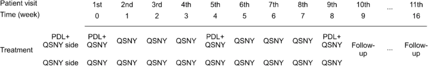
Fig. 1. Schematized summary of treatment schedule. PDL: pulsed-dye laser, QSNY: Q-switched Nd:YAG laser.
mide laser, oral and topical tranexamic acid are effective in the treatment of melasma7-11.
Dermoscopy is a useful tool for detecting clinically in- visible or neglectable vessel changes effectively. We thought these findings may be helpful for the candidate se- lection for treatment including vascular-target therapy.
Several studies have been reported the effect of PDL in melasma treatment, but there has been no comparative study on the therapeutic effect of PDL between melasma patients who show dermoscopic finding of visibly wid- ened capillaries and who do not. Therefore, this study was conducted to investigate whether PDL provides additional improvements when combined with QSNY for melasma treatment, using dermoscopy to find subtle vascular struc- tures and evaluated whether these findings showed differences.
MATERIALS AND METHODS
Study subjects
We conducted a randomized, single-blind, split-face pro- spective study between December 2015 and April 2016.
Seventeen Korean women with melasma aged 18 years or above were included in the present study. The Fitzpatrick skin type was determined for all patients. The exclusion criteria included underlying skin diseases, use of iso- tretinoin or contraceptive pills during the past six months, pregnancy or breastfeeding, topical bleaching agent usage within one month before recruitment, employment of chemical peels, laser therapy or intense pulsed light treat- ment also within six months at the study’s beginng, and sunlight or UV light exposures. This clinical study was ap- proved by the Institutional Review Board of Ulsan University Hospital (Ulsan, Korea) (IRB no. 2014-03-006) and written informed consent was obtained from all subjects.
Treatment methods
Fig. 1. shows a schematized summary of the treatment schedule. A total of 17 patients were treated, and each side of the face was randomly allocated to either QSNY or
PDL+QSNY treatment. All subjects were treated with a total of nine full-face QSNY treatment sessions (Cosjet TRⓇ; Won Technology, Daejeon, Korea) at one-week intervals.
On the PDL+QSNY side of the face, additional PDL treat- ments (Cynergy MultiplexTM; Cynosure, Westford, MA, USA) were performed with a total of three sessions at baseline, week 4, and week 8. The patients were treated first with low-fluence QSNY on the entire face and sub- sequent PDL treatment was performed immediately after QSNY treatment on the half of the face with a 10% over- lap of treatment spots to target the vessels. Both laser ther- apy were stopped when mild erythema appeared around the melasma lesion. No anesthesia was necessary prior to the treatment. The assignment to treatments was made on- ly when a subject attended the first therapy session. Laser parameters for QSNY settings were a 1,064 nm wave- length, 7 mm spot size, and a fluence of 1.2∼2.0 J/cm2 at 10 Hz (five to seven passes), whereas the PDL settings were of a 595 nm wavelength, seven mm spot size, a 20-milisecond pulse duration, as well as a 7∼8 J/cm2 flu- ence (two to three passes). The patients were not allowed to use any other form of treatment or functional cosmetics and they were also instructed to avoid sun exposure and to apply a broad-spectrum sunscreen.
Efficacy assessments
Before each session, one week after the last treatment (week 9), and follow-up visits after eight weeks since the last treatment (week 16), digital photographic documen- tation (Nikon D7000; Nikon Corporation, Tokyo, Japan) was obtained under the same conditions. Dermoscopic images were also obtained using dermoscopy equipment (X20) (DermLite II Pro; 3Gen, San Juan Capistrano, CA, USA). Calibration of the different parameters (luminosity, contrast, white balance, color balance) was performed pri- or to starting a series of images. To visualize vessels, the glass lens was carefully placed on the lesion; also, mini- mal pressure was applied. Based on the fact whether the visibly widened capillaries were detected or not in dermo- scopic images, estimations were made. Two independent dermatologists evaluated the melasma area and severity
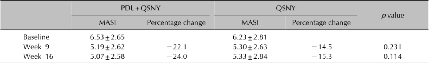
Table 1. The changes of the MASI scores during the study period (n=17)
PDL+QSNY QSNY
p-value
MASI Percentage change MASI Percentage change
Baseline 6.53±2.65 6.23±2.81
Week 9 5.19±2.62 −22.1 5.30±2.63 −14.5 0.231
Week 16 5.07±2.58 −24.0 5.33±2.84 −15.3 0.114
Values are presented as mean±standard deviation or percentage. MASI: melasma area and the severity index, PDL: pulsed-dye laser, QSNY: Q-switched Nd:YAG laser.
Fig. 2. Dermoscopic image of a patient who showed visibly widened capillaries along with pigmentation of a pseudonetwork pattern at the first visit.
index (MASI) scores, using the photographs taken at base- line, week 9 and 16. The mean value of the data obtained from each half of the face was calculated and the mean percentage change was compared. Hemi-MASI score was used because of the study design of split-face, and it was calculated based on the percentage of the involved area (A=0∼6: 0=0%, 1=1%∼9%, 2=10%∼29%, 3=30%∼
49%, 4=50%∼69%, 5=70%∼89%, 6=90%∼100%), darkness of pigment (D=0∼4: 0=absent or normal skin color without evidence of hyperpigmentation, 1=slight visible hyperpigmentation, 2=mild visible, 3=marked, 4=severe), and homogenicity of the hyperpigmentation (H=0∼4: 0=minimal, 1=slight, 2=mild, 3=marked, 4=severe).
Hemi-MASI score=forehead, 0.15×(D+H)×A+malar, 0.30×(D+H)×A+chin, 0.05×(D+H)×A.
Safety assessments
At each follow-up visit, adverse events were also evaluated.
Patients were asked to reports side effects such as eryth- ema, burning, swelling, blistering, crust, hyperpigmen- tation or hypopigmentation, atrophy, and others during and after treatment. The dermatologist categorized the de- gree of erythema and burning into four grades: none, mild, moderate, and severe.
Statistical analysis
All analyses were performed using SPSS Statistics ver. 21.0 (IBM Co., Armonk, NY, USA). Continuous variables were described as means and standard deviations (SD). Data were analyzed using Mann-Whitney U-test. p<0.05 was considered statistically significant.
RESULTS
Demographics
Seventeen melasma patients were enrolled in this study.
The Fitzpatrick skin type was III in eight, IV in five pa- tients, and V in four patients. The mean age was 41 years (range, 31∼53 years). The disease duration ranged from 3
to 15 years (mean±SD, 12.47±5.62 years).
Treatment efficacy assessments
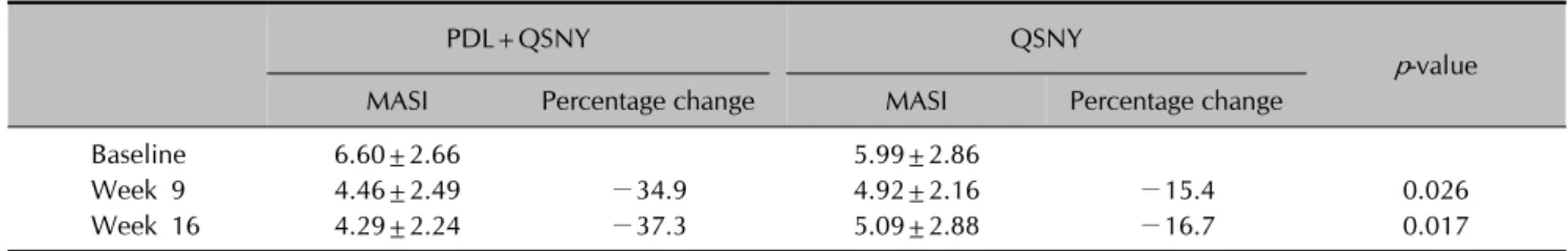
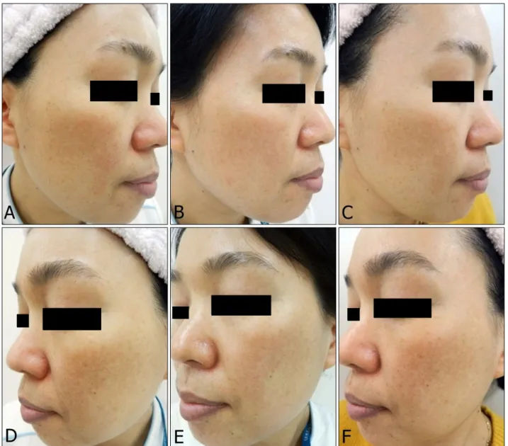
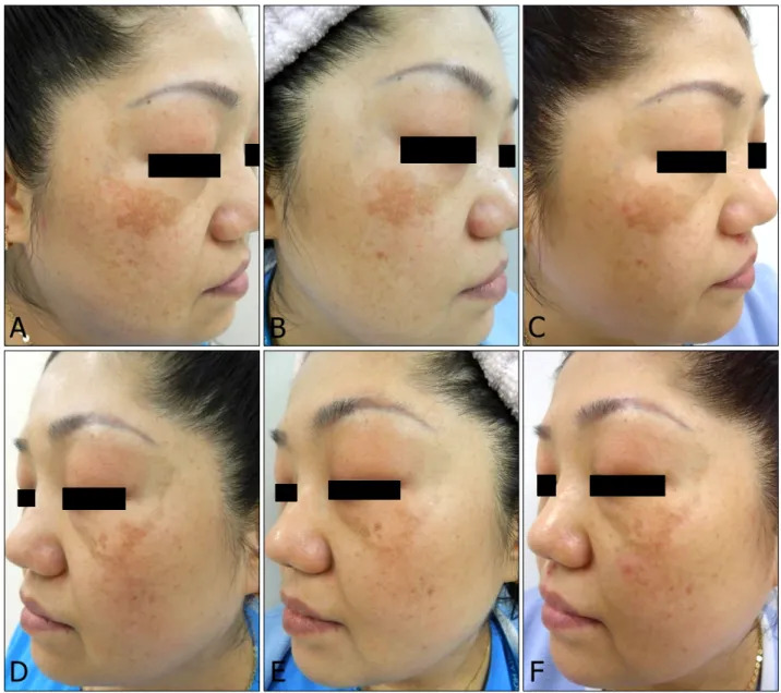
MASI scores on the PDL+QSNY and QSNY side de- creased significantly during the study period. There was no significant difference in the MASI score change be- tween both sides in all periods (Table 1). Among 17 pa- tients, seven (41.2%) showed visibly widened capillaries in dermoscopic images (Fig. 2) whose MASI scores of both PDL+QSNY and QSNY sides were decreased significantly during the study period. These patients showed significant difference in both sides in terms of changes in the MASI score during treatment (Table 2). However, patients who did not show visibly widened capillaries showed no sig- nificant difference in the MASI score change between both sides (Table 3). Fig. 3 illustrated the clinical appearance of one case among the seven patients; this subject showed visibly widened capillaries in dermoscopic images at baseline.
Safety assessments
A majority of the patients had a mild burning sensation
Table 2. The changes of the MASI scores of the patients who showed visibly widened capillaries on dermoscopy (n=7)
PDL+QSNY QSNY
p-value
MASI Percentage change MASI Percentage change
Baseline 6.60±2.66 5.99±2.86
Week 9 4.46±2.49 −34.9 4.92±2.16 −15.4 0.026
Week 16 4.29±2.24 −37.3 5.09±2.88 −16.7 0.017
Values are presented as mean±standard deviation or percentage. MASI: melasma area and the severity index, PDL: pulsed-dye laser, QSNY: Q-switched Nd:YAG laser.
Table 3. The changes of the MASI scores of the patients who didn’t show visibly widened capillaries on dermoscopy (n=10)
PDL+QSNY QSNY
p-value
MASI Percentage change MASI Percentage change
Baseline 6.48±2.80 6.40±2.91
Week 9 5.71±2.72 −13.3 5.57±3.00 −13.8 0.971
Week 16 5.62±2.77 −14.7 5.49±2.96 −14.3 0.853
Values are presented as mean±standard deviation or percentage. MASI: melasma area and the severity index, PDL: pulsed-dye laser, QSNY: Q-switched Nd:YAG laser.
tolerable during laser therapy. A mild erythema was ob- served after the treatment, lasting for approximately one to two hours in most patients. Pigmentary adverse events as- sociated with the PDL+QSNY side were observed in two out of 17 subjects. One patient whose Fitzpatrick skin type was IV showed focal purpura observed in the PDL+QSNY side at the last treatment, which disappeared spontaneously after 1 week. Postinflammatory hyperpig- mentation (PIH) developed after two months since the last treatment. Another patient whose Fitzpatrick skin type was V exhibited a rebound hyperpigmentation (RH) without purpura on PDL+QSNY side at the follow-up visit after 8 weeks since the last treatment (Fig. 4).
DISCUSSION
Melasma is a chronic distressing condition and often ther- apeutically challenging. Various treatments, including whitening or bleaching agents (e.g., hydroquinone, aze- laic acid), chemical peels (e.g., glycolic, β-hydroxyl, and trichloroacetic acids), topical steroids, as well as laser treatment demonstrated some therapeutic effect but were often unsuccessful with regard to refractory melasma12,13. A large-spot-size, low-energy-mode of the 1,064 nm QSNY is a popular method of melasma therapy in Asia.
The QSNY has a longer wavelength that can penetrate deeper to target both mixed and dermal melasma. It can also target lesional melanocytes and causes of subcellular damage to its melanin particles precisely14,15. Moreover,
QSNY may also produce nonspecific dermal wounds and induce inflammation, facilitating a migration of melano- phages16. However, these effects were transient and rever- sible, with a high recurrence rate9-11.
The pathogenesis of melasma is not completely under- stood, which renders treatment more complicated. There are many possible contributing factors which include ge- netic influences, exposure to UV radiation, female sex hormones, thyroid dysfunction, cosmetics, and drugs1. Although the cause of melasma has not been fully com- prehended, it has recently been suggested that vascular endothelial growth factor (VEGF) and skin vascularization might play a role in the melasma pigmentation2,3. Human melanocytes may respond to angiogenic factors because normal human melanocytes show functional VEGF receptors.
Therefore, VEGF may have a direct influence on melano- cyte behavior through its receptors2,3. It is also known to stimulate the release of arachidonic acid as well as the phosphorylation and activation of cytosolic phospholipase A2. It is possible that the resulting metabolites from the arachidonic acid pathway affect melanogenesis2,3.
Cho et al.2 reported that only the area affected by mel- asma presents a pronounced vascular change, showing significant increases in the number and size of dermal blood vessels. Moreover, they considered that the number of vessels is positively related to the degree of pigmen- tation. Park et al.17 demonstrated that the degree of eryth- ema is positively correlated with that of pigmentation in a melasma lesion. Thus, vascularity increases may play an
Fig. 3. Serial clinical photographs of a melasma patient with visibly widened capillaries on dermoscopy. Comparing the melasma area and severity index (MASI) score reduction from the baseline, pulsed-dye laser (PDL)+Q-switched Nd:YAG laser (QSNY) side (A, B, and C) revealed a significantly greater reduction of the MASI scores at week 9 and 16 than the QSNY side (D, E, and F).
The PDL+QSNY side maintained a MASI score reduction at week 16 (C), while it increased for the QSNY side (F).
important role in the melasma pathogenesis and the cause of PIH after laser therapy by an increase of cytokines and soluble factors released from proliferated vessels17. From this point of view, laser targeting blood vessel might decrease the stimulation of melanocytes and prevent relapse. PDL targets oxyhemoglobin within cutaneous vas- culatures and it has been widely employed to treat diverse vascular disorders. By targeting not only melanin but also vascularization, PDL might provide—in combination with QSNY—an effective and successful therapeutic approach for melasma.
In our split-face study, the MASI scores of both sides de- creased significantly during the study period. There was
no significant difference in the MASI score change be- tween both sides in all periods. Some of the patients ex- hibited a greater improvement and all of them had visibly widened capillaries on dermoscopic image. These seven patients showed significant difference in both sides in terms of changes in the MASI score during treatment (p=0.026).
It is reasonable to assume that the efficacy of PDL+QSNY is greater in a melasma with obvious vascular lesions.
Moreover, not only the PDL+QSNY reveals an effective- ness in MASI score reduction of during the treatment peri- od, but there was also a tendency for relapse prevention after treatment, while only-side QSNY revealed an in-
Fig. 4. The patient who exhibited a rebound hyperpigmentation without purpura on pulsed-dye laser (PDL)+Q-switched Nd:YAG laser (QSNY) side at week 16. PDL+QSNY side (A, B, and C) revealed a great reduction of the melasma area and severity index score at week 9 (B) from the baseline (A), but rebound hyperpigmentation occurred at week 16 (C). QSNY side (D, E, and F) showed no rebound hyperpigmentation.
crease of the MASI score during the follow-up period.
Laser parameter settings for low fluence QSNY are rela- tively well established in the Asian melasma treatments.
However, in most previous studies which used QSNY at 2.5∼4.0 J/cm2, there were adverse events such as post- inflammatory hypopigmentation and punctate leuko- derma2-4. Thus, we used lower fluence of 1.2∼2.0 J/cm2 than previous studies to minimize the possibility of these unwanted outcomes.
Geddes et al.7 and Passeron9 investigated the effect of vas- cular targeted therapy of melasma and they chose PDL set- ting as energy fluence of 7.5∼8 J/cm2 and 10 J/cm2, respectively. These laser settings were chosen to minimize
thermal damage and risk of rebound melasma. In addition to these studies, in our preliminary study for determining the parameter of PDL, we observed several cases of pur- pura after PDL treatment using short pulse width. Thus, we used long pulse width to prevent purpura. Also, we as- sumed that even though ten patients did not show vas- cular prominence at dermoscopy finding, they may get subclinical improvement after PDL treatment. Indeed, PDL+QSNY treatment reduced MASI scores of the lesion among these 10 patients (Table 3).
Even with our conservative PDL parameters (7 mm spot size, a 20-millisecond pulse duration, and a fluence of 7∼
8 J/cm2), two patients who both had visibly widened capil-
laries on dermoscopic images presented with adverse ef- fects such as PIH and RH. Hence, an increase in vascu- larity may be potentially associated with increased in- flammatory cytokines and enhanced melanogenesis. Along with excessive cumulative laser energy of PDL+QSNY, this might be a factor with regard to adverse effects such as PIH and RH in our study.
We judge that it can be difficult to clinically find a capil- lary underlying the melasma lesions. Dermoscopy aids in the visualization of the epidermis and dermis, which is usually used to diagnose pigmented lesions. Recently, it has also increasingly been used to find vascular lesions.
The skin’s structure can be observed on an enlarged scale, which helps effectively in identifying even subtle vascular structures. We used dermoscopy to detect subtle te- langiectasia within melasma lesions. This may also allow the best candidates’ selection for the addition of a vas- cular-targeted therapy and for monitoring a patient’s treat- ment progress.
The study limitations included its small sample size and a relatively short follow-up period. Melasma relapses are common after a certain period despite proper treatments.
We could not evaluate long-term results of efficacy in our melasma patients. Additionally, the characteristics of the split-face study make it difficult to detect improvement.
In conclusion, PDL combined with QSNY may be consid- ered as an effective treatment for melasma patients who show visibly widened capillaries on dermoscopy. By tar- geting the vascular lesions in melasma, PDL may partially inhibit melanocyte activation. These findings could pro- vide physicians with a melasma treatment concept, target- ing multiple components. We believe that additional PDL treatment can be effective in managing melasma with a te- langiectatic component. Also, dermoscopy may be a help- ful diagnostic tool for the candidate selection for PDL treatment. Further prospectively oriented research with ad- ditional subjects, skin types, and a long-term follow-up is needed.
CONFLICTS OF INTEREST
The authors have nothing to disclose.
REFERENCES
1. Grimes PE. Melasma. Etiologic and therapeutic consi- derations. Arch Dermatol 1995;131:1453-1457.
2. Cho SB, Kim JS, Kim MJ. Melasma treatment in Korean women using a 1064-nm Q-switched Nd:YAG laser with low pulse energy. Clin Exp Dermatol 2009;34:e847-e850.
3. Wattanakrai P, Mornchan R, Eimpunth S. Low-fluence Q-switched neodymium-doped yttrium aluminum garnet (1,064 nm) laser for the treatment of facial melasma in Asians. Dermatol Surg 2010;36:76-87.
4. Chan NP, Ho SG, Shek SY, Yeung CK, Chan HH. A case series of facial depigmentation associated with low fluence Q-switched 1,064 nm Nd:YAG laser for skin rejuvenation and melasma. Lasers Surg Med 2010;42:712-719.
5. Kim EH, Kim YC, Lee ES, Kang HY. The vascular characteristics of melasma. J Dermatol Sci 2007;46:111-116.
6. Kim EJ, Park HY, Yaar M, Gilchrest BA. Modulation of vascular endothelial growth factor receptors in mela- nocytes. Exp Dermatol 2005;14:625-633.
7. Geddes ER, Stout AB, Friedman PM. Long-pulsed dye laser of 595 nm in combination with pigment-specific modalities for a patient exhibiting increased vascularity within lesions of melasma. Dermatol Surg 2016;42:556-559.
8. Lee HI, Lim YY, Kim BJ, Kim MN, Min HJ, Hwang JH, et al.
Clinicopathologic efficacy of copper bromide plus/yellow laser (578 nm with 511 nm) for treatment of melasma in Asian patients. Dermatol Surg 2010;36:885-893.
9. Passeron T. Long-lasting effect of vascular targeted therapy of melasma. J Am Acad Dermatol 2013;69:e141-e142.
10. Passeron T, Fontas E, Kang HY, Bahadoran P, Lacour JP, Ortonne JP. Melasma treatment with pulsed-dye laser and triple combination cream: a prospective, randomized, single-blind, split-face study. Arch Dermatol 2011;147:
1106-1108.
11. Na JI, Choi SY, Yang SH, Choi HR, Kang HY, Park KC.
Effect of tranexamic acid on melasma: a clinical trial with histological evaluation. J Eur Acad Dermatol Venereol 2013;27:1035-1039.
12. Grimes PE. The safety and efficacy of salicylic acid che- mical peels in darker racial-ethnic groups. Dermatol Surg 1999;25:18-22.
13. Pathak MA, Fitzpatrick TB, Kraus EW. Usefulness of retinoic acid in the treatment of melasma. J Am Acad Dermatol 1986;15:894-899.
14. Mun JY, Jeong SY, Kim JH, Han SS, Kim IH. A low fluence Q-switched Nd:YAG laser modifies the 3D structure of melanocyte and ultrastructure of melanosome by subcellular- selective photothermolysis. J Electron Microsc (Tokyo) 2011;60:11-18.
15. Kim JH, Kim H, Park HC, Kim IH. Subcellular selective photothermolysis of melanosomes in adult zebrafish skin following 1064-nm Q-switched Nd:YAG laser irradiation. J Invest Dermatol 2010;130:2333-2335.
16. Na SY, Cho S, Lee JH. Intense pulsed light and low-fluence Q-switched Nd:YAG laser treatment in melasma patients.
Ann Dermatol 2012;24:267-273.
17. Park GH, Lee JH, Choi JR, Chang SE. The degree of erythema in melasma lesion is associated with the severity of disease and the response to the low-fluence Q-switched 1064-nm Nd:YAG laser treatment. J Dermatolog Treat 2013;24:297-299.