Isolation and Identification of Respiratory Cells from Human Amniotic Fluid
Eun Jung Kima, M.S., Yong Won Parkb, M.D., Young Han Kimb, M.D., Yu Seun Kima,c, M.D., Jung-Tak Oha,*, M.D.
aDepartment of Surgery, Yonsei University College of Medicine, Seoul, Korea
bDepartment of Obstetrics and Gynecology, Yonsei University College of Medicine, Seoul, Korea
cThe Research Institute for Transplantation, Yonsei University College of Medicine, Seoul, Korea
Submission : 09 / 4 / 30 Acceptance : 09 / 5 / 21 Correspondence: Jung-Tak Oh, M.D.
Department of Surgery, Yonsei University College of Medicine 250 Seongsan-no (134 Sinchon-dong), Seodaemun-gu Seoul 120-752, Korea
Tel : 02)2228-2124, Fax : 02)313-8289 E-mail: [email protected]
Recently, amniotic fluid has gained attention as one of the potential sources for cell therapy and tissue engineering because it has characteristics of multipotent stem cells. However, current knowledge about what types of cells are naturally found in amniotic fluid is still limited. In this study, we aimed to investigate whether human amniotic fluid contains cells that have characteristics of respiratory cells. Samples of human amniotic fluid (5 mL per sample) obtained from amniocenteses were cultured with small airway growth medium (SAGM). Cells were grown until the third passage and the presence of type II alveolar cells were characterized by inverted microscopy, immunofluorescence, and reverse transcription polymerase chain reaction (RT-PCR). On inverted microscopy, cultured cells showed typical polygonal and cobblestone-like epithelial morphology.
The morphology of cells was not changed after selection and passing.
Immunofluorescence analysis demonstrated that the isolated cells stained positive for surfactant protein C (SPC), specific marker for type II alveolar cells. Cells also stained positive for TTF-1 protein but negative for CD 31 and vimentin. RT-PCR analysis of cells showed expression of SPC mRNA. This study has demonstrated that respiratory cells can be isolated and identified from human amniotic fluid cultured in SAGM medium. Our results may provide the basis for further investigations of amniotic fluid.
Index Words:Amniotic fluid, Respiratory cell, Isolation, Human
INTRODUCTION
Amniotic fluid serves to cushion the
fetus, protects it from trauma, allows for musculoskeletal development, and main- tains a constant temperature in the womb during pregnancy1. It is also used for prenatal diagnosis of a wide range of fetal abnormalities2,3. In addition to these important functions, amniotic fluid has gained attention as one of the potential
sources for cell therapy and tissue engineering because it contains hetero- geneous populations of fetal cells of all 3 germ layers4,5. Recently, studies revealing that cells isolated from human amniotic fluid have characteristics of multipotent stem cells have been reported6,7.
However, although use of human amniotic fluid is a well-established and popular method in routine prenatal diagnosis and human amniotic fluid stem cells can be successfully isolated, current data about what types of cells are naturally found in amniotic fluid are still limited4. Previous studies of amniotic fluid cell types were usually focused on the morphological aspects with limited biochemical criteria, not the characteri- zation of specific cell types2,8,9. Research on amniotic fluid stem cells has also focused on the isolation of multipotent stem cells, not the identification of cells found in amniotic fluid6,7,10,11. Identification of specific cell types from amniotic fluid would be helpful to understand its characteristics and guide further inves- tigations of amniotic fluid.
It would be interesting and important to determine which types of cells can be isolated and cultured from amniotic fluid.
In this study, we aimed to investigate whether human amniotic fluid contains cells that have characteristics of res- piratory cells.
MATERIALS AND METHODS
1.1 Cell isolation and culture
Ten samples of human amniotic fluid (5 mL per sample) were obtained from amniocenteses that were performed for routine prenatal diagnosis at gestational ages ranging from 17 to 20 weeks. This study was approved by the Institutional Review Board of Yonsei University College of Medicine (4-2007-0280).
Amniotic fluid samples were centrifuged at 2000 rpm for 10 min and cell pellets were removed and resuspended in 5 mL of Dulbecco’s Modified Eagle Medium (GIBCO, Grand Island, NY). Cells were plated on collagen type I-coated 60-mm tissue culture dishes (IWAKI; Asahi Techno Glass, Japan).
All cells were fed with small airway growth medium (SAGM; BioWhittaker, Walkersville, MD) supplemented with 0.5 mg/mL of bovine serum albumin, 5 µg/mL of insulin, 10 µg/mL of transferrin, 30 µg/
mL of bovine pituitary extract, 0.5 µg/mL of epinephrine, 6.5 ng/mL of triiodo- thyronine, 0.1 ng/mL of retinoic acid, 0.5 µg/mL of hydrocortisone, and 0.5 ng/mL of human epidermal growth factor. Cultures were maintained at 37 ℃ with 5 % CO2 in a humidified incubator. Culture medium was changed every 3-4 days. After 10-14 days, cultured cells were observed using an Olympus IX 70 inverted microscope,
and colonies were selected using cloning cylinders (Sigma-Aldrich, St. Louis, MO) with 0.25 % Trypsin-EDTA (Invitrogen, Carlsbad, Canada), and trypsinized cells were seeded in collagen type I-coated 60-mm tissue culture dishes. Cells were grown until the third passage and analyzed by immunofluorescence analysis and RT-PCR.
1.2 Immunofluorescence analysis
After the third passages, cells were plated on LabTek tissue culture chamber slides (Fischer Scientific, Pittsburgh, PA).
After 2 days of growth, cells were washed with PBS and fixed in 4 % formaldehyde/PBS for 10 min. Cells were incubated in 0.2 % TritonX-100 in PBS for 10 min on ice, followed by 15 % fetal bovine serum at room temperature for 10 min to ensure permeabilization. Cells were then incubated overnight at 4 ℃ with primary rabbit polyclonal anti-prosurfactant protein C (proSP-C) antibodies (1:100, Chemicon, Temecula, CA) and mouse monoclonal anti-thyroid transcription factor-1 (TTF-1) antibodies (1:100, Chemicon) for alveolar type II cells, monoclonal anti- vimentin-Cy3 antibody produced in mouse clone V9 (1:50, Sigma) for mesenchymal derived cells, and monoclonal anti-CD31 (PECAM-1)-FITC produced in mouse clone WM-59 (1:50, Sigma) for endothelial cells. Slides were then incubated with
Alexa Fluor 594 goat anti-rabbit Ig (1:500, Molecular Probes, Eugene, OR) and Alexa Fluor 594 goat anti-mouse Ig (1:500, Molecular Probes). As positive controls for each antibody, A549 cells (ATCC, Rockville, MD) for ProSP-C and for TTF-1, HUVEC cells (Clonetics, San Diego, CA) for CD31, and fibroblast cells (from human healthy gingival tissue) for vimentin were used. Slides were treated with an anti-fade substance (Molecular Probes) and observed using Carl Zeiss LSM510 confocal laser-scanning microscopy.
1.3 RT-PCR
Total RNA was isolated from cells grown in SAGM after the third passage using an RNeasy kit (QIAGEN, Germany), and 0.5 µg RNA was reverse-transcribed using AMV reverse transcriptase (Promega, Madison, WI). The cDNAs were generated using random hexamers (Promega) at 45
℃ for 1 hr. Two-step nested RT-PCR was employed to amplify SPC mRNA12. The first PCR amplification was carried out in a 20 µL AccuPower PCR PreMix (Bioneer, Korea) containing 100 ng of DNA template under the following conditions: 5 min hot start at 95 ℃, 30 cycles at 95 ℃ for 30 sec, 57 ℃ for 30 sec, 72 ℃ for 30 sec, and a final extension for 5 min at 72 ℃. Twenty pmol of primers were used in each reaction. The first PCR
Table 1. RT-PCR Primer Sequences and Expected Product Sizes
Gene Primer Sequence Product
size (bp)
SPC
First primer Forward 5’-AAAGAGGTCCTGATGGAGAGC-3’
Reverse 5’-TAGATGTAGTAGAGCGGCACCT-3’ 456
Nested primer Forward 5’-AACGCCTTCTTATCGTGGTG-3’
Reverse 5’-GTGAGAGCCTCAAGACTGG-3’ 313
GAPDH Forward 5’-GAAGGTGAAGGTCGGAGT-3’
Reverse 5’-GAAGATGGTGATGGGATTTC-3’ 226
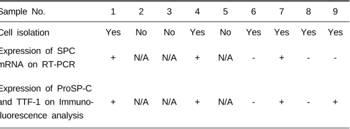
Table 2. Summary of Cell Isolation, RT-PCR and Immunofluorescence
Sample No. 1 2 3 4 5 6 7 8 9
Cell isolation Yes No No Yes No Yes Yes Yes Yes
Expression of SPC
mRNA on RT-PCR + N/A N/A + N/A - + - -
Expression of ProSP-C and TTF-1 on Immuno- fluorescence analysis
+ N/A N/A + N/A - + - +
N/A, not available
product was diluted 1:50 with distilled water. One µL of diluted first PCR product was used as a template for the nested reaction with the same cycling conditions and primer concentrations outlined above. RT-PCR amplification of the housekeeping gene, GAPDH, was used to monitor the quality of the mRNA and control for the efficiency of the RT step.
The oligonucleotide primers are described in Table 1. The nested PCR products were loaded onto 2 % agarose gel and visualized by ethidium bromide staining.
Product sizes were compared to a 100-bp molecular size marker. A549 cells were used as a positive control for SPC.
RESULTS
Among 10 samples of amniotic fluid, 1 was discarded because of contamination.
From 9 samples, 6 had cell colonies and the isolation of cells was possible. Positive results from RT-PCR and immuno- fluorescence analysis were obtained in 3
Fig. 1. Morphology of cells by inverted microscopy (100×). Cultured cells displayed a typical polygonal and cobblestone-like epithelial appearance.
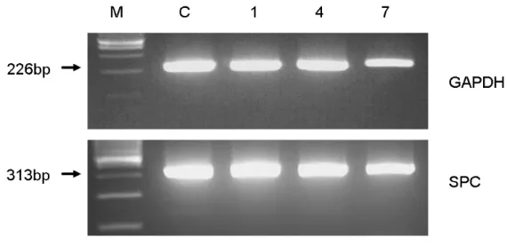
Fig. 2. RT-PCR of SPC mRNA extracted from cells cultured in SAGM. Lane M, 100-bp ladder molecular size marker; lane C, control (A549 cell); lanes 1, 4, 7, Cells from sample Nos. 1, 4, and 7. Expression of SPC mRNA was markedly augmented by nested PCR amplification. The cDNA was obtained from A549 cells that were used as a positive control and also expressed SPC mRNA.
(sample No. 1, 4, 7) and 4 (sample No. 1, 4, 7, 9) of these 6 samples (Table 2).
2.1 Cell culture and isolation
Ten to 14 days after initial plating, polygonal and cobblestone-like cells were observed with few colony formation. Cell
morphology did not change after selection and passaging (Fig. 1). Selected cells were maintained and expanded until the third passage but did not reach confluence.
2.2 RT-PCR
Expression of SPC mRNA was not detected in the first PCR reaction but was markedly augmented by nested PCR amplification. The cDNA obtained from
A549 cells was used as a positive control and also expressed SPC mRNA (Fig. 2).
2.3 Immunofluorescence analysis
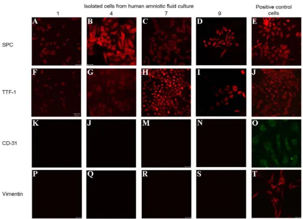
Isolated cells expressed type II alveolar cell-specific surfactant protein proSP-C.
Cells also stained positive for TTF-1, a
Fig. 3. Immunofluorescence staining. Isolated cells from different human amniotic fluid culture (sample Nos. 1, 4, 7, and 9) expressed SPC (A-D) and TTF-1(F-I) but negatively expressed CD31 (K-N) and vimentin (P-S). Positive control cells for SPC and TTF-1 (A549 cells; E, J), CD31 (HUVEC cells; O), and vimentin (fibroblast cells; T) were clearly positive.
specific marker for type II alveolar cell differentiation. However, no cells immuno- reactive to CD31 or vimentin were detected. Positive control cells for each antibody were clearly positive (Fig. 3).
DISCUSSION
This study demonstrates that respiratory cells could be isolated from human amniotic fluid cultivated in SAGM medium in vitro. Most of these cells had an appearance of epithelial cell-like mor- phology13,14 and expressed type II alveolar
cell markers SPC (mRNA and protein) as well as TTF-1. To the best of our knowledge, this is the first report of the successful isolation of respiratory cells from human amniotic fluid.
The possibility of amniotic fluid having respiratory cells can be speculated because amniotic fluid is composed of hetero- geneous fetal cells including hematopoietic, mesenchymal, epithelial, trophoblastic, and possibly more primitive stem cells5,15.
The composition of the amniotic fluid cell is thought to be correlated with the production and turnover of amniotic fluid.
The significant pathways involved in the regulation of amniotic fluid are excretion of fetal urine, swallowing of amniotic fluid and gastrointestinal excretion, secretion of fluid by fetal lung, water transport across the skin of the fetus, amniotic membrane, and fetal vessels in the umbilical cord16. As a result of such fluid dynamics, different types of cells can be identified in amniotic fluid cultures2,9,17. Among them, the secretion of large volumes of fluid by fetal lung is one of the major sources of amniotic fluid, especially during the second half of gestation. According to this physiology, it could be expected that respiratory cells are found in amniotic fluid.
Among the various types of respiratory cells, type II alveolar cells are responsible for surfactant production and are crucial to the natural regenerative process of the alveoli. In response to injury, type II alveolar cells are able to proliferate and differentiate to type I alveolar cells and are considered to be the alveolar epithelial stem cell18,19. In our study, the identi- fication of type II alveolar cells is based on amplification of SPC mRNA by RT-PCR and the presence of Pro-SPC and TTF-1 by immunofluorescence staining. SPC is an integral membrane protein that is expressed only in type II alveolar cells and is known to be a specific marker for type II alveolar
cells20,21. TTF-1 is essential for branching morphogenesis of the lung and enhances expression of surfactant proteins by type II alveolar cells22. Identification of SPC and its corresponding mRNA and of TTF-1 serves to confirm the presence of type II alveolar cells.
Type II alveolar cells are difficult to culture in vitro because of their low rate of division and tendency to differentiate into type I alveolar cells23. In this study, we chose a serum-free medium designed for in vitro maintenance of mature alveolar epithelial cells, SAGM medium, from among the few published des- criptions of type II alveolar cell culture techniques24-27. This medium contains factors known to enhance respiratory cell differentiation and has been successfully used in previous studies of type II alveolar cell cultures from different cell sources12,14. However, the effectiveness of SAGM has not been fully evaluated and a recent study reported conflicting effects of SAGM28. Cell yield of our study was low, and further investigations of cell culture methods are required to get a sufficient number of type II alveolar cells.
Recent experimental research suggests that amniotic fluid is a potential source of stem cells. These studies successfully demonstrated that human amniotic fluid stem cells can be differentiated into specialized cell populations such as neural,
hepatic, osteogenic, renal, and lung epithelial lineages6,7,10,11. However, those studies were mainly concentrated on the isolation and differentiation of amniotic fluid stem cells with limited supportive data rather than what types of cells are normally found in amniotic fluid. Iden- tification of specific cell types of amniotic fluid would be important in understanding the characteristics of amniotic fluid.
In conclusion, we have shown that respiratory cells can be isolated and identified from human amniotic fluid cultured in SAGM medium. Our results may provide the basis for further investigations.
ACKNOWLEDGEMENT
This study was supported by a faculty research grant of Yonsei University College of Medicine (6-2004-1003).
REFERENCES
1. Cunningham, FG, Leveno, KJ, Bloom, SL, Hauth, JC, Gilstrap III, LC, Wenstrom, KD: Fetal growth and development. In: Cunningham FG, Levono KJ, et al, editors. Williams Obstetrics.
22nd ed. New York (NY): McGraw Hill;
2005. Pp. 91-120.
2. Gosden, CM: Amniotic fluid cell types and culture. Br Med Bull 39:348-354, 1983
3. Harman, CR: Amniotic fluid abnormal-
ities. Semin Perinatol 32: 288-294, 2008 4. Prusa, AR, Hengstschlager, M: Amniotic
fluid cells and human stem cell research:
a new connection. Med Sci Monit 8:
RA253-257, 2002
5. Fauza, D: Amniotic fluid and placental stem cells. Best Pract Res Clin Obstet Gynaecol 18:877-891, 2004
6. De Coppi, P, Bartsch, G, Jr., Siddiqui, MM, Xu, T, Santos, CC, Perin, L, Mostoslavsky, G, Serre, AC, Snyder, EY, Yoo, JJ, Furth, ME, Soker, S, Atala, A:
Isolation of amniotic stem cell lines with potential for therapy. Nat Biotechnol 25:
100-106, 2007
7. Kim, J, Lee, Y, Kim, H, Hwang, KJ, Kwon, HC, Kim, SK, Cho, DJ, Kang, SG, You, J: Human amniotic fluid- derived stem cells have characteristics of multipotent stem cells. Cell Prolif 40:
75-90, 2007
8. Megaw, JM, Priest, JH, Priest, RE, Johnson, LD: Differentiation in human amniotic fluid cell cultures: II: Secretion of an epithelial basement membrane glycoprotein. J Med Genet 14:163-167, 1977
9. Priest, RE, Marimuthu, KM, Priest, JH:
Origin of cells in human amniotic fluid cultures: ultrastructural features. Lab Invest 39:106-109, 1978
10. Carraro, G, Perin, L, Sedrakyan, S, Giuliani, S, Tiozzo, C, Lee, J, Turcatel, G, De Langhe, SP, Driscoll, B, Bellusci, S, Minoo, P, Atala, A, De Filippo, RE, Warburton, D: Human amniotic fluid stem cells can integrate and differentiate into epithelial lung lineages. Stem Cells 26:
2902-2911, 2008
11. Perin, L, Giuliani, S, Jin, D, Sedrakyan, S, Carraro, G, Habibian, R, Warburton, D, Atala, A, De Filippo, RE: Renal differentiation of amniotic fluid stem cells. Cell Prolif 40:936-948, 2007
12. Berger, MJ, Adams, SD, Tigges, BM,
Sprague, SL, Wang, XJ, Collins, DP, McKenna, DH: Differentiation of um- bilical cord blood-derived multilineage progenitor cells into respiratory epithelial cells. Cytotherapy 8:480-487, 2006
13. Endres, M, Leinhase, I, Kaps, C, Wentges, M, Unger, M, Olze, H, Ringe, J, Sittinger, M, Rotter, N: Changes in the gene expression pattern of cytokeratins in human respiratory epithelial cells during culture. Eur Arch Otorhinolaryngol 262:390-396, 2005
14. Samadikuchaksaraei, A, Cohen, S, Isaac, K, Rippon, HJ, Polak, JM, Bielby, RC, Bishop, AE: Derivation of distal airway epithelium from human embryonic stem cells. Tissue Eng 12:867-875, 2006
15. Torricelli, F, Brizzi, L, Bernabei, PA, Gheri, G, Di Lollo, S, Nutini, L, Lisi, E, Di Tommaso, M, Cariati, E: Identification of hematopoietic progenitor cells in human amniotic fluid before the 12th week of gestation. Ital J Anat Embryol 98:119-126, 1993
16. Brace, RA, Resnik, R: Dynamics and disorders of amniotic fluid. In: Creasy RK, Renik R, editors. Maternal-Fetal Medicine. Philadelphia (Pa): WB Saunders;
1999. Pp. 632-43
17. Prusa, AR, Marton, E, Rosner, M, Bettelheim, D, Lubec, G, Pollack, A, Bernaschek, G, Hengstschlager, M:
Neurogenic cells in human amniotic fluid.
Am J Obstet Gynecol 191:309-314, 2004 18. Otto, WR: Lung stem cells. Int J Exp
Pathol 78:291-310, 1997
19. Reddy, R, Buckley, S, Doerken, M, Barsky, L, Weinberg, K, Anderson, KD, Warburton, D, Driscoll, B: Isolation of a putative progenitor subpopulation of alveolar epithelial type 2 cells. Am J Physiol Lung Cell Mol Physiol 286:
L658-667, 2004
20. Glasser, SW, Eszterhas, SK, Detmer, EA, Maxfield, MD, Korfhagen, TR: The murine SP-C promoter directs type II
cell-specific expression in transgenic mice. Am J Physiol Lung Cell Mol Physiol 288:L625-632, 2005
21. Weaver, TE, Conkright, JJ: Function of surfactant proteins B and C. Annu Rev Physiol 63:555-578, 2001
22. Kolla, V, Gonzales, LW, Gonzales, J, Wang, P, Angampalli, S, Feinstein, SI, Ballard, PL: Thyroid transcription factor in differentiating type II cells: regulation, isoforms, and target genes. Am J Respir Cell Mol Biol 36:213-225, 2007
23. Archer, F, Jacquier, E, Lyon, M, Chastang, J, Cottin, V, Mornex, JF, Leroux, C: Alveolar type II cells isolated from pulmonary adenocarcinoma: a model for JSRV expression in vitro. Am J Respir Cell Mol Biol 36:534-540, 2007 24. Ali, NN, Edgar, AJ, Samadikuchaksaraei,
A, Timson, CM, Romanska, HM, Polak, JM, Bishop, AE: Derivation of type II alveolar epithelial cells from murine embryonic stem cells. Tissue Eng 8:
541-550, 2002
25. Qin, M, Tai, G, Collas, P, Polak, JM, Bishop, AE: Cell extract-derived differ- entiation of embryonic stem cells. Stem Cells 23:712-718, 2005
26. Rippon, HJ, Polak, JM, Qin, M, Bishop, AE: Derivation of distal lung epithelial progenitors from murine embryonic stem cells using a novel three-step differ- entiation protocol. Stem Cells 24:1389- 1398, 2006
27. Van Vranken, BE, Romanska, HM, Polak, JM, Rippon, HJ, Shannon, JM, Bishop, AE: Coculture of embryonic stem cells with pulmonary mesenchyme: a microenvironment that promotes differ- entiation of pulmonary epithelium. Tissue Eng 11:1177-1187, 2005
28. Samadikuchaksaraei, A, Bishop, AE:
Effects of growth factors on the differentiation of murine ESC into type II pneumocytes. Cloning Stem Cells 9:407- 416, 2007
사람 양수에서 호흡기세포의 분리
연세대학교 의과대학 외과학교실1, 산부인과학교실2, 장기이식연구소3
김은정1․박용원2․김영한2․김유선1,3․오정탁1
최근에 양수에 존재하는 세포들은 다양한 세포분화의 능력을 가지고 있으며 세포 치료와 조직공 학의 세포원으로서의 가능성이 높다는 연구 결과가 많이 보고되고 있다. 그러나 양수 내에 어떤 종 류의 세포가 정상적으로 존재하는 지에 대해서는 제한적으로 알려져 있다. 본 연구에서는 사람의 양수 내에 호흡기세포를 분리 배양하는 것을 목표로 하였다.
임신 17주에서 20주 사이의 산모 10명에서 각각 5 mL의 양수를 획득하여 small airway growth medium (SAGM) 배양액에서 계대 배양을 시행하였으며 type II alveolar cells이 존재하는 지에 대 하여 면역형광염색 및 RT-PCR을 시행하였다. 배양된 세포는 광학현미경 상 상피세포와 유사한 다 면체의 모양이었으며 계대 배양 시에 변화 없이 동일한 형체를 보였다. 배양된 세포는 면역형광염 색 상 type II alveolar cell의 특이표지자인 surfactant protein C (SPC) 및 TTF-1 protein에 대해 서는 양성이었으나 CD 31 및 vimentin에 대해서는 음성이었으며, RT-PCR 상 SPC mRNA 가 발 현되었다.
이상의 결과로 사람의 양수를 특이 배양액에 배양하면 호흡기세포를 분리, 확인할 수 있음을 알 았으며 이러한 결과가 향후 양수세포의 추가적인 연구에 활용될 수 있다고 생각된다.
(J Kor Assoc Pediatr Surg 15(1):1~10), 2009.
Index Words:Amniotic fluid, Respiratory cell, Isolation, Human
교신저자:오정탁
서울특별시 서대문구 성산로 250 (신촌동 134), 120-752 연세대학교 의과대학 외과학교실 소아외과 Tel : 02)2228-2124, Fax : 02)313-8289
E-mail: [email protected]