ISSN 0378-6471 (Print)⋅ISSN 2092-9374 (Online)
https://doi.org/10.3341/jkos.2017.58.10.1169
Original Article
간헐외사시로 외직근후전술을 시행한 환자군과 경과관찰한 환자군에서의 사시각의 변화량 비교
Comparison of Exodrift between Natural Group and Postoperative Group in Intermittent Exotropia Patients
이유미⋅이명원⋅경성은
Yu Mi Lee, MD, Myung Won Lee, MD, Sung Eun Kyung, MD, PhD
단국대학교 의과대학 안과학교실
Department of Ophthalmology, Dankook University College of Medicine, Cheonan, Korea
Purpose: To compare the exodrift between unilateral lateral rectus (ULR) recession and observation groups in moderate angle intermittent exotropia (IXT).
Methods: A retrospective study was performed in 769 patients who were diagnosed with IXT from 2005 to 2015. Seventy-six pa- tients were enrolled in this study that presented with IXT of 20 to 25 prism diopters (PD) on their first visit and were observed for more than 6 months without or after operation. The observation group (group 1) was composed of 29 patients who had regular examination without operation. The surgery group (group 2) was composed of 47 patients with ULR recession that were ob- served for deviation changes since surgery.
Results: The mean age was 71.8 ± 22.0 months at first visit in group 1 and 91.1 ± 18.9 months before surgery in group 2 (p <
0.01). The distant exodeviation was 22.9 ± 2.5 PD at first visit in group 1 and 22.9 ± 2.4 PD before surgery in group 2 (p = 0.89).
During follow-up, mean exodrift was 0.6 ± 9.0 PD in group 1 and 10.0 ± 7.4 PD in group 2 (p < 0.01). Exodrift up to postoperative 6 months in group 2 was 3.2 ± 4.0 PD and exodrift from postoperative 6 months to 2 years in group 2 was 7.1 ± 6.9 PD. More exo- drift was noticed after post-operative 6 months (p = 0.04).
Conclusions: Comparing the exodrift between the groups in moderate angle IXT, patients in the observation group showed less exodrift. Patients who had a ULR recession presented more exodrift after postoperative 6 months. Even though they were ortho- tropic at postoperative 6 months when the operation was thought to be stabilized, an increase in exodrift after postoperative 6 months could not be excluded.
J Korean Ophthalmol Soc 2017;58(10):1169-1175
Keywords: Exodrift, Intermittent exotropia, Lateral rectus muscle recession
■Received: 2017. 3. 30. ■ Revised: 2017. 9. 6.
■Accepted: 2017. 9. 24.
■Address reprint requests to Sung Eun Kyung, MD, PhD Department of Ophthalmology, Dankook University Hospital,
#201 Manghyang-ro, Dongnam-gu, Cheonan 31116, Korea Tel: 82-41-550-6497, Fax: 82-41-550-7050
E-mail: [email protected]
*Conflicts of Interest: The authors have no conflicts to disclose.
ⓒ2017 The Korean Ophthalmological Society
This is an Open Access article distributed under the terms of the Creative Commons Attribution Non-Commercial License (http://creativecommons.org/licenses/by-nc/3.0/) which permits unrestricted non-commercial use, distribution, and reproduction in any medium, provided the original work is properly cited.
간헐외사시는 우리나라 소아에서 흔한 사시로 주된 치료 방법은 수술이지만, 수술 전 사시각이 20프리즘디옵터(prism diopters, PD) 정도로 크지 않고 외형적인 문제가 두드러지 지 않으며 양안시 기능에 문제가 없을 때는 재발에 대한 우 려 때문에 수술을 권하기 힘들 때가 있다.1,2 수술적 치료방 법으로 사시각의 크기와 사시의 분류에 따라 양안 혹은 단 안 외직근후전술이나 한눈 외직근후전술과 내직근절제술, 양안 내직근절제술 등을 시행한다.3,4 주로 양안의 외직근후
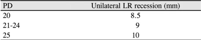
Table 1. Surgical table for moderate angle intermittent exo-
tropiaPD Unilateral LR recession (mm)
20 8.5
21-24 9
25 10
PD = prism diopters; LR = lateral rectus.
전술을 시행하지만,5,6 사시각이 25PD 이하로 크지 않은 경 우에는 단안 외직근후전술만으로도 양안 외직근후전술과 비슷한 성공률을 보인다는 연구들이 있다.7-11 하지만, 단안 외직근후전술 후 과도한 후전으로 인한 안구운동장애의 가 능성이 있으며9 장기간 추적관찰할 경우 부족교정이 많아 진다는 보고가 있다.12
간헐외사시 환자를 경과관찰 하였을 때 시간이 지나도 사시각 변화량이 크지 않다는 보고들이 있다.13-16 간헐외사 시로 후전술을 시행하는 경우, 수술 전 사시각과 수술 후 사시각을 합쳐보게 되면 전체 사시량이 수술을 안하고 관 찰한 군에 비해 많아 보이기도 한다. 이는 재발로 인해 수 술을 하는 경우 전체 사시량이 오히려 증가한 것 같아 보이 기 때문이다. 따라서 저자들은 간헐외사시로 정기적인 경 과관찰을 한 환자군과 간헐외사시로 수술 후 경과관찰한 환자군을 대상으로 사시각 변화를 후향적으로 분석하여 사 시각 변화량을 비교해 보고자 하였다.
대상과 방법
2005년 3월부터 2015년 5월까지 본원 안과를 내원한 환자 들 중에서 20PD 이상 25PD 이하의 간헐외사시를 보이는 환 자를 대상으로 후향적 연구를 진행하였다. 본 연구는 단국대 학교병원 임상연구심사위원회(Institutional Review Board, IRB) 승인을 통해 진행되었으며, 헬싱키선언(Declaration of Helsinki)을 준수하였다. 수술하지 않은 환자(1군) 29명은 6 개월 이상 외래에서 경과관찰한 환자를 대상으로 모집하였 고, 수술 후 경과관찰한 환자(2군) 47명은 한 명의 수술자 에 의해 수술한 환자 중에서 수술 후 6개월 이상 경과관찰 한 군을 대상으로 모집하였다. 대상 환자의 나이, 성별을 조사하고, 초진 시 사시각, 굴절이상, 근거리 입체시, 원거 리 융합 여부를 검사하였다. 제일안위에서 프리즘 가림 검 사를 통해 근거리(33 cm), 원거리(5 m)의 사시각을 측정하 였고, 원거리에서 상하좌우 주시 시 사시각을 각각 측정하여 AV형 사시와 외측불일치 여부를 조사하였다. 입체시 검사는 근거리에서 티트무스원(Stereo optical Co., Inc., Chicago, IL, USA)을 이용하여 33 cm 거리에서 측정하였다. 2군의 환자들에서 단안 외직근후전술을 시행하였는데, 수술 전
측정한 원거리 사시각을 기준으로 20PD일 경우 8.5 mm, 21-24PD는 9 mm, 25PD는 10 mm를 후전하였다(Table 1).
사시 수술의 기왕력이 있는 경우, 마비사시, 제한사시, 영 아외사시, 사근 수술을 함께 한 경우, 신경계 이상, 발달이 상, 사시 외 안과 질환이 있는 경우는 본 연구에서 제외하 였다. 1군에서는 초진 시 사시각과 마지막 내원 시 측정한 사시각의 차이를 총 사시각 변화량(total exodrift)으로 정의 하였고, 2군에서는 수술 직후 측정한 사시각과 마지막 내원 시 측정한 사시각의 차이를 총 사시각 변화량으로 정의하 였다. 또한 사시각 변화량을 1년 단위로 보았는데, 1군에서 는 초진 시부터 1년까지의 사시각 변화를 사시각 변화량 1 (exodrift 1)로 정의하였고, 초진 시부터 2년까지의 사시각 변화를 사시각 변화량 2 (exodrift 2)로 정의하였다. 2군에 서는 수술 후 안정화를 보이는 시간을 6개월로 생각하여,17 수술 후 6개월부터 수술 후 18개월까지의 사시각의 변화를 사시각 변화량 1 (exodrift 1)로 정의하였고, 수술 후 6개월 부터 수술 후 30개월까지 사시각 변화를 사시각 변화량 2 (exodrift 2)로 정의하였다.
2군의 수술 후 결과에 대하여 5PD 이하의 내사위부터 10PD 이하의 외사위까지를 성공적인 교정, 6PD 이상의 내 사위를 보이는 경우 과교정, 10PD 초과의 외사위를 보이는 경우 재발로 정의하였다.18 총 사시각 변화량에 대하여 5PD 이하로 사시각이 줄었거나 10PD 이하로 사시각이 늘었을 경우에는 사시각이 유지된다고 정의하였으며, 10PD 초과 로 사시각이 늘었을 경우는 간헐외사시가 진행되었다고 정 의하였고, 사시각이 6PD 이상 줄었을 경우에는 호전되었다 고 정의하였다. 통계처리는 SPSS Statistics 22 (IBM, Corp., Armonk, NY, USA)을 이용하였고 p값이 0.05 미만일 때를 통계학적으로 유의한 것으로 간주하였다.
결 과
수술하지 않은 환자(1군)는 29명이었으며, 수술 후 경과 관찰한 환자(2군)는 47명이었다. 두 군의 초진 시 평균나이 는 1군은 71.8 ± 22.0 (44-131)개월, 2군은 84.3 ± 23.5 (21-126)개월로 두 군 간에 통계학적으로 유의한 차이를 보 였다(p=0.02, t-test). 2군에서의 수술 시 평균나이는 91.3 ± 18.9 (41-134)개월이었으며, 1군의 초진 나이와 비교하였을 때는 통계학적으로 유의한 차이를 보였다(p<0.01, t-test). 1군 에서 남자 환자 17명, 여자 환자가 12명이었으며, 2군에서 는 남자 환자가 19명, 여자 환자가 28명으로 두 군 간의 성별 의 유의한 차이는 없었다(p=0.16, Fisher’s exact test). 1군 에서 초진 시 원거리 사시각은 22.9 ± 2.5PD였으며, 2군에 서 초진 시 원거리 사시각은 23.1 ± 3.8PD로 두 군 간에 통
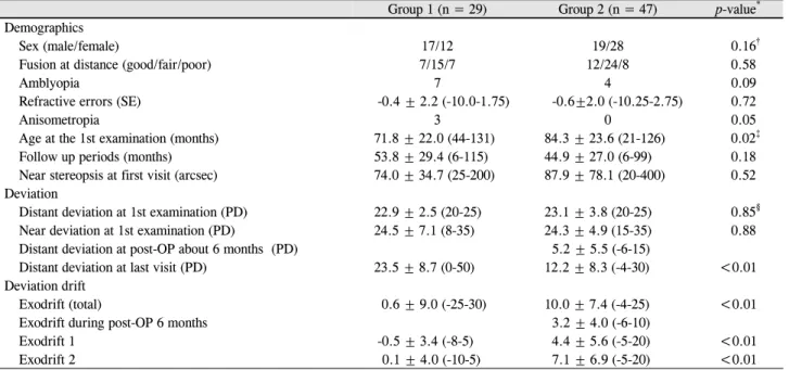
Table 2. Clinical characteristics of Group 1 and 2
Group 1 (n = 29) Group 2 (n = 47) p-value* Demographics
Sex (male/female) 17/12 19/28 0.16†
Fusion at distance (good/fair/poor) 7/15/7 12/24/8 0.58
Amblyopia 7 4 0.09
Refractive errors (SE) -0.4 ± 2.2 (-10.0-1.75) -0.6±2.0 (-10.25-2.75) 0.72
Anisometropia 3 0 0.05
Age at the 1st examination (months) 71.8 ± 22.0 (44-131) 84.3 ± 23.6 (21-126) 0.02‡
Follow up periods (months) 53.8 ± 29.4 (6-115) 44.9 ± 27.0 (6-99) 0.18
Near stereopsis at first visit (arcsec) 74.0 ± 34.7 (25-200) 87.9 ± 78.1 (20-400) 0.52 Deviation
Distant deviation at 1st examination (PD) 22.9 ± 2.5 (20-25) 23.1 ± 3.8 (20-25) 0.85§
Near deviation at 1st examination (PD) 24.5 ± 7.1 (8-35) 24.3 ± 4.9 (15-35) 0.88
Distant deviation at post-OP about 6 months (PD) 5.2 ± 5.5 (-6-15)
Distant deviation at last visit (PD) 23.5 ± 8.7 (0-50) 12.2 ± 8.3 (-4-30) <0.01
Deviation drift
Exodrift (total) 0.6 ± 9.0 (-25-30) 10.0 ± 7.4 (-4-25) <0.01
Exodrift during post-OP 6 months 3.2 ± 4.0 (-6-10)
Exodrift 1 -0.5 ± 3.4 (-8-5) 4.4 ± 5.6 (-5-20) <0.01
Exodrift 2 0.1 ± 4.0 (-10-5) 7.1 ± 6.9 (-5-20) <0.01
Values are presented as mean ± SD unless otherwise indicated. ‘Group 1’ is ‘the patients who had regular examination about the deviation with- out operation’ and ‘Group 2’ is ‘the patients who had a unilateral lateral rectus recession’. ‘Exodrift (total)’ means ‘difference of PD between last visit and first visit in group 1 and between last visit and post-operative day 1 in group 2’. ‘Exodrift 1’ means ‘difference of PD during first 1 year in group 1 and from postoperative 6 months to postoperative 18 months in group 2’. ‘Exodrift 2’ means ‘difference of PD during 2 years in group 1 and from postoperative 6 months to postoperative 30 months in group 2’.
SE = spherical equivalent; PD = prism diopters; post-OP = postoperative.
*p-value by student t-test; †p-value by Fisher’s exact test; ‡The difference of the age between group 1 (71.8 ± 22.0) at first examination and group 2 (91.1 ± 18.9) at operation was also significantly efficient (p < 0.01, t-test); §The difference of distant deviation between group 1 (22.9
± 2.5) at first examination and group 2 (22.9 ± 2.4) at operation was also not significantly efficient (p = 0.89, t-test).
계학적으로 유의한 차이는 보이지 않았다(p=0.85, t-test). 2 군에서 수술 전 측정한 원거리 사시각은 22.9 ± 2.4PD로 1 군의 초진 시 원거리 사시각과 비교하였을 때 역시 두 군 간 의 유의한 차이는 없었다(p=0.89, t-test). 굴절부등(p=0.05, t-test)을 제외한 약시, 굴절이상, 융합여부 및 근거리 입체 시의 차이를 비교한 결과 두 군 간에 통계학적으로 유의한 차이를 보이는 변수는 없었다(Table 2).
경과관찰 기간은 1군에서 초진부터 마지막 내원일까지 평균 53.8 ± 29.4 (6-115)개월이었고, 2군에서는 수술 직후 부터 마지막 내원일까지 평균 44.9 ± 27.0 (6-99)개월이었으 며, 두 군에서 차이는 통계적으로 유의하지 않았다(p=0.18, t-test). 수술 결과 47명 중 성공적인 교정을 보인 환자는 20 명(42.6%)에 해당하였고, 재발을 보인 환자는 27명(57.4%) 이었으며 과교정된 환자는 없었다.
1군에서 마지막으로 측정한 사시각의 평균은 23.5 ± 8.7 (0-50)PD였으며, 2군에서 수술 직후 측정한 사시각의 평균 은 2.2 ± 3.6 (-10-8)PD였고, 마지막으로 측정한 사시각의 평균은 12.2 ± 8.3 (-4-30)PD로 수술 후 마지막으로 측정한 사시각은 경과관찰한 군보다 통계적으로 유의하게 작았다 (p<0.01, t-test). 총 사시각 변화량은 1군에서는 평균 0.6 ±
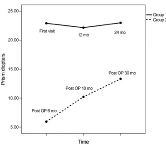
9.0PD의 외사시 변화를 보였고 2군에서는 평균 10.0 ± 7.4PD로 수술 후 외사시로의 진행이 더욱 많이 나타났으 며, 나이를 보정하였을 때 사시각 변화량은 두 군 간에 유 의한 차이를 보였다(F=22.63, p<0.01, analysis of covariance [ANCOVA]). 2군의 환자 중 6개월에 추적관찰 하였을 때 사시각 변화량은 3.2 ± 4.0PD였으며, 사시각 변화량 2는 7.1
± 6.9PD로 사시각 변화량 1인 4.4 ± 5.6PD에 비하여 사시 각 변화량이 통계적으로 유의하게 더 컸다(p=0.01, paired t-test) (Fig. 1).
총 사시각 변화량에 대하여 1군과 2군의 환자들은 유지되 는 군, 진행하는 군, 호전되는 군으로 분류하였다(Table 3).
1군에서는 대부분이 사시각에 변화를 보이지 않았으며, 4 명(13.8%)에서는 6PD 이상 사시각이 감소하는 소견을 보 였고, 단 1명(3.4%)에서만 30PD의 사시각 변화량을 보였 다. 반면 2군에서는 유지되는 경우는 21명(44.7%)이었고, 진행한 환자는 26명(55.3%)이었다.
Pearson 상관분석을 이용하여 사시각 변화량과 상관관계 가 있는 요소를 찾아보았다. 1군에서는 초진 시 나이, 굴절 이상, 추적관찰 기간, 입체시, 초진 시 사시각 등은 사시각 변화량과 상관관계를 나타내지 않았다. 2군에서는 초진 시
Figure 1. Exodrift 1 and exodrift 2 in each group. In group 2,
exodrift 2 was 7.1 ± 6.9 PD which is more than exodrift 1 of 4.1 ± 5.7 (p = 0.01, paired t-test). Exodrift 1 was -0.5 ± 3.4 PD in group 1 and 4.4 ± 5.6 PD in group 2 and there was sig- nificant difference (p < 0.01, t-test). Exodrift 2 was 0.1 ± 4.0 PD in group 1 and 7.1 ± 6.9 PD in group 2 and there was sig- nificant difference (p < 0.01, t-test). Exodrift 1: difference of PD during first 1 year in group 1 and from postoperative 6 months to postoperative 18months in group 2. Exodrift 2: dif- ference of PD during first 2 years in group 1 and from post- operative 6 months to postoperative 30 months in group 2.Post-OP = postoperative; mo = months.
Table 3. The number of patient according to amount of total
exodrift in group 1 and group 2Group 1 Group 2 Total exodrift* ≤ -6† PD 4 (13.8) 0 (0) Total exodrift > -6 PD and ≤ 10 PD 24 (82.8) 21 (44.7) Total exodrift > 10 PD 1 (3.4) 26 (55.3) Values are presented as n (%). ‘Group 1’ is ‘the patients who had reg- ular examination about the deviation without operation’ and ‘Group 2’ is ‘the patients who had a unilateral lateral rectus recession’.
PD = prism diopters.
*Total exodrift was defined by subtraction; difference between final deviation and first visit deviation in group 1, difference between final deviation and postoperative first deviation in group 2; †Esotropia was calculated as minus number. The negative number of exodrift means decrease of exotropia deviation.
사시각, 근거리 입체시, 굴절이상은 사시각 변화량과 상관 관계가 없었으나, 초진 시 나이와 추적관찰 기간은 사시각 변화량과 통계적으로 유의한 상관관계를 나타내었다(γ
=-0.3, p=0.05, γ=0.5, p<0.01). 다중회귀분석 결과 2군에서 추적관찰 기간만이 사시각 변화량에 영향을 미치는 것으로 나타났다(Y=8.88+0.13X, p<0.01). 나이를 보정한 공분산분 석의 결과 1군에서 초진 시 원거리 융합 정도는 사시각 변 화량과 통계학적으로 유의한 관련성을 보이지 않았으며
(F=0.28, p=0.76), 2군에서도 수술 전 원거리 융합 정도는 사시각 변화량과 유의한 관련성을 보이지 않았다(F=0.39, p=0.68).
고 찰
기본간헐외사시는 시간이 지날수록 사시각과 양안시 기 능이 나빠진다고 알려져 있다.19 von Noorden20은 수술 받 지 않은 5-10세 사이의 간헐외사시 환자를 평균 3.5년간 추 적 관찰한 결과 75%에서 진행하였다고 하였다. 간헐외사 시 환자는 정상인에 비해 원거리 입체시 기능이 떨어져있
으나,21,22 수술 후에는 향상되며,23-25 근거리 입체시의 경우
는 수술에 의한 영향이 적은 것으로 알려졌다.21,26,27 술 후 원거리 입체시 향상에 대해 Roh et al23은 술 전 216초각에 서 133초각으로, Suh et al24은 305초 각에서 221초각으로 향상되는 결과를 보고하였다. 하지만 사시가 간헐성이기 때문에 환자나 보호자가 이러한 기능적인 면을 자각하기 어렵고, 대부분 보호자의 미용적 목적으로 치료를 결정하 게 된다. Kim et al28이 시행한 사시 환자 및 보호자를 대상 으로 한 설문조사에 따르면 77%의 환자 및 보호자가 수술 전 한 번의 수술로 사시가 완전히 교정될 것이라고 기대하 고 있었고 수술 전 60%의 환자 및 보호자는 사시의 재발에 대하여, 26%의 환자가 과교정에 대하여 가장 두려워하고 있었다. 이처럼 간헐외사시의 수술의 과교정 및 높은 재발 률의 가능성이 걱정되어 환자 및 보호자뿐만 아니라 의사 역시 수술을 망설이게 된다.29 국내 연구에서 4세 이전에 간헐외사시 교정수술을 할 경우 속발 내사시의 발생 빈도 가 더 높았다는 보고가 있고,30 간헐외사시의 수술 후 단기 결과는 만족스러운 것으로 알려졌으나, 장기 관찰할 경우 초기에는 성공을 보이다가 시간이 지날수록 외편위 되어 경과관찰 기간의 정도에 따라 재발률이 높다고 보고하였 다.5,31,32
한편, 간헐외사시 환자를 경과관찰 하였을 때 시간이 지 나도 사시각 변화량이 크지 않다는 보고들이 있다. Hiles et al13에 의하면 48명의 간헐외사시 환자에서 평균 11.7년 동 안 경과관찰 한 결과 81%에서 10프리즘디옵터 이내의 변 화를 보였다고 하였으며, Sanfilippo and Clahane14은 31명 에 대하여 5년 6개월 동안 경과관찰 하였을 때 사시각이 증 가한 경우가 없다고 하였다. Kii and Nakagawa15는 10명의 환자에서 6.2년 동안 경과관찰 하였을 때 처음 측정한 사시 각이 16.6PD이며 마지막에 측정한 사시각 15.7PD로써 유 의한 차이를 보이지 않는다고 하였으며, Romanchuk et al16 은 109명의 간헐외사시 환자를 대상으로 평균 9년간 경과 관찰 하였을 때 사시각 변화량은 평균 0.3PD를 보였고,
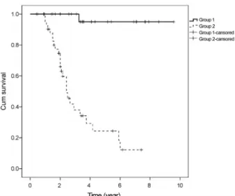
Figure 2. Kaplan-meier survival curve. Group 1 is the ob-
servation group who didn’t have a surgery and group 2 is the surgery group who had a unilateral lateral recession. Exodrift more than 10 PD was plotted in the survival curve. These sur- vival curve indicated that the estimated mean times to exodrift more than 10 PD were 8.3 ± 3.1 years in group 1 and 3.7 ± 3.8 years in group 2. The cumulative probability of exodrift was sinificantly higher in group 2 than group 1 (p < 0.01, log rank test).10PD 이상 사시각 변화를 보인 환자는 단 23%에 해당하였 다. 본 연구에서도 치료하지 않고 54개월 정도 경과관찰한 환자 중 82.8%에서 10PD 이하의 사시각 변화를 보였다 (Fig. 2).
최근에는 25PD 미만의 사시각의 경우 단안 외직근 수술 이 보편화되고 있다.7-11 단안 외직근후전술의 장점은 수술 및 마취 시간의 단축과 합병증 발생 기회의 감소, 이차 수 술 선택의 다양함, 과교정 위험이 적은 것 등이지만,33.34 과 도한 후전으로 안구 운동 장애로 인한 복시를 유발할 수 있
고,34,35 추적기간이 길어지면 부족교정이 많다는 단점이 있
다.35 부족교정에 대하여 Cho et al36은 20-25PD의 간헐외사 시 환자에서 단안 외직근후전술의 성공률은 63%로 두 눈 외직근후전술의 성공률보다 낮다고 보고하였고, 본 연구에 서 단안 외직근후전술의 성공률은 42.6% (47명 중 20명)였 으며 55.3%에서 수술 이후 10PD 초과의 사시각 변화를 나 타내었다(Fig. 2).
수술 직후 과교정 정도가 재발과 상관관계가 있을 것이 라는 많은 연구가 있었는데, Rabb and Parks37, Knapp38은 수술 직후 약 11-20PD, Scott et al5은 4-14PD의 과교정이 좋은 결과를 얻을 수 있다고 하였고, Roh and Paik의 연구39 에서도 술 후 1일째 10-20PD의 과교정을 보였던 군에서 술 후 1년째 재발률이 유의하게 낮음을 확인할 수 있었다. Lee and Lee40 연구에서는 양안 외직근후전술에서는 수술 직후
11-20PD의 과교정이 단안 외직근후전술 및 내직근절제술 을 시행한 군에서는 1-10PD의 과교정이 최종적으로 수술 성공률이 가장 높다고 보고하였다. 즉 수술 직후 10-20PD 정도의 과교정을 보였던 환자에서 시간이 지났을 때 재발 률이 낮다는 것은 약 10-20PD 정도의 외편위로의 움직임이 있어 최종 사시각이 수술의 성공 범위 안에 유지된 것을 말 한다. 이것은 본 연구에서 수술 후 사시각의 변화량 10.0 ± 7.4PD와 비슷한 변화량일 것을 추측해 볼 수 있다. 하지만 본 연구의 술자는 수술 초기의 과교정을 목표로 하는 것을 피하려 하였는데 그 이유는 수술 후 사시각 변화량을 예측 하여 정확히 예측량 만큼만 과교정하기가 어려울 뿐만 아 니라, 어린 소아에서는 수술 후 과교정으로 인한 속발내사 시로 초래된 복시를 피하기 위해 한 눈에 약시나 억제가 생 기고 이로 인해 입체시 상실 등의 감각기능 이상을 초래할 수 있기 때문에41 현실에서 치료에 적용하기 어려운 부분이 있다고 생각하였기 때문이다. 본 연구결과에서도 사시각 변화량이 유지되는 21명(44.7%)에 대하여 보았을 때에는 수술 직후부터 총 사시각 변화량이 3.2 ± 3.9PD에 해당하 며, 이 환자군에서 수술 직후 과교정을 목표로 했다면 최종 적으로 내사시가 발생하였을 수도 있다.
간헐외사시환자에서 일차 수술로 단안 혹은 양안 외직근 후전술을 시행 후 재발한 환자에 관하여 내직근절제술 후 의 사시각 변화에 대하여 연구한 논문이 있다.17 일차 수술 후 1일째부터 수술 후 6개월까지의 사시각 변화량이 수술 6개월 이후 사시각 변화량보다 많았으며 내직근 절제술로 재수술했을 때도 술 후 6개월까지의 사시각 변화량이 더 컸으며 총 경과관찰 기간은 1차 수술 후 20.2 ± 9.2개월, 재 수술 후 22.2 ± 9.9개월이었다. 하지만 본 연구에서는 6개 월까지의 사시각 변화량보다 수술 후 6개월 이후 2년 동안 사시각 변화량이 더 크게 나타났는데 이는 앞선 연구보다 경과관찰이 길었기 때문에 관찰할 수 있었다.
간헐외사시의 병인은 아직 정확하게 밝혀져 있지 않지만 Duane42이 주장한 신경분포의 불균형에 의해 능동적인 눈 모음과 눈벌림 운동 간의 상호작용의 와해가 원인이 된다 는 설과 Bielschowsky43가 주장한 기계적 해부학적 요인에 의한다는 설이 제시되고 있다. 본 연구에서 수술 후의 사시 각의 변화량이 자연 관찰 시 사시각 변화량보다 큰 것 역시 명확히 설명하긴 어렵지만, 재발된 간헐외사시에서 양안의 내직근절제술보다 단안의 많은 양의 내직근절제술 시행한 후 6개월 뒤 사시각의 변화가 적다는 보고17가 있었고, 양안 외직근후전술 후의 사시각 변화량이 단안 외직근후전술 및 내직근절제술 후의 사시각 변화량보다 컸다는 연구결과41 를 고려한다면 본 연구에서 6개월 이후 사시각 변화량이 더 많은 군에서는 외직근후전술의 효과가 시간이 지나면서
감소되는 것이 원인이 아닐까 추측해 본다. 따라서 수술 후 결과 예측에 어려워서 첫 번째 수술로 시행하지 않는 단안 내직근절제술을 중등도 간헐 외사시의 첫 번째 수술 방법 으로 먼저 시도해 보는 것도 한 방법이 아닐까 생각한다.
또한 외직근후전술 시행 후 6개월 이후에도 사시각 변화량 이 적어도 1년간은 증가할 수 있기 때문에 외직근후전술 후 6개월까지 정위를 유지하더라도 사시각이 증가할 가능 성을 배제하기는 어려울 것으로 생각된다.
본 연구를 통하여 단안 외직근후전술 후 약 6개월 동안 3.2 ± 4.0PD의 사시각 변화를 보이며, 수술 6개월 이후의 사시각 변화량이 더 큰 것을 알 수 있었다. 또한 경과관찰 만 하는 경우에는 사시각 변화량이 크지 않으며 사시각 측 면에서만 보았을 때는 크게 호전되지도 악화되지도 않는 양상을 보였다. 초진 시 융합 정도는 두 군에서 모두 사시 각 변화량과 관련성이 없었다. 외직근후전술 후 사시각 변 화량에 영향을 주는 요소는 다중회귀분석 결과 경과관찰 기간뿐이었으며, 이는 곧 수술한 환자에서는 추적기간이 길수록 사시각 변화량이 많아지고, 나아가 부족교정이 많 아진다는 것을 의미한다. 외직근후전술 6개월 이후에도 사 시각 변화가 있었으며, 시간에 따른 외직근후전술 효과의 약화 여부가 수술 후 사시각 변화량에 영향을 미치는지 알 아보기 위해 내직근절제술만을 시행한 경우와의 비교 등의 추가적인 연구가 필요할 것이라 생각한다.
REFERENCES
1) Jenkins R. Demographics: geographic variations in the prevalence and management of exotropia. Am Orthopt J 1992;42:82-7.
2) Park DG, Moon, SH, Noh DH, Kim MM. Comparison between 20 and 25 prism diopters in bilateral rectus muscle recession for inter- mittent exotropia. J Korean Ophthalmol Soc 2014;55:1669-73.
3) von Noorden GK, Campos EC. Binocular vision and ocular mo- tility: theory and management of strabismus, 6th ed. St. Louis:
Mosby, 2002; 631.
4) Burian HM. Exodeviations: their classification, diagnosis, and treatment. Am J Ophthalmol 1966;62:1161-6.
5) Scott WE, Keech R, Mash AJ. The postoperative results and stabil- ity of exodeviations. Arch Ophthalmol 1981;99:1814-8.
6) Ruttum MS. Initial versus subsequent postoperative motor align- ment in intermittent exotropia. J AAPOS 1997;1:88-91.
7) Reynolds JD, Hiles DA. Single lateral rectus muscle recession for small angle exotropia. In: Reinecke RD, ed. Strabismus 2: Proceedings of the Fourth Meeting of the International Strabismological Association, October 25-29, 1982, Asilomar, California (Pt. 2). New York: Grune & Stratton, 1984; 247-53.
8) Kushner BJ. Selective surgery for intermittent exotropia based on distance/near differences. Arch Ophthalmol 1998;116:324-8.
9) Lee SH, Kim JY, Kwon JY. The effect of unilateral lateral rectus re- cession for the treatment of moderate-angle exotropia. J Korean Ophthalmol Soc 2005;46:2045-9.
10) Spierer O, Spierer A, Glovinsky J, Ben-Simon GJ. Moderate-an- gles exotropia: a comparison of unilateral and bilateral rectus mus- cle recession. Ophthalmic Surg Lasers Imaging 2010;41:355-9.
11) Menon V, Singla MA, Saxena R, Phulijele S. Comparative study of unilateral and bilateral surgery in moderate exotropia. J Pediatr Ophthalmol Strabismus 2010;47:288-91.
12) Moon KJ, Choi WC, Park C. The long-term effect of unilateral lat- eral rectus muscle recession for moderate angle exotropia. J Korean Ophthalmol Soc 1998;39:1885-90.
13) Hiles DA, Davies GT, Costenbader FD. Long-term observations on unoperated intermittent exotropia. Arch Ophthalmol 1968;80:
436-42.
14) Sanfilippo S, Clahane AC. The effectiveness of orthoptics alone in selected cases of exodeviation: the immediate results and several years later. Am Orthopt J 1970;20:104-17.
15) Kii T, Nakagawa T. Natural history of intermittent exotropia--stat- istical study of preoperative strabismic angle in different age groups. Nippon Ganka Gakkai Zasshi 1992;96:904-9.
16) Romanchuk KG, Dotchin SA, Zurevinsky J. The natural history of surgically untreated intermittent exotropia-looking into the distant future. J AAPOS 2006;10:225-31.
17) Yang HK, Hwang JM. Bilateral vs unilateral medial rectus re- section for recurrent exotropia after bilateral lateral rectus recession. Am J Ophthalmol 2009;148:459-65.
18) Cho SC, Yang HK, Hwang JM. Three-year surgical outcome of exotropia. J Korean Ophthalmol Soc 2012;53:1674-9.
19) Lee JC, Lee YC, Lee SY. Comparison of postoperative outcomes according to deviation angle in moderate-angle intermittent exo- tropia of basic type. J Korean Ophthalmol Soc 2013;54:475-8.
20) von Noorden GK. Binocular vision and ocular motility: theory and management of strabismus, 4th ed. St. Louis: CV Mosby, 1990;
330-9.
21) Stathacopoulos RA, Rosenbaum AL, Zanoni D, et al. Distance stereoacuity. Assessing control in intermittent exotropia. Ophthal- mology 1993;100:495-500.
22) Zanoni D, Rosenbaum AL. A new method for evaluating distance stereo aciuty. J Pediatr Ophthalmol Strabismus 1991;28:255-60.
23) Roh YB, Kim CM, Oum BS, Lee JS. Distance stereoacuity in chil- dren with intermittent exotropia using B-VAT II video acuity tester.
J Korean Ophthalmol Soc 1998;39:578-82.
24) Suh WJ, Lee UK, Kim MM. Change of postoperative distance ster- eoacuity in intermittent exotropic patients. J Korean Ophthalmol Soc 2000;41:758-63.
25) O’Neal TD, Rosenbaum AL, Stathacopoulos RA. Distance stereo acuity improvement in intermittent exotropic patients following strabismus surgery. J Pediatr Ophthalmol Strabismus 1995;32:
353-7; discussion 358.
26) Beneish R, Flanders M. The role of stereopsis and early post- operative alignment in long-term surgical results of intermittent exotropia. Can J Ophthalmol 1994;29:119-24.
27) Baker JD, Davies GT. Monofixational intermittent exotropia. Arch Ophthalmol 1979;97:93-5.
28) Kim JH, Kim SH, Cho YA. A Study of patient concerns and return to daily life after strabismus surgery. J Korean Ophthalmol Soc 2012;53:440-5.
29) Baek SU, Lee JY. Long-term outcome of surgery for intermittent exotropia. J Korean Ophthalmol Soc 2013;54:1079-85.
30) Cho YA, Lee JK. Early surgery before 4 years of age in intermittent
= 국문초록 =
간헐외사시로 외직근후전술을 시행한 환자군과 경과관찰한 환자군에서의 사시각의 변화량 비교
목적: 중등도의 간헐외사시에서 외직근후전술 여부가 경과관찰만 시행한 군과 외직근후전술을 시행한 군에서 사시각 변화에 영향을 미치는지 알아보고자 한다.
대상과 방법: 2005년부터 2015년까지 간헐외사시로 진단된 769명 중 초진 시 20프리즘디옵터(prism diopters, PD) 이상 25PD 이하의 사시각으로 6개월 이상 경과관찰하거나 외직근후전술 후 6개월 이상 경과관찰하였던 환자 76명을 대상으로 후향적으로 분석하였다.
수술을 하지 않고 정기적인 경과관찰을 한 환자군(1군) 29명에 대하여 시간에 따른 사시각의 변화량을 관찰하였고, 단안 외직근후전술 을 받은 환자군(2군) 47명에 대하여 수술 직후부터 사시각의 변화 양상을 관찰하였다.
결과: 1군의 환자들의 초진 시 평균연령은 71.8 ± 22.0개월이었고, 2군의 환자의 수술 전 평균연령은 91.1 ± 18.9개월이었다(p<0.01).
1군의 초진 시 원거리 사시각은 22.9 ± 2.5PD였으며, 2군의 수술 전 원거리 사시각은 22.9 ± 2.4PD로 차이를 보이지 않았다 (p=0.89). 경과관찰 기간 동안 평균 사시각 변화량은 1군에서 0.6 ± 9.0PD였으며, 2군에서 10.0 ± 7.4PD로 나타났다(p<0.01). 2군에 서 수술 후 6개월 전까지의 사시각 변화량의 평균은 3.2 ± 4.0PD, 수술 후 6개월 이후 2년 동안 사시각 변화량의 평균은 7.1 ± 6.9PD로 수술 6개월 이후 사시각 변화량이 더 많았다(p=0.04).
결론: 중등도의 간헐외사시 환자를 대상으로 외직근후전술을 시행한 군과 수술을 시행하지 않고 관찰한 군에 대하여 사시각 변화를 비교한 결과 수술을 시행하지 않은 군에서 사시각 변화량이 적었다. 외직근후전술을 시행한 군에서 수술 6개월 이후 사시각 변화량이 더 많아 술 후 안정이 되었다고 생각되는 6개월에 정위 소견을 보이더라도 이후 사시각 증가의 가능성을 배제할 수 없을 것으로 생각 된다.
<대한안과학회지 2017;58(10):1169-1175>
exotropia. J Korean Ophthalmol Soc 2004;45:620-5.
31) Ing MR, Nishimura J, Okino L. Outcome study of bilateral lateral rectus recession for intermittent exotropia in children. Ophthalmic Surg Lasers 1999;30:110-7.
32) Kim MM, Cho ST. Long-term surgical results of intermittent exotropia. J Korean Ophthalmol Soc 1994;35:1321-6.
33) Nelson LB, Bacal DA, Burke MJ. An alternative approach to the surgical management of exotropia--the unilateral lateral rectus recession. J Pediatr Ophthalmol Strabismus 1992;29:357-60.
34) Weakley DR Jr, Stager DR. Unilateral lateral rectus recessions in exotropia. Ophthalmic Surg 1993;24:458-60.
35) Roh YB, Choi HY. Surgical results of unilateral and bilateral lateral rectus recessions in exotropia under 25 prism diopter. J Korean Ophthalmol Soc 1997;38:474-8.
36) Cho HJ, Ma YR, Park YG. Comparison of surgical results between bilateral and unilateral lateral rectus recession in 20~ 25 prism di- opters intermittent exotropia. J Korean Ophthalmol Soc 2002;43:
1993-9.
37) Rabb EL, Parks MM. Recession of the lateral recti. Early and late postoperative alignments. Arch Ophthalmol 1969;82:203-8.
38) Knapp P. Symposium on strabismus: transactions of the New Orleans Academy of Ophthalmology. St. Louis: CV Mosby, 1971;
233-41.
39) Roh JH, Paik HJ. Clinical study on factors associated with re- currence and reoperation in intermittent exotropia. J Korean Ophthalmol Soc 2008;49:1114-9.
40) Lee S, Lee YC. Relationship between motor alignment at post- operative day 1 and at year 1 after symmetric and asymmetric sur- gery in intermittent exotropia. Jpn J Ophthalmol 2001;45:167-71.
41) Elsas FJ. Consecutive esotropia. Am Orthopt J 1992;42:94-7.
42) Duane A. A New Classification of the Motor Anomalies of the Eye: Based upon Physiological Principles, Together with Their Symptoms, Diagnosis and Treatment. New York: J.H. Vail & Co., 1897; 49-51.
43) Bielschowsky A. Divergence excess. Arch Ophthalmol 1934;12:
157-66.