© 2016 The Korean Ophthalmological Society
This is an Open Access article distributed under the terms of the Creative Commons Attribution Non-Commercial License (http://creativecommons.org/licenses /by-nc/3.0/) which permits unrestricted non-commercial use, distribution, and reproduction in any medium, provided the original work is properly cited.
Original Article
Visual Outcomes, Patient Satisfaction and Spectacle Independence with a Trifocal Diffractive Intraocular Lens
Florian Tobias Alwin Kretz1,2,3, Chul Young Choi1,4, Matthias Müller2,3, Matthias Gerl2,3, Ralf Helmar Gerl2,3, Gerd Uwe Auffarth1,2
1International Vision Correction and Research Centre (IVCRC) & David J Apple International Laboratory of Ocular Pathology, Department of Ophthalmology, University of Heidelberg, Heidelberg, Germany
2International Vision Correction Research Network (IVCRC.net), Heidelberg, Germany
3Augenklinik Ahaus-Raesfeld-Rheine, Gerl Group, Ahaus, Germany
4Department of Ophthalmology, Kangbuk Samsung Hospital, Sungkyunkwan University College of Medicine, Seoul, Korea
Purpose: To evaluate visual outcomes following implantation of a trifocal diffractive intraocular lens (IOL) and to analyze their correlation with patient satisfaction and ease of performing daily tasks.
Methods: This was a prospective study enrolling 100 eyes of 50 patients undergoing cataract surgery with im- plantation of trifocal IOL AT LISA tri 839MP. Visual and refractive outcomes were evaluated during a 3-month follow-up. Postoperatively, a questionnaire was used to evaluate patient satisfaction with regard to surgical outcome, spectacle independence, perception of photic phenomena, and ease of performing some vision-re- lated activities.
Results: A total of 91%, 87%, and 79% of eyes achieved a monocular uncorrected distance, near, and interme- diate visual acuity of 0.1 logarithm of the minimum angle of resolution or better, respectively. After the surgery, 96% of the patients could perform their daily activities without problems. The mean spectacle independence scores for reading, doing computer work, and for distance were 10.33 ± 12.47, 5.71 ± 11.90, and 3.92 ± 9.77, respectively (scale: 0 = no spectacles needed; 40 = spectacles always needed). No correlation was found between spectacle independence and visual outcome (-0.101 ≤ r ≤ 0.244, p ≥ 0.087). Mean scores (0 = no symptoms; 40 = strong symptoms) for glare at night, ghost images, and halos were 15.15 ± 12.02, 4.49 ± 7.92, and 13.34 ± 10.82, respectively. No correlation was found between photic phenomena and visual outcome (-0.199 ≤ r ≤ 0.209, p ≥ 0.150). A total of 80% of patients reported satisfaction with the surgery outcome, and 86% would recommend the surgery to friends and family.
Conclusions: Implantation of the AT LISA tri 839MP IOL after cataract surgery provides effective visual resto- ration associated with a minimal level of photic phenomena, a positive impact on the performance of vision-re- lated daily activities, and a high level of postoperative patient satisfaction.
Key Words: Cataract, Presbyopia, Surveys and questionnaire
Received: May 8, 2015 Accepted: September 21, 2015
Corresponding Author: Gerd Uwe Auffarth, MD, PhD, FEBO. International Vision Correction and Research Centre (IVCRC) & David J Apple Interna- tional Laboratory of Ocular Pathology, Department of Ophthalmology, University of Heidelberg, Im Neuenheimer Feld 400, Heidelberg 69120, Germany.
Tel: 49-6221-56 34763, Fax: 49-6221-56 5422, E-mail: [email protected]
Trifocal diffractive intraocular lenses (IOLs) distribute light to distant, intermediate, and near foci without a clini- cally significant deterioration in visual quality [1,2]. Con- sequently, trifocal IOLs provide significantly better inter- mediate vision compared to conventional bifocal diffractive IOLs [3].One of the currently available trifocal IOLs is the AT LISA tri 839MP IOL from Carl Zeiss Med- itec (Jena, Germany), which combines a central 4.3-mm trifocal area with a bifocal diffractive surface between 4.3 and 6 mm in diameter [4]. Previous studies have demon- strated that this IOL is able to provide good visual contrast sensitivity and optical quality outcomes [3-6]. Likewise, Law et al. [6] evaluated a sample of 60 eyes of 30 patients implanted with the AT LISA tri 839MP IOL to determine the level of patient satisfaction with regard to surgical out- come and the impact of the surgery on daily vision-related activities. These authors found that 76.7% of patients were entirely satisfied with the visual outcome and had no diffi- culties in watching TV or driving 6 months after surgery [6]. Moderate difficulties were reported by a limited per- centage of patients in this same study for performing some near and intermediate vision tasks, such as reading the newspaper or working on the computer [6]. Multifocal IOLs have been demonstrated to have an impact on patient quality of life, and this impact has been shown to depend on visual outcome [7,8]. The current study aimed to inves- tigate the correlations between visual outcome obtained with the trifocal diffractive IOL AT LISA tri 839MP and patient satisfaction, spectacle independence, and ease in performing vision-related tasks.
Materials and Methods
Patients
In this prospective study, 100 eyes of 50 patients were enrolled. The included patients had cataract or presbyopia/
pre-presbyopia suitable for refractive lens exchange and were seeking spectacle independence. Excluded were pa- tients had a history of glaucoma or retinal detachment, corneal disease, irregular corneal astigmatism, abnormal iris, macular degeneration or retinopathy, neurophthalmic disease, history of ocular inflammation, or previous ocular surgery or corneal astigmatism of 1.25 diopter (D) or greater. In all cases, bilateral cataract surgery was per-
formed with implantation of the trifocal IOL AT LISA tri 839MP (Carl Zeiss Meditec). All patients were adequately informed about the study and signed a consent form. The study adhered to the tenets of the Declaration of Helsinki and was approved by the local ethics committee.
Examination protocol
A complete preoperative ophthalmological examination was performed, including manifest refraction, keratome- try, monocular uncorrected distance visual acuity (UDVA) and corrected distance visual acuity (CDVA), Goldmann applanation tonometry, slit lamp examination, corneal to- pography, biometry (IOL Master v.4.3, Carl Zeiss Meditec), and funduscopy. In all cases, the SRK-T formula was used to calculate the IOL power for implantation based on mea- surements of corneal power, axial length (AL), and anteri- or chamber depth, obtained with the IOL Master system.
Target refraction was emmetropia in all cases.
Patients were examined the day after surgery and at 1 and 3 months after surgery. At 3 months postoperatively, the examination protocol was identical to the preoperative protocol with the additional evaluation of binocular UDVA, monocular and binocular uncorrected intermediate visual acuity (UIVA) (66 cm), and monocular and binocu- lar uncorrected near visual acuity (UNVA) (40 cm). In ad- dition, all patients completed a self-developed question- naire including questions about the level of patient satisfaction with regard to surgical outcome, level of spec- tacle independence, and difficulty performing vision-relat- ed activities after surgery. Specifically, patients were asked about difficulty performing the following activities (scale:
1 = no difficulty; 2 = moderately difficult; 3 = difficult;
and 4 = unable to perform): reading the newspaper, read- ing a book, watching TV, driving a car during the day, driving a car at night, shopping, doing computer work, working in the house or garden, and doing handicraft work. In addition, patients were asked about the intensity of the following symptoms using a scale from 0 (not at all) to 40 (very strong): glare during the day; glare at night; oc- ular burning or pain; halos; ghost images; visual problems under bright, normal, and low light conditions; irritation from halos in traffic situations; and irritation from halos during daily activities. The spectacle independence scores for reading, doing computer work, and for distance were also evaluated on a 0 to 40 scale (not at all/complete depen-
dence). Finally, the following questions about satisfaction with the surgery were asked: “Have you been able to per- form your daily activities since surgery?” (yes/partly/no),
“Are you happy with the outcome of the surgery?” (yes/
partly/no), “Would you choose the same IOL model again?” (yes/no), “Were you able to complete this question- naire without glasses?” (yes/no), and “Would you recom- mend the surgery and IOL?” (yes/no).
Surgery
Experienced surgeons performed all surgeries using a standard, sutureless, micro-coaxial 2.2-mm phacoemulsifi- cation technique. All incisions were made at the steep axis of the cornea. Topical anesthesia and mydriatic drops were instilled prior to the surgical procedure in all cases. After capsulorrhexis creation and phacoemulsification, the IOLs were inserted into the capsular bag through the main inci- sion using the BLUEMIXS 180 injector (Carl Zeiss Med- itec). A postoperative topical therapy of combined antibiot- ic and steroid (tobramycin 0.3%, dexamethasone 0,1%;
Tobradex, Alcon, Fort Worth, TX, USA) was prescribed to be applied four times daily for 1 week.
Statistical analysis
SPSS ver. 15.0.1 (SPSS Inc., Chicago, IL, USA) was used for statistical analysis. The Kolmogorov-Smirnov test was used to determine the normality of the data distribution.
When parametric analysis was possible, Student’s t-test for paired data was performed for all parameter comparisons between preoperative and postoperative examinations, and Student’s t-test for unpaired data was performed for com- parisons between independent groups. When parametric analysis was not possible, the Wilcoxon rank sum test was applied to assess the differences between consecutive ex- aminations, and the Mann-Whitney test was used to evalu- ate differences between independent groups. The correla- tions between different clinical variables were investigated with the Pearson or Spearman correlation coefficient de- pending on whether the normality condition could be as- sumed. In all cases, a p-value <0.05 was considered statis- tically significant. The chi-square test was used to assess differences in categorical variables.
Results
The study enrolled a total of 100 eyes of 50 patients with a mean age of 57.5 years. The mean preoperative AL was 23.92 mm, ranging from 21.43 to 28.59 mm, and the mean preoperative anterior chamber depth was 3.24 mm, rang- ing from 2.28 to 3.95 mm. The mean preoperative flattest (K1) and steepest (K2) corneal radii of curvature were 7.91 mm (range, 7.30 to 9.11 mm) and 7.75 mm (range, 7.12 to 8.94 mm), respectively. The mean IOL power was 21.3 D and ranged from 5 to 29 D. During the follow-up period, no significant posterior capsule opacification was detected.
Visual acuity and refractive outcomes
Table 1 summarizes the preoperative and postoperative visual and refractive data obtained from our study sample.
As shown, significant improvement was observed in mon- ocular logarithm of the minimum angle of resolution (log- MAR) UDVA and CDVA (p < 0.001). Likewise, significant change was observed in manifest sphere (p < 0.001) and cylinder (p = 0.001) after surgery. A total of 86% and 99%
of eyes had postoperative spherical equivalents within 0.50 and 1.00 D of emmetropia, respectively.
Fig. 1 displays the distribution of postoperative monocu- lar and binocular uncorrected, intermediate and near dis- tance visual outcomes in the analyzed sample. As shown, a total of 91%, 87%, and 79% of eyes had monocular UDVA, UNVA, and UIVA of 0.1 logMAR or better, respectively.
Likewise, a total of 99%, 99% and 94% of eyes achieved UDVA, UNVA, and UIVA of 0.20 logMAR or better, re- spectively (Fig. 1). As expected, binocular values of UDVA (p = 0.036), UNVA (p < 0.001), and UIVA (p = 0.001) were significantly better when measured under binocular condi- tions compared to monocular conditions.
No significant correlation was found between IOL power and postoperative spherical equivalent (r = 0.131, p = 0.193).
There were no significant correlations between postopera- tive spherical equivalent and AL (r = 0.148, p = 0.141), K1 (r
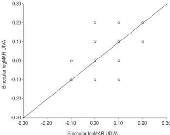
= -0.066, p = 0.514), K2 (r = 0.014, p = 0.887), or anterior chamber depth (r = 0.147, p = 0.144). In contrast, a statisti- cally significant correlation was found between binocular UDVA and UIVA (r = 0.635, p < 0.001) (Fig. 2), as well as between binocular UIVA and UNVA (r = 0.453, p = 0.001).
Preoperative spherical equivalent did not correlate signifi- cantly with any measures of uncorrected postoperative vi-
sual acuity (UDVA: r = 0.169, p = 0.094; UIVA: r = 0.126, p
= 0.210; UNVA: r = -0.027, p = 0.793). Likewise, no signifi- cant correlation was found between preoperative and post- operative spherical equivalents (r = -0.171, p = 0.089). Fur- thermore, no significant correlations were found between postoperative spherical equivalent and visual outcomes at different distances (UDVA: r = -0.155, p = 0.100; UIVA: r
= -0.101, p = 0.317; UNVA: r = 0.156, p = 0.122).
Concerning the level of impact of residual astigmatism on vision, postoperative manifest cylinder did not correlate significantly with postoperative monocular UDVA (r =
-0.058, p = 0.575), UIVA (r = 0.056, p = 0.590), or UNVA (r
= 0.104, p = 0.312). It should be noted that only four eyes (4%) showed a postoperative cylinder greater than 1 D.
Preoperative cylinder showed a poor but significant cor- relation with monocular postoperative UNVA (r = -0.224, p = 0.037), and it showed a correlation with monocular postoperative at the limit of statistical significance UIVA (r
= -0.206, p = 0.05). No significant correlation was found between preoperative cylinder and postoperative monocu- Table 1. Summary of the preoperative and postoperative visual and refractive data in the analyzed sample
Preoperative Postoperative
p-value Mean (range) / median (IQA) Mean (range) / median (IQA)
Monocular logMAR UDVA 0.72 (0.10 to 2.00) / 0.60 (0.70) 0.06 (-0.10 to 0.30) / 0.10 (0.10) <0.001
Binocular logMAR UDVA - 0.04 (-0.10 to 0.20) / 0.00 (0.10) -
Sphere (D) -0.05 (-13.25 to 6.25) / +1.25 (2.25) +0.20 (-0.75 to 1.00) / +0.25 (0.50) <0.001 Cylinder (D) -0.89 (-4.00 to 0.00) / -0.75 (0.50) -0.58 (-1.75 to 0.00) / -0.50 (0.31) 0.001 Spherical equivalent (D) -0.44 (-14.00 to 5.88) / +0.81 (2.66) -0.08 (-1.38 to 0.75) / 0.00 (0.53) 0.001 Monocular logMAR CDVA 0.16 (0.00 to 1.30) / 0.10 (0.30) 0.04 (-0.20 to 0.30) / 0.00 (0.10) <0.001
Monocular logMAR UNVA - 0.06 (-0.10 to 0.30) / 0.05 (0.10) -
Binocular logMAR UNVA - 0.01 (-0.10 to 0.20) / 0.00 (0.19) -
Monocular logMAR UIVA - 0.09 (-0.10 to 0.30) / 0.10 (0.10) -
Binocular logMAR UIVA - 0.04 (-0.10 to 0.20) / 0.00 (0.10) -
IQA = interquartile amplitude; logMAR = logarithm of the minimum angle of resolution; UDVA = uncorrected distance visual acuity; D
= diopter; CDVA = corrected distance visual acuity; UNVA = uncorrected near visual acuity; UIVA = uncorrected intermediate visual acuity.
% of eyes
100 90 80 70 60 50 40 30 20 10
0 UDVA
Monocular Binocular
Visual outcome
UIVA UNVA UDVA UIVA UNVA
20 / 20 or better 20 / 25 or better 20 / 30 or better 20 / 40 or better
Fig. 1. Distribution of monocular and binocular postoperative un- corrected distant, intermediate, and near visual outcomes in the analyzed sample. UDVA = uncorrected distance visual acuity;
UIVA = uncorrected intermediate visual acuity; UNVA = uncor- rected near visual acuity.
0.30 0.20 0.10 0.00 -0.10 -0.20 -0.30
-0.30 -0.20 -0.10 0.00 0.10 0.20 0.30
Binocular logMAR UIVA
Binocular logMAR UDVA
Fig. 2. Scatterplot showing the relationship between postoper- ative binocular logarithm of the minimum angle of resolution (logMAR) uncorrected distance visual acuity (UDVA) and un- corrected intermediate visual acuity (UIVA).
lar UDVA (r = -0.102, p = 0.348).
Ability to perform daily activities
After surgery, a total of 96% of patients were able to perform their daily activities without any problems. Only two patients (4%) reported that they could only partly per- form their daily activities following surgery. Fig. 3 shows the distribution of the answers when patients were asked about difficulty in performing daily activities. No statisti- cally significant differences in binocular logMAR UNVA were found between patients reporting no difficulty or partial difficulty in reading a newspaper (p = 0.290), read- ing a book (p = 0.755), doing computer work (p = 0.084), or doing handicraft work (p = 0.385). A difference near the limit for statistical significance was found in binocular logMAR UIVA between patients reporting no difficulty or partial difficulty in doing computer work (0.01 ± 0.07 vs.
0.06 ± 0.08, p = 0.060). No statistically significant differ- ence in binocular logMAR UDVA was found between pa- tients reporting no difficulty or partial difficulty in driving at night (p = 0.346). When the results were compared based on preoperative refractive status, no statistically sig- nificant differences were found with regard to the difficul- ty levels of performing daily activities evaluated among myopes and hyperopes (p ≥ 0.299).
Spectacle independence
Mean spectacle independence scores (range, 0 to 40) for reading, doing computer work, and for distance were 10.33
± 12.47, 5.71 ± 11.90, and 3.92 ± 9.77, respectively. Binocu- lar UDVA did not correlate significantly with spectacle in- dependence score for reading (r = -0.073, p = 0.619), doing computer work (r = 0.097, p = 0.505), or for distance (r = -0.101, p = 0.484). A similar finding was obtained for bin- ocular UIVA (reading: r = 0.172, p = 0.237; computer work:
r = 0.244, p = 0.091; distance: r = 0.016, p = 0.910) and UNVA (reading: r = 0.005, p = 0.974; computer work: r = 0.247, p = 0.087; distance: r = 0.084, p = 0.561). A total of 43 patients (86%) were able to complete the questionnaire without the use of any additional optical correction. No significant differences among myopes and hyperopes were found in the levels of spectacle independence for reading (p = 0.597), doing computer work (p = 0.947), or for dis- tance (p = 0.265).
Photic phenomena and other visual disturbances The mean intensity scores were: for glare during the day 10.15 ± 9.08 (range, 0 to 30), glare at night 15.15 ± 12.02 (range, 1 to 39), ocular burning or pain 6.51 ± 9.28 (range, 0 to 37), halos 13.34 ± 10.82 (range, 0 to 37), ghost images 4.49 ± 7.92 (range, 0 to 36), visual problems under bright light 10.34 ± 10.36 (range, 0 to 37), visual problems under normal light 4.88 ± 7.73 (range, 0 to 30), visual problems under dim light 13.90 ± 10.58 (range, 0 to 39), irritation from halos in traffic 12.32 ± 11.21 (range, 0 to 39), and irri- tation from halos during daily activities 7.24 ± 8.69 (range, 0 to 35), respectively. No significant correlations between binocular UDVA and the subjective grades of evaluated disturbances were found (glare during the day: r = 0.037, p
= 0.803; glare at night: r = 0.008, p = 0.957; ocular burning or pain: r = -0.022, p = 0.882; halos: r = -0.109, p = 0.166;
ghost images: r = -0.199, p = 0.166; visual problems under bright light: r = -0.133, p = 0.359; visual problems under normal light: r = 0.003, p = 0.981; visual problems under dim light: r = -0.113, p = 0.433; irritation from halos in traf- fic: r = 0.097, p = 0.518; irritation from halos during daily activities: r = -0.191, p = 0.203). Likewise, binocular UIVA (glare during the day: r = 0.045, p = 0.760; glare at night: r
= 0.108, p = 0.469; ocular burning or pain: r = 0.030, p = 0.837; halos: r = -0.013, p = 0.930; ghost images: r = -0.169, p = 0.241; visual problems under bright light: r = -0.003, p
= 0.984; visual problems under normal light: r = 0.113, p = 0.435; visual problems under dim light: r = -0.133, p = 0.356; irritation from halos in traffic: r = 0.108, p = 0.472;
Activity
% of patients
NoPartly YesNo assesible
Read a newspaper Read a bookWatch
TV
Drive a car during dayDrive a car at night Go shopping
Do computer work Do work in house / garden
Do handicraft 10090
8070 6050 4030 2010 0
Fig. 3. Distribution of patient responses regarding difficulty per- forming the following activities (scale: 1 = yes, 2 = partly, 3 = no, and 4 = no assessable): reading the newspaper, reading a book, watching TV, driving a car during the day, driving a car at night, shopping, doing computer work, working in the house or garden, and doing handicraft work.
irritation from halos during daily activities: r = 0.040, p = 0.790) and UNVA (glare during the day: r = 0.182, p = 0.211; glare at night: r = 0.083, p = 0.579; ocular burning or pain: r = -0.043, p = 0.770; halos: r = 0.209, p = 0.150; ghost images: r = -0.141, p = 0.328; visual problems under bright light: r = 0.046, p = 0.749; visual problems under normal light: r = 0.128, p = 0.374; visual problems under dim light:
r = -0.022, p = 0.881; irritation from halos in traffic: r = 0.083, p = 0.578; irritation from halos during daily activi- ties: r = 0.145, p = 0.337) did not correlate with the subjec- tive grade of evaluated symptoms. No significant postop- erative differences in the levels of the different photic phenomena were found between myopes and hyperopes (p
≥ 0.073).
Patient overall satisfaction
A total of 80% of patients reported to be completely happy with the surgical outcome, and 16% reported to be moderately happy. Only two patients (4%) reported to be unhappy with the results of cataract surgery. A total of 44 patients (88%) would choose the same IOL again, and 43 patients (86%) would recommend the surgery and IOL. No significant differences between myopic and hyperopic pa- tients were found with regard to patient satisfaction with the surgery (p = 0.120).
Discussion
The trifocal IOL evaluated in the current study com- bines a bifocal and trifocal diffractive pattern on the ante- rior surface of the lens in order to provide an asymmetrical light distribution generating distant, intermediate, and near foci [3-6].Several studies have confirmed the good perfor- mance of this IOL in terms of visual and refractive out- comes [3-6], but there is still limited evidence on the im- pact of this multifocal IOL on patient satisfaction, spectacle independence, and patient ability to perform common daily activities, such as reading or driving. For this reason, this study has evaluated these aspects by means of a self-developed questionnaire and evaluation of visual and refractive outcomes.
In our sample, mean monocular postoperative logMAR UDVA ranged from -0.10 to 0.30, with a mean value of 0.06. This confirms the efficacy of the evaluated IOL in
successfully restoring distant vision after lensectomy.
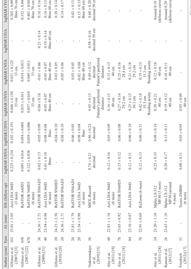
Likewise, an excellent binocular logMAR UDVA was found in our sample, with a mean value of 0.04 and a range from -0.10 to 0.20. Our results are consistent with those re- ported by most authors evaluating the same and other tri- focal IOLs [3-6,9-15]. Table 2 summarizes the visual and refractive outcomes reported by other authors evaluating trifocal IOLs and other types of multifocal IOLs [3,4,6,7,10- 13,15-24]. Somewhat worse UDVA outcomes have been re- ported by Alio et al. [10] and Sheppard et al. [11] and for trifocal IOLs that combine two bifocal diffractive patterns (FineVision IOL from PhysIOL). The studies reported mean postoperative logMAR UDVA values of 0.19 ± 0.09 and 0.18 ± 0.13, respectively. Differences in sample size, patient characteristics, follow-up, and visual acuity testing can explain the differences between the same types of IOL. Voskresenskaya et al. [15] also reported slightly worse postoperative UDVA outcomes with another trifocal IOL model (mean, 0.13) compared to those obtained in our se- ries (Table 2). In comparison with other types of diffrac- tive multifocal IOLs, our results were either comparable [16-22] or better (Table 2)[23]. When our results were com- pared to those obtained with other refractive multifocal IOLs, the superiority of the evaluated diffractive IOL was obvious [7,18,24].This can be easily explained by the great- er amount of higher-order aberrations induced by refrac- tive multifocal IOLs compared to diffractive models [7,24].
Specifically, rotationally asymmetric refractive multifocal IOLs are known to induce significant amounts of primary coma, which can limit the visual acuity achieved with the implant [7,24].
The good uncorrected visual outcome found in our se- ries was consistent with the significant changes in magni- tude of the manifest refraction components. A mean post- operative spherical equivalent of +0.08 D (range, -1.38 to +0.75 D) was found in our series, which is similar to values reported by other authors evaluating the same trifocal IOL [3-6] and other trifocal [9,12-14] and multifocal IOLs (Table 2) [16-23].In contrast, a myopic residual refractive error trend was reported with the trifocal MIOL-Record, which exhibited a mean postoperative spherical equivalent of -0.41 ± 0.49 D [15].This might be due to the use of a non-optimized A-constant for this specific type of trifocal IOL or to the selection of an inadequate refractive target. It should be noted that some residual myopia is intended when monofocal IOLs are implanted; however, this does
Table 2. Comparison of our monocular outcomes to those obtained by previous authors using different multifocal IOL modalities Author (year)EyesAL (mm)IOL (follow-up)logMAR UDVAlogMAR CDVAlogMAR UNVAlogMAR CNVAlogMAR UIVAlogMAR CIVA Alfonso et al. (2007)[23]16225.01 ± 3.05Acri.LISA 366D (3 mon)0.225 ± 0.2340.102 ± 0.1910.048 ± 0.150 33 cm0.031 ± 0.125 33 cm- 0.265 ± 0.099 Bino 70 cm
Alfonso et al. (2007) [22]650-ReSTOR A60D30.095 ± 0.0160.054 ± 0.0050.015 ± 0.0110.014 ± 0.013- 0.352 ± 0.040 670-ReSTOR N60D3 (6 mon)0.122 ± 0.0380.039 ± 0.0060.057 ± 0.010 33 cm0.049 ± 0.011 33 cm-
0.401 ± 0.042 Bino 70 cm
Alfonso et al. (2009)[21]3624.36 ± 2.71ReSTOR SN6AD30.02 ± 0.13-0.05 ± 0.09-0.04 ± 0.18-0.01 ± 0.060.21 ± 0.140.16 ± 0.16 4023.54 ± 0.98Acri.LISA 366D (6 mon)0.01 ± 0.18 Bino-0.08 ± 0.08 Bino
-0.05 ± 0.07 Bino 40 cm -0.05 ± 0.07 Bino 40 cm 0.19 ± 0.14 Bino 60 cm 0.18 ± 0.13 Bino 60 cm
Alfonso et al. (2010) [20]2024.21 ± 2.56ReSTOR SN60D3--0.04 ± 0.10- 0.03 ± 0.05- 0.38 ± 0.14 2024.36 ± 2.71ReSTOR SN6AD3--0.08 ± 0.10--0.05 ± 0.06-0.14 ± 0.17 2023.81 ± 1.79ReSTOR SN6AD1--0.06 ± 0.05-0.03 ± 0.05-0.02 ± 0.13 2023.54 ± 0.98Acri.LISA 366D (6 mon)--0.08 ± 0.04 Bino--0.02 ± 0.08 Bino 40 cm-0.15 ± 0.15 Bino 60 cm Voskresenskaya et al. (2010)
[15]
36-MIOL-Record (6 mon)
0.74 ± 0.21 decimal 0.86 ± 0.23 decimal 0.85 ± 0.13 decimal
Patient’s preferred distance
0.89 ± 0.12 decimal
Patient’s preferred distance
0.58 ± 0.16 decimal 50 cm0.60 ± 0.20 decimal 50 cm Alio et al. (2011) [19]4822.83 ± 1.18Acri.LISA 366D (6 mon)0.12 ± 0.160.03 ± 0.090.16 ± 0.13 40 cm0.13 ± 0.13 40 cm- - Alio et al. (2011) [18]7823.03 ± 0.92ReSTOR SN60D30.15 ± 0.120.06 ± 0.08
0.27 ± 0.14 29.2 cm 0.25 ± 0.16 28.4 cm
- - 8423.10 ± 0.87Acri.LISA 366D0.12 ± 0.110.06 ± 0.10
0.25 ± 0.15 30.1 cm 0.21 ± 0.18 29.2 cm
-- 7022.91 ± 0.69ReZoom (6 mon)0.12 ± 0.130.06 ± 0.11
0.42 ± 0.18 35.3 cm
Reading acuity
0.19 ± 0.15 33.3 cm
Reading acuity
-- Alio et al. (2011) [24]2423.20 ± 1.18Mplus LS-312 MF30 (3 mon)0.25 ± 0.330.09 ± 0.180.30 ± 0.21 40 cm0.12 ± 0.18 40 cm-
Around 0.10 (defocus curve)
Ramon et al. (2012) [7]2623.63 ± 1.18Mplus LS-312 MF30 (around 6 mon)
0.20 ± 0.170.13 ± 0.130.19 ± 0.12 40 cm0.18 ± 0.11 40 cm-
Around 0.29 (defocus curve)
Friedrich (2012)[17]31-Tecnis ZMB00 (6 mon)0.08 ± 0.110.03 ± 0.050.03 ± 0.09 40 cm--- (Continuing)
Table 2. Continued Author (year)EyesAL (mm)IOL (follow-up)logMAR UDVAlogMAR CDVAlogMAR UNVAlogMAR CNVAlogMAR UIVAlogMAR CIVA Cochener et al. (2012) [13]
9423.13 ± 1.17FineVision (6 mon)0.08 ± 0.120.03 ± 0.160.01 ± 0.06 35 cm0.00 ± 0.05 35 cm0.08 ± 0.12 65 cm0.08 ± 0.10 65 cm Vryghem et al. (2013) [12]
5023.70 ± 1.08FineVision (6 mon)0.06 ± 0.090.00 ± 0.080.1
1 ± 0.12 35 cm
0.09 ± 0.12 35 cm0.05 ± 0.19 70 cm0.06 ± 0.19 70 cm Alio et al. (2013) [10]40-FineVision (6 mon)0.18 ± 0.13 0.05 ± 0.06 0.26 ± 0.15 40 cm -0.20 ± 0.11 80 cm - Sheppard et al. (2013) [11]
30-FineVision (2 mon)0.19 ± 0.090.08 ± 0.08---- Schmickler et al. (2013) [16]
104-Tecnis ZMB00 (4 to 6 mon)0.02 ± 0.10-0.05 ± 0.080.15 ± 0.30 Patient’s preferred distance
0.15 ± 0.33 Patient’s preferred distance
- Around 0.22 (defocus curve) Mojzis et al. (2014)[4]60-AT LISA tri 839 MP (6 mon)-0.03 ± 0.09-0.05 ± 0.080.20 ± 0.12 33 cm0.13 ± 0.10 33 cm0.08 ± 0.10 66 cm0.06 ± 0.11 66 cm Law et al. (2014) [6]6020.96 ± 25.88 AT LISA tri 839 MP (6 mon)0.05 ± 0.07-0.02 ± 0.050.16 ± 0.07 40 cm (binocular)
0.12 ± 0.07 40 cm (binocular)
-0.16 ± 0.07 70 cm (binocular) Mojzis et al. (2014)[3]3023.26 ± 1.29AT LISA 8010.00 ± 0.13-0.03 ± 0.110.21 ± 0.120.16 ± 0.110.24 ± 0.160.06 ± 0.10 3023.21 ± 0.90AT LISA tri 839 MP (3 mon)-0.05 ± 0.08-0.06 ± 0.070.07 ± 0.09 33 cm0.06 ± 0.07 33 cm0.03 ± 0.08 80 cm0.03 ± 0.08 80 cm Our study (2015)10023.92 ± 1.29AT LISA tri 839 MP (3 mon)0.06 (-0.10 to 0.30)0.04 (-0.20 to 0.30)0.06 (-0.10 to 0.30) 40 cm
-0.09 (-0.10 to 0.30) 66 cm
- IOL = intraocular lens; AL = axial length; logMAR = logarithm of the minimum angle of resolution; UDVA = uncorrected distance visual acuity; CDVA = corrected distance vi- sual acuity; UNVA = uncorrected near visual acuity; CNVA = corrected near visual acuity; UIVA = uncorrected intermediate visual acuity; CIVA = corrected intermediate visual acuity.
not apply to trifocal IOLs, which are very susceptible to minimal residual refractive error. In our sample, a signifi- cant correlation was found between binocular postopera- tive UDVA and UIVA and also between UIVA and UNVA. Specifically, the worse was the postoperative bin- ocular UDVA, the worse was the postoperative binocular UIVA, and vice versa. This confirms that the presence of residual refractive error significantly contributes to simul- taneous degradation of distant, intermediate, and near vi- sual foci. Furthermore, a trend of worse near visual out- come in cases of higher preoperative astigmatism was found, which confirms the need for a toric trifocal IOL in such cases.
The near visual outcomes obtained in our series were excellent, with 87% of eyes achieving a logMAR UNVA of 0.1 or better, 99% of eyes achieving 0.2 or better, and all eyes achieving 0.3 or better. The mean postoperative log- MAR UNVA was 0.06 when measured at 40 cm. This re- sult is equivalent to that reported by Mojzis et al. [4] with the same type of trifocal IOL (0.07 ± 0.09 measured at 33 cm) and slightly better than the UNVA outcome reported by the same authors in another study [3] (0.20 ± 0.12 mea- sured at 33 cm) and by Law et al. [6] (0.16 ± 0.07 binocu- larly measured at 40 cm). Several factors might account for this discrepancy, such as differences in patient samples (e.g., age, AL), surgical procedure, or examination proto- col. The results of various authors evaluating the FineVi- sion IOL also showed some discrepancies with regard to UNVA outcome. Mean logMAR values of 0.00 ± 0.04 (measured at 35 cm), 0.26 ± 0.15 (measured at 40 cm), 0.11
± 0.12 (measured at 35 cm), and 0.01 ± 0.06 (measured at 35 cm) have been reported by Cochener et al. [9],Alio et al.
[10],Vryghem and Heireman [12], and Cochener et al. [13], respectively. As previously stated, this variability in evalu- ation of the same type of IOL might be attributed to dis- crepancies in clinical protocol and sample selection. In any case, all reported mean values showed that trifocal IOLs are a good option for restoration of near visual function after cataract surgery. With the binary in phase trifocal IOL MIOL-Record, a mean logMAR UNVA of 0.07 was measured at the patient’s preferred distance [15]. In com- parison with bifocal diffractive IOLs, the evaluated trifo- cal IOL provides similar near visual outcomes (Table 2) [16-23]. In contrast, worse UNVA outcomes have been re- ported with rotationally asymmetric refractive multifocal IOLs [7,24]. Ramon et al. [7] and Alio et al. [24] reported
mean UNVA values of 0.19 ± 0.42 (measured at 40 cm) and 0.30 ± 0.21 (measured at 40 cm), respectively. As shown for distant visual outcomes, the increase in high- er-order aberrations with the Mplus IOL limits the near vi- sual acuity that can be achieved [7,24].
The intermediate visual outcomes obtained in our series were also very good, with 79% of eyes achieving a log- MAR UIVA of 0.1 or better, 94% of eyes achieving 0.2 or better, and all eyes achieving 0.3 or better. The mean post- operative logMAR UIVA was 0.09 when measured at 66 cm. Similar UIVA outcomes have been reported by other authors for the same trifocal IOL [3-6] and for other types of trifocal IOLs (Table 1) [9,12-14]. Only a slightly worse mean UIVA (0.20 ± 0.11, measured at 80 cm) was obtained in the study by Alio et al. [10]. As previously stated, differ- ences in sample size, patient characteristics, follow-up, and visual acuity testing might have contributed to this dis- crepancy. In a comparative study of the bifocal and trifocal AT LISA IOLs conducted by Mojzis et al. [3], a significant- ly better UIVA was obtained in the group of eyes implant- ed with the trifocal IOL (bifocal 0.24 ± 0.16 vs. 0.03 ± 0.08, 80 cm, p < 0.01). With regard to the binary in phase trifo- cal MIOL-Record, a poorer UIVA was reported by Voskresenskaya et al. [15] (mean value of approximately 0.20 logMAR measured at 50 cm) when compared to the FineVision and AT LISA trifocal IOLs. As expected, the outcomes reported by different authors evaluating diffrac- tive bifocal IOLs were more limited than those obtained by our research group for the AT LISA trifocal IOL and by other authors for the FineVision and AT LISA trifocal IOLs [3,20-23].
The level of spectacle independence achieved with im- plantation of the evaluated trifocal IOL was high, exhibit- ing low mean spectacle dependence scores. Specifically, the mean spectacle independence scores for reading, doing computer work, and for distance were 10.33 ± 12.47, 5.71 ± 11.90, and 3.92 ± 9.77, respectively. This result is consistent with that obtained by Sheppard et al. [11] using the Near Activity Visual Questionnaire to assess patient satisfaction with uncorrected near vision after cataract surgery and implantation of the trifocal IOL FineVision. The authors found that a good mean Near Activity Visual Question- naire Rasch score for near vision satisfaction (15.9 ± 10.7 logits) [11].In our series, no correlation was found between level of spectacle independence and binocular distant, in- termediate, or near visual outcome. This can be easily ex-
plained, as most patients achieved binocular UDVA, UNVA, and UIVA close to 0.00 logMAR with a lack of significant residual refractive errors. This confirms the ex- cellent performance of the trifocal IOL in terms of visual rehabilitation. Indeed, most patients reported no difficulty in performing tasks such as reading a newspaper, reading a book, driving a car at night, doing computer work, or do- ing handicraft work. Law et al. [6] used a self-developed questionnaire to evaluate the level of difficulty in perform- ing vision-related tasks after cataract surgery with implan- tation of the same trifocal IOL that was evaluated in our series. They found no patients reporting difficulties in watching TV or driving [6].Likewise, as in our study, moderate difficulty was reported by a limited percentage of patients for performing some near and intermediate vi- sual tasks, such as reading the newspaper or working with the computer [6].In our series, 96% of postoperative pa- tients were able to perform their daily activities without any problems, and only two patients reported partial abili- ty in performing their daily activities. As can be expected, a non-significant trend of a more limited binocular UIVA was found in patients reporting partial difficulty in doing computer work. According to these findings, the evaluated trifocal IOL has a positive impact on vision-related daily activities and allows them to be performed with minimal or no difficulty. Alio et al. [8] concluded in a comparative study that patients with bifocal full diffractive IOLs were able to better perform several daily tasks at near and inter- mediate distances compared to patients with apodized multifocal or monofocal IOLs.
Patients also graded the levels of photic phenomena us- ing a subjective scale ranging from 0 (not at all) to 40 (very strong). Mean scores between 5 and 15 were obtained for glare during day, glare at night, halos, ghost images, both- ersome halos in traffic, and bothersome halos during the day. This confirms a low occurrence of such potentially disabling disturbances. Furthermore, the perceptions of photic phenomena or visual disturbances did not correlate with binocular distant, intermediate, or near visual out- come. This seems logical, as the presence of photic phe- nomena with diffractive multifocal IOLs seems to be relat- ed to the diffractive component rather than residual refractive error. According to theoretical simulations, a double-halo formation due to two non-focused powers can be expected with the trifocal IOL evaluated in the current study [25].Law et al. [6] found that the halos associated
with this IOL were well-tolerated, and their perception was reduced over time. Therefore, difficulties associated with perception decreased from 80% at 1 month to 40% at 6 months after IOL implantation. These authors also con- firmed gradual reduction in the difficulties associated with glare perception. The percentage of patients experiencing these difficulties decreased from 73.3% at 1 month to 13.3% at 6 months after surgery [6]. Vryghem and Heire- man [12] investigated the rate of halo perception after im- plantation of the FineVision trifocal IOL and concluded that 68% of patients did not perceive them as significant or disabling. Sheppard et al. [11] used halometry to measure the angular sizes of monocular and binocular photopic scotomas arising from a glare source after FineVision tri- focal IOL implantation and found a mean scotoma size similar to that of previous studies on multifocal and ac- commodating IOLs.
The good visual and refractive outcomes, high level of spectacle independence, and low level of visual distur- bances obtained in our series led to high levels of patient satisfaction. We found that 80% of patients reported com- plete satisfaction with the surgical outcome, and 16% re- ported to be moderately satisfied. Likewise, 88% of pa- tients would choose the same IOL again, and 86% of patients would recommend the surgery and IOL to others.
These results are consistent with those reported for other types of multifocal IOLs. Lubinski et al. [26] found that mean overall patient satisfaction was 9.19 ± 1.20 (scale from 1 to 10, with 10 being the best score) at 6 months af- ter implantation of an aspheric bifocal diffractive IOL.
As with any scientific study, our clinical study has sev- eral limitations that should be acknowledged. First, cor- rected near and intermediate visual acuity were not mea- sured or recorded. These parameters are important for determining the potential interference of residual refrac- tive error on final visual outcome. Another limitation was the use of a non-validated questionnaire to evaluate patient satisfaction, spectacle independence, and photic phenome- na [6].Future studies should be conducted using validated questionnaires in order to assess these parameters. This study can be considered an initial approach in evaluating the impact of AT LISA trifocal IOL implantation on pa- tient daily activities.
In conclusion, cataract surgery with implantation of the AT LISA tri 839 MP IOL provides excellent distant, inter- mediate, and near visual outcomes. It is also associated
with a high level of refractive correction predictability, a minimal level of disturbing photic phenomena, a positive impact on performance of vision-related daily activities, and high levels of postoperative patient satisfaction.
Conflict of Interest
The members of the International Vision Correction and Research Centre receive research grants, travel costs, and lecture honoraria from Carl Zeiss Meditech (Jena, Germa- ny).
Acknowledgements
The study was supported by a research grant from the Klaus Tschira Foundation.
References
1. Gatinel D, Pagnoulle C, Houbrechts Y, Gobin L. Design and qualification of a diffractive trifocal optical profile for intraocular lenses. J Cataract Refract Surg 2011;37:2060-7.
2. Valle P, Oti J, Canales V, Cagigal M. Visual axial PSF of diffractive trifocal lenses. Opt Express 2005;13:2782-92.
3. Mojzis P, Kukuckova L, Majerova K, et al. Comparative analysis of the visual performance after cataract surgery with implantation of a bifocal or trifocal diffractive IOL. J Refract Surg 2014;30:666-72.
4. Mojzis P, Pena-Garcia P, Liehneova I, et al. Outcomes of a new diffractive trifocal intraocular lens. J Cataract Re- fract Surg 2014;40:60-9.
5. Marques EF, Ferreira TB. Comparison of visual outcomes of 2 diffractive trifocal intraocular lenses. J Cataract Re- fract Surg 2015;41:354-63.
6. Law EM, Aggarwal RK, Kasaby H. Clinical outcomes with a new trifocal intraocular lens. Eur J Ophthalmol 2014;24:501-8.
7. Ramon ML, Pinero DP, Perez-Cambrodi RJ. Correlation of visual performance with quality of life and intraocular ab- errometric profile in patients implanted with rotationally asymmetric multifocal IOLs. J Refract Surg 2012;28:93-9.
8. Alio JL, Plaza-Puche AB, Pinero DP, et al. Quality of life evaluation after implantation of 2 multifocal intraocular
lens models and a monofocal model. J Cataract Refract Surg 2011;37:638-48.
9. Cochener B, Vryghem J, Rozot P, et al. Clinical outcomes with a trifocal intraocular lens: a multicenter study. J Re- fract Surg 2014;30:762-8.
10. Alio JL, Montalban R, Pena-Garcia P, et al. Visual out- comes of a trifocal aspheric diffractive intraocular lens with microincision cataract surgery. J Refract Surg 2013;
29:756-61.
11. Sheppard AL, Shah S, Bhatt U, et al. Visual outcomes and subjective experience after bilateral implantation of a new diffractive trifocal intraocular lens. J Cataract Refract Surg 2013;39:343-9.
12. Vryghem JC, Heireman S. Visual performance after the implantation of a new trifocal intraocular lens. Clin Oph- thalmol 2013;7:1957-65.
13. Cochener B, Vryghem J, Rozot P, et al. Visual and refrac- tive outcomes after implantation of a fully diffractive tri- focal lens. Clin Ophthalmol 2012;6:1421-7.
14. Lesieur G. Outcomes after implantation of a trifocal dif- fractive IOL. J Fr Ophtalmol 2012;35:338-42.
15. Voskresenskaya A, Pozdeyeva N, Pashtaev N, et al. Initial results of trifocal diffractive IOL implantation. Graefes Arch Clin Exp Ophthalmol 2010;248:1299-306.
16. Schmickler S, Bautista CP, Goes F, et al. Clinical evalua- tion of a multifocal aspheric diffractive intraocular lens. Br J Ophthalmol 2013;97:1560-4.
17. Friedrich R. Intraocular lens multifocality combined with the compensation for corneal spherical aberration: a new concept of presbyopia-correcting intraocular lens. Case Rep Ophthalmol 2012;3:375-83.
18. Alio JL, Grabner G, Plaza-Puche AB, et al. Postoperative bilateral reading performance with 4 intraocular lens mod- els: six-month results. J Cataract Refract Surg 2011;37:842- 52.
19. Alio JL, Plaza-Puche AB, Pinero DP, et al. Optical analy- sis, reading performance, and quality-of-life evaluation af- ter implantation of a diffractive multifocal intraocular lens.
J Cataract Refract Surg 2011;37:27-37.
20. Alfonso JF, Fernandez-Vega L, Puchades C, Montes-Mico R. Intermediate visual function with different multifocal intraocular lens models. J Cataract Refract Surg 2010;36:
733-9.
21. Alfonso JF, Puchades C, Fernandez-Vega L, et al. Visual acuity comparison of 2 models of bifocal aspheric intraoc- ular lenses. J Cataract Refract Surg 2009;35:672-6.
22. Alfonso JF, Fernandez-Vega L, Baamonde MB, Mon- tes-Mico R. Prospective visual evaluation of apodized dif- fractive intraocular lenses. J Cataract Refract Surg 2007;
33:1235-43.
23. Alfonso JF, Fernandez-Vega L, Senaris A, Montes-Mico R.
Prospective study of the Acri.LISA bifocal intraocular lens.
J Cataract Refract Surg 2007;33:1930-5.
24. Alio JL, Pinero DP, Plaza-Puche AB, Chan MJ. Visual out- comes and optical performance of a monofocal intraocular
lens and a new-generation multifocal intraocular lens. J Cataract Refract Surg 2011;37:241-50.
25. Alba-Bueno F, Vega F, Millan MS. Halos and multifocal intraocular lenses: origin and interpretation. Arch Soc Esp Oftalmol 2014;89:397-404.
26. Lubinski W, Gronkowska-Serafin J, Podboraczynska-Jod- ko K. Clinical outcomes after cataract surgery with im- plantation of the Tecnis ZMB00 multifocal intraocular lens. Med Sci Monit 2014;20:1220-6.