© 2020 The Korean Ophthalmological Society
This is an Open Access article distributed under the terms of the Creative Commons Attribution Non-Commercial License (http://creativecommons.org/licenses /by-nc/3.0/) which permits unrestricted non-commercial use, distribution, and reproduction in any medium, provided the original work is properly cited.
Original Article
Central retinal artery occlusion (CRAO) is an ocular vascular occlusive disease with very poor prognosis. In
previous studies, Varma et al. [1] reported that only 10% of patients with spontaneous reperfusion experienced mean- ingful vision recovery. CRAO is the ocular analogue of ce- rebral stroke. The same atherosclerotic risk factors that predispose to cardiovascular, peripheral vascular, and cere- brovascular disease are present in CRAO, and these must be actively evaluated to prevent further medical comorbid- ities [2]. Effective treatment of CRAO must target acute Purpose: To investigate the clinical manifestations and prognosis of eyes with cilioretinal artery sparing central
retinal artery occlusion (CRAO)
Methods: A retrospective study was conducted on 90 eyes diagnosed with complete CRAO, including 16 cases of cilioretinal artery sparing CRAO. Clinical features, visual outcome, papillomacular bundle involvement, and remnant visual field were analyzed according to cilioretinal artery sparing.
Results: Among eyes with complete CRAO, the proportion of cilioretinal artery sparing CRAO was 17.8% (16 / 90). Mean initial best-corrected visual acuities (BCVAs) (2.04 ± 0.69 vs. 2.34 ± 0.47, p = 0.039) and final BCVAs (1.65 ± 0.87 vs. 2.22 ± 0.84, p = 0.001) were significantly better in eyes of the cilioretinal artery sparing group than the non-sparing group. The proportion with poor visual outcome (final BCVA <20 / 200) was 81.3%
in the cilioretinal artery sparing group and 97.3% in the non-sparing group (p = 0.01). In sub-group analysis within cilioretinal artery sparing CRAO eyes, ischemic involvement of the papillomacular bundle at disease on- set was significantly more frequent in the poor vision group (BCVA <20 / 200, 12 / 13 [92.3%]) than in the good vision group (BCVA ≥20 / 200, 1 / 3 [33.3%], p = 0.016) and it was associated with preserved central visual field.
Conclusions: Although cilioretinal artery sparing is common in CRAO and has a better prognosis than complete CRAO, the visual outcome is generally poor and only a small proportion of eyes has preserved small central visual field. Ischemic injury of the papillomacular bundle at the acute stage of CRAO correlates with poor visu- al outcome and could be a prognostic sign.
Key Words: Ciliary arteries, Optical coherence tomography, Retinal artery occlusion
Received: August 8, 2019 Final revision: September 20, 2019 Accepted: September 25, 2019
Corresponding Author: Se Joon Woo, MD, PhD. Department of Oph- thalmology, Seoul National University Bundang Hospital, 82 Gumi-ro 173beon-gil, Bundang-gu, Seongnam 13620, Korea. Tel: 82-31-787-7377, Fax: 82-31-787-4057, E-mail: [email protected]
Clinical Manifestations and Visual Prognosis of Cilioretinal Artery Sparing Central Retinal Artery Occlusion
Yong Hoon Kim1,2, Kyu Hyung Park1, Se Joon Woo1
1Department of Ophthalmology, Seoul National University Bundang Hospital, Seoul National University College of Medicine, Seongnam, Korea
2Department of Ophthalmology, Seoul National University Hospital, Seoul National University College of Medicine, Seoul, Korea
reperfusion of the CRAO and prevention of ocular compli- cations and further end-organ ischemia [1].
CRAO can be classified into 4 distinct clinical entities:
non-arteritic CRAO, non-arteritic CRAO with cilioretinal artery sparing, arteritic CRAO associated with giant cell arteritis, and transient non-arteritic CRAO [3]. Although the inner half of the sensory retina derives its blood supply from the central retinal artery, in many cases a cilioretinal artery may supply a small portion of the retina around the optic disc. Cilioretinal arteries have been histologically shown to originate from short posterior ciliary arteries and, in rare instances, directly from the choroidal vessels [4]. The temporal cilioretinal artery may spare the fovea in some cases of CRAO. Therefore, cilioretinal artery sparing is important for protecting the macula and preserving good visual prognosis in CRAO [5]. It is generally believed that visual outcome after CRAO is better in the presence of a patent cilioretinal artery, which bypasses the occlusion site in the central retinal artery [4]. However, there have only been several case reports of cilioretinal artery sparing CRAO [6,7]. The incidence, clinical features, and visual outcomes of cilioretinal artery sparing CRAO have not been well studied and knowledge about cilioretinal artery sparing CRAO is currently insufficient.
The purpose of this study was to investigate the clinical manifestations and visual prognosis of cilioretinal artery sparing CRAO.
Materials and Methods
This study was approved by the institutional review board of Seoul National University Bundang Hospital (B- 1905/540-110). Informed consent was waived due to the retrospective nature of the study. Initially, 209 eyes of pa- tients with CRAO patients who visited our Ophthalmology Outpatient Clinic between 1 January 2003 and 31 March 2019 were reviewed. Patients diagnosed as acute non-arte- ritic complete CRAO with a follow-up period of ≥1 month were included in this study as cilioretinal artery sparing was only found in eyes with complete CRAO. Cases with iatrogenic causes (e.g., filler injection, intraocular surger- ies; n = 27), combined ocular pathologies (e.g., proliferative diabetic retinopathy, age-related macular degeneration; n = 29), trauma (n = 3), follow-up period <1 month (n = 21), and incomplete CRAO (n = 39) were excluded. In total, 90
eyes form 90 patients with complete CRAO were finally included for the analysis.
Data pertaining to patient age, sex, and best-corrected visual acuity (BCVA) was obtained. Snellen visual acuity measurements were converted into logarithmic minimum angle of resolution (logMAR) equivalent values for statis- tical analysis. The ophthalmic examination included slit- lamp biomicroscopy, indirect ophthalmoscopy, fundus photography, fluorescein angiography, optical coherence tomography (OCT; Spectralis OCT, Heidelberg Engineer- ing, Heidelberg, Germany), and Goldmann visual field pe- rimetry. Initial OCT images were compared with final OCT images to assess quantitative and qualitative changes in the retinal structure. If patients showed abrupt improve- ment in visual acuity at the follow-up visit, Goldmann vi- sual field and BCVA were carefully re-measured to rule out the possibility of extra-foveal fixation.
The papillomacular bundle is a collection of retinal nerve fibers that carries information from the macula (central reti- na) to the optic nerve. To analyze the inner retinal structure, OCT images of horizontal scans of the foveal center includ- ing the papillomacular bundle area (nasal macula) and the temporal macula were analyzed. With these OCT images, inner retinal structural changes were evaluated by assessing inner retinal thickness, inner retinal hyper-reflectivity, and loss of layer-by-layer integrity. Inner retinal thickening was defined as increased thickness compared with correspond- ing areas of the contralateral unaffected eye. Inner retinal hyper-reflectivity was defined as increased reflectivity com- pared with adjacent normal retinal areas. Loss of lay- er-by-layer integrity was defined as indistinguishable bor- ders or loss of layers due to ischemic injury [8].
All statistical analyses were performed using IBM SPSS Statistics ver. 22 (IBM Corp., Armonk, NY, USA). Fisher’s exact test was used to examine between group differences in non-continuous variables (given unequal sample size).
Mann-Whitney U-test was used for continuous variables, which were expressed as mean ± standard deviation. A p-value of <0.05 was considered statistically significant in all tests.
Results
Of 90 eyes with complete CRAO, 16 eyes showed cilio- retinal artery sparing (17.8%). A representative case with
cilioretinal artery sparing CRAO is shown in Fig. 1A-1D and 2A-2C. Table 1 presents demographic and clinical
characteristics of study subjects (16 cases of cilioretinal ar- tery sparing CRAO and 74 cases of non-sparing complete CRAO). Mean age (67.00 ± 11.34 vs. 63.01 ± 16.09) and sex ratio (male ratio, 56.2% vs. 58.1%) were not different be- tween the 2 groups. Mean initial BCVA and mean final BCVA were significantly different between cilioretinal ar- tery sparing CRAO and non-sparing complete CRAO groups (2.04 ± 0.69 vs. 2.34 ± 0.47, p = 0.039; 1.65 ± 0.87 vs. 2.22 ± 0.84, p = 0.001) (Fig. 3). The proportion with poor visual outcome (final BCVA <20 / 200) was 81.3% in the cilioretinal artery sparing group and 97.3% in the non-sparing group (p = 0.01). Mean initial macular thick- ness was significantly different between groups and was thicker in the non-sparing complete CRAO group (355.19 ± 115.11 vs. 454.86 ± 152.56, p = 0.018). Mean final central macular thickness was greater in the cilioretinal artery sparing group than in the non-sparing group, but without statistical significance (233.73 ± 39.64 vs. 212.56 ± 32.78 µm, p = 0.075). Systemic characteristics associated with CRAO, such as diabetes mellitus, hypertension, stroke, ca- rotid stenosis, and cardiac disease, were not significantly different between the two groups.
Demographic and clinical features of the 16 cilioretinal artery sparing CRAO cases are shown in detail in Table 2.
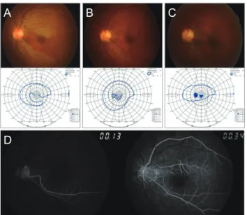
Among all 16 patients, 10 patients had hypertension, three patients had previous stroke history, and six patients had carotid stenosis. Two patients had coronary artery disease, two patients had hyperlipidemia, two patients had angina, and two patients had atrial fibrillation. Seven patients were Fig. 1. Clinical manifestation of cilioretinal artery sparing
central retinal artery occlusion in one patient (fundus photo, Goldmann perimetry) (A) at initial visit, (B) at 1 month, and (C) at final visit. (D) Initial fundus fluorescein angiography reveals central retinal arterial filling delay and arterio-venous transit time delay except the retinal area which is perfused by the cilio- retinal artery (case 8).
A
D
B C
Fig. 2. Fundus photography (left column) and fluorescein angio- graphs (right column) of a patient with cilioretinal artery sparing central retinal artery occlusion (A) at initial examination, (B) at 1 day, and (C) after 1 month. Initial and final visual acuities of this patient were hand motion (case 7).
A
B
C
Fig. 3. Graphs showing initial and final best-corrected visual acuity (BCVA) between eyes with cilioretinal artery sparing and non-sparing central retinal artery occlusion (CRAO). logMAR = logarithmic minimum angle of resolution.
BCVA (logMAR)
1 2 4
3
0
Initial visit Final visit
p = 0.039 p = 0.001
Cilio-retinal artery sparing CRAO Non-sparing CRAO
treated with intra-arterial thrombolysis (one patient failed intra-arterial thrombolysis because of internal carotid ar-
tery obstruction), one patient was treated with panretinal photocoagulation, one patient was treated with intravitreal Table 1. Comparison between demographic and clinical characteristics of cilioretinal artery sparing CRAO and complete CRAO groups
Characteristics Cilioretinal artery sparing CRAO (n = 16) Non-sparing CRAO (n = 74) p-value
Age (yr) 67.00 ± 11.34 63.01 ± 16.09 0.350
Sex, male : female 9 (56.2) : 7 (43.8) 43 (58.1) : 31 (41.9) 0.893
Initial BCVA (logMAR) 2.04 ± 0.69 2.34 ± 0.47 0.039
Final BCVA (logMAR) 1.65 ± 0.87 2.22 ± 0.84 0.001
Initial central macular thickness 355.19 ± 115.11 454.86 ± 152.56 0.018
Final central macular thickness 233.73 ± 39.64 212.56 ± 32.78 0.075
Systemic characteristic
Diabetes mellitus 1 (6.3) 17 (23.0) 0.132
Hypertension 9 (56.3) 41 (55.4) 0.952
Stroke 2 (12.5) 25 (33.8) 0.094
Carotid stenosis 6 (37.5) 12 (16.2) 0.054
Cardiac disease 4 (25.0) 11 (14.9) 0.329
Values are presented as mean ± standard deviation or number (%).
CRAO = central retinal artery occlusion; BCVA = best-corrected visual acuity; logMAR = logarithmic minimum angle of resolution.
Table 2. Demographic and systemic characteristics of cases with cilio-retinal artery sparing central retinal artery occlusion Case Age Sex DM HTN Stroke Carotid stenosis Other risk factors Management Initial VA
(Snellen) Final VA
(Snellen) PMB involvement
1 73 M - + - + Coronary artery disease PRP 0.06 FC +
2 59 M - + - + None IAT HM 0.02 +
3 71 M - - - - Hyperlipidemia None HM HM +
4 69 M - + - + None IAT fail* HM HM +
5 78 M + + - - None Intravitreal
Avastin injection HM HM +
6 74 M - + - + Hyperlipidemia IAT HM HM +
7 67 M - - - - Coronary artery disease IAT FC HM -
8 56 F - + - - None IAT 0.1 0.9 -
9 75 F - + - - Angina None HM 0.02 +
10 80 M - - + + Atrial fibrillation, angina PRP, intravitreal
Avastin injection 0.8 0.5 -
11 70 F - - + + None None NLP NLP +
12 82 F - + - - None None HM 0.04 +
13 69 F - + - - None None HM HM +
14 36 F - - - - None None 0.1 0.4 +
15 63 F - + - - Hyperlipidemia IAT HM 0.08 +
16 60 F - - + - None IAT HM 0.1 +
DM = diabetes mellitus; HTN = hypertension; VA = visual acuity; PMB = papillomacular bundle; PRP = panretinal photocoagulation;
FC = finger count; IAT = intra-arterial thrombolysis; HM = hand motion; NLP = no light perception.
*IAT failed because of internal carotid artery obstruction.
bevacizumab injection, and one patient was treated with combined panretinal photocoagulation and intravitreal bevacizumab injection. Papillomacular bundle involve- ment was observed in 13 eyes (81.3%) with cilioretinal ar- tery sparing CRAO. Table 3 shows comparative analysis between the good vision group (BCVA ≥20 / 200, n = 3) and the poor vision group (BCVA <20 / 200, n = 13) of pa- tients with cilioretinal artery sparing CRAO according to initial BCVA. BCVA of 20 / 200 was set as the criteria to define good or poor visual outcome groups in reference to prior studies on the visual outcome of eyes with CRAO [9].
In a prior prospective study of non-arteritic CRAO with cilioretinal artery sparing, initial visual acuity was grouped as 20/30 or better in 29% of eyes, 20 / 60 to 20 / 100 in 14% of eyes, 20 / 200 in 6% of eyes, counting fin- gers in 20% of eyes, and hand motion in 26% of eyes [10].
Mean age (57.33 ± 22.03 vs. 69.23 ± 7.14, p = 0.103), sex ra- tio (male ratio, 33.3% vs. 61.5%, p = 0.409), initial central macular thickness, and final central macular thickness (357.67 ± 172.02 vs. 354.62 ± 107.84, p = 0.969; 246.67 ± 13.65 vs. 257.85 ± 86.61, p = 0.831) were not significantly different. BCVAs at the initial and final visit were signifi-
cantly different between the two groups (1.13 ± 1.11 vs 2.25
± 0.38, p = 0.006; 0.25 ± 0.18 vs 1.98 ± 0.57, p < 0.001). On Goldmann perimetry, all three (100%) patients of the good vision group showed intact central visual fields (within 5 degrees), and only two (15.4%) patients of the poor vision group showed intact central visual fields (p = 0.002). In analysis of the inner retinal structure, features of the pap- illomacular bundle area on horizontal OCT foveal scans were significantly different in inner retinal thickening and inner retinal hyper-reflectivity between the two groups (1 [33.3%] vs. 12 [92.3%], p = 0.016). Layer-by-layer integrity loss was greater in the poor vision group than in the good vision group, but without statistical significance (1 [33.3%]
vs. 9 [69.2%], p = 0.277). Representative images of OCT features and Goldmann perimetry are shown in Fig. 4A- 4C, 5A-5D, and 6.
Discussion
Our study showed that 17.8% (16 / 90) of complete CRAO showed cilioretinal artery sparing CRAO. Visual Table 3. Comparative analysis between good vision group (initial BCVA ≥20 / 200) and poor vision group (initial BCVA <20 / 200)
Characteristics Good vision group (n = 3) Poor vision group (n = 13) p-value
Age (yr) 57.33 ± 22.03 69.23 ± 7.14 0.103
Sex, male : female 1 (33.3) : 2 (66.7) 8 (61.5) : 5 (38.5) 0.409
Initial BCVA (logMAR) 1.13 ± 1.11 2.25 ± 0.38 0.006
Final BCVA (logMAR) 0.25 ± 0.18 1.98 ± 0.57 <0.001
Initial central macular thickness 357.67 ± 172.02 354.62 ± 107.84 0.969
Final central macular thickness 246.67 ± 13.65 257.85 ± 86.61 0.831
Intact central visual field (<5 degrees) 3 (100) 2 (15.4) 0.002
Types of visual field defect
Central island 2 (66.7) 0 <0.001
Paracentral scotoma 1 (33.3) 1 (7.7) 0.255
Central and cecocentral scotoma 0 1 (7.7) 0.647
Temporal island 0 10 (76.9) 0.010
No visual field 0 1 (7.7) 0.647
Papillomacular bundle area (at initial exam)
Inner retinal thickening 1 (33.3) 12 (92.3) 0.016
Inner retinal hyper-reflectivity 1 (33.3) 12 (92.3) 0.016
Loss of layer-by-layer integrity 1 (33.3) 9 (69.2) 0.277
Values are presented as mean ± standard deviation or number (%).
BCVA = best-corrected visual acuity; logMAR = logarithmic minimum angle of resolution.
outcome was generally poor, and only 18.7% of cases showed VA better than 20/200. Good visual outcome was associated with sparing of the whole papillomacular bun- dle (fovea to disc) from ischemic damage.
Previous studies reported a prevalence of cilioretinal ar- tery sparing CRAO from 14.0% to 26.0% [3]. In normal eyes, the prevalence of one or more cilioretinal arteries has previously been reported to be 49.5% of individuals and 32.1% of eyes [11]. The incidence of cilioretinal artery was 35.0% in all subjects and 18.5% in all eyes in the Han pop- ulation of north China. Men and women have an equal distribution of cilioretinal arteries [5].Although there is no research data from Koreans, we infer from this study that about half of cilioretinal arteries showed impaired perfu- sion in eyes with complete CRAO. This suggests that CRAO is caused by emboli at various locations, such as the central retinal artery only, ophthalmic artery, or multi- ple branches including the central retinal artery and ciliary arteries.
All cilioretinal arteries ran through the temporal side of the central retinal artery trunk and thus, may possibly per- fuse the central fovea. In accordance, initial and final BCVAs and initial central macular thickness were signifi- cantly different between the cilioretinal artery sparing group and the non-sparing complete CRAO group. In our previous study, structural changes including initial inner and outer retinal thickening, baseline macula edema, final retinal thinning, and central macular thickness at initial and final presentation differed significantly according to the severity of retinal ischemia or CRAO stages [12]. The degree of macular edema in the acute phase and retinal thinning at final examination correlated significantly with final visual acuity. Therefore, OCT examination may be useful in evaluating retinal ischemic damage and predict- ing visual prognosis.We previously showed that papillo- macular bundle involvement in branch retinal artery oc- Fig. 4. Representative photographic images of fundus photogra-
phy and images from horizontal foveal scan of spectral-domain optical coherence tomography (OCT). Lines in fundus photogra- phy indicate horizontal OCT scans covering papillomacular bun- dle area. OCT images show morphologic changes to inner retinal layer of papillomacular bundle area (inner retinal thickening, inner retinal hyper-reflectivity [HR], loss of layer-by-layer integ- rity). (A) Case 10, (B) case 5, and (C) case 4.
A
B
C
Fig. 5. (A) Optical coherence tomography images showing inner retinal structural changes in papillomacular bundle area and (B) fundus photography, (C) fluorescein angiography, (D) Goldmann perimetry of eye in good vision group (left column, case 10) and eye in poor vision group (right column, case 2).
A B
C
D
clusion—demonstrated by inner retinal thickening, inner retinal hyper-reflectivity, and loss of layer-by-layer integri- ty on OCT—could be a meaningful indicator of visual acuity prognosis [8]. In the present study, inner retinal thickening and inner retinal hyper-reflectivity of the papil- lomacular bundle were significantly different between the good vision group (BCVA ≥20 / 200) and the poor vision group (BCVA <20 / 200) of patients with cilioretinal ar- tery sparing CRAO. These factors could be prognostic markers of visual acuity and visual field defects. Our study confirmed that papillomacular bundle involvement is im- portant for visual prognosis in CRAO, consistent with our previous study on branch retinal artery occlusion [8].
We recently reported five characteristic types of visual field defects that were associated with CRAO stages: pe- ripheral constriction only, paracentral scotoma, central and cecocentral scotoma, temporal island, and no visual field.
We also reported that worse stages of CRAO, poor base- line BCVA, thick baseline central macular thickness, and poor baseline OCT morphologic features showed statisti- cally significant associations with baseline severe visual field defects [13]. In the good vision group, all patients showed intact central visual field, namely two central is- lands and one paracentral scotoma. But in the poor vision group, there was one paracentral scotoma (7.7%), one cen- tral and cecocentral scotoma (7.7%), 10 temporal islands (76.9%), and one case with no visual field (7.7%). Papillo- macular bundle sparing by cilioretinal artery sparing is as- sociated with central visual field preservation, leading to good visual outcome.
Our study has several limitations including its retrospec- tive nature (possibilities of selection bias) and the small number (n = 16) of patients with cilioretinal artery sparing CRAO. However, there were only several case reports of Fig. 6. Visual field features on final Goldmann perimetry of eyes with cilioretinal artery sparing central retinal occlusion. All eyes in good vision group (best-corrected visual acuity ≥20 / 200) showed preserved central visual fields, but most (84.6%) of the poor vision group (best-corrected visual acuity <20 / 200) had impaired central visual fields.
Good vision group
Poor vision group
cilioretinal artery sparing CRAO in the past and there has been no large-scale clinical study. To our knowledge, our study includes the largest number of cases with cilioretinal artery sparing CRAO to date. In that sense, our study is the first report of the incidence of cilioretinal artery spar- ing CRAO and visual outcomes.
In conclusion, although eyes with cilioretinal artery sparing are common in CRAO and have better prognosis than those with complete CRAO, the visual outcome is generally poor and only a small proportion of cases have preserved small central visual field. Ischemic injury of the papillomacular bundle at the acute stage correlates with poor visual outcome and could be a prognostic factor.
Conflict of Interest
No potential conflict of interest relevant to this article was reported.
Acknowledgements
This research was partly supported by the Bio & Medi- cal Technology Development Program of the National Re- search Foundation (NRF) funded by the Korean govern- ment (MSIT) (No. 2018M3A9B5021319).
References
1. Varma DD, Cugati S, Lee AW, Chen CS. A review of cen- tral retinal artery occlusion: clinical presentation and man- agement. Eye (Lond) 2013;27:688-97.
2. Cugati S, Varma DD, Chen CS, Lee AW. Treatment options
for central retinal artery occlusion. Curr Treat Options Neurol 2013;15:63-77.
3. Hayreh SS. Acute retinal arterial occlusive disorders. Prog Retin Eye Res 2011;30:359-94.
4. Brown GC, Shields JA. Cilioretinal arteries and retinal ar- terial occlusion. Arch Ophthalmol 1979;97:84-92.
5. Liu L, Liu LM, Chen L. Incidence of cilioretinal arteries in Chinese Han population. Int J Ophthalmol 2011;4:323-5.
6. Doguizi S, Sekeroglu MA, Anayol MA, Yilmazbas P. Cen- tral retinal artery occlusion with double cilioretinal artery sparing. Retin Cases Brief Rep 2019;13:75-8.
7. Bansal N, Bansal RK. A middle hyper-reflective band on spectral domain optical coherence tomography in a case of acute nonarteritic central retinal artery occlusion with sparing of cilioretinal artery. Indian J Ophthalmol 2019;67:1345.
8. Cho KH, Ahn SJ, Jung C, et al. Ischemic injury of the pap- illomacular bundle is a predictive marker of poor vision in eyes with branch retinal artery occlusion. Am J Ophthal- mol 2016;162:107-20.
9. Chen CS, Lee AW. Management of acute central retinal ar- tery occlusion. Nat Clin Pract Neurol 2008;4:376-83.
10. Hayreh SS, Zimmerman MB. Central retinal artery occlu- sion: visual outcome. Am J Ophthalmol 2005;140:376-91.
11. Justice J Jr, Lehmann RP. Cilioretinal arteries. A study based on review of stereo fundus photographs and fluores- cein angiographic findings. Arch Ophthalmol 1976;94:1355- 8.
12. Ahn SJ, Woo SJ, Park KH, et al. Retinal and choroidal changes and visual outcome in central retinal artery occlu- sion: an optical coherence tomography study. Am J Oph- thalmol 2015;159:667-76.
13. Kim HM, Park YJ, Park KH, Woo SJ. Visual field defects and changes in central retinal artery occlusion. PLoS One 2019;14:e0209118.