• 검색 결과가 없습니다.

Predisposing factors for external apical root resorption associated with orthodontic treatment

N/A
N/A
Protected

Academic year: 2022

Share "Predisposing factors for external apical root resorption associated with orthodontic treatment"

Copied!
9
0
0

로드 중.... (전체 텍스트 보기)

전체 글

(1)

Predisposing factors for external apical root

resorption associated with orthodontic treatment

Objective: This study aimed to identify possible risk factors for external apical root resorption (EARR) in the maxillary incisors after orthodontic treatment.

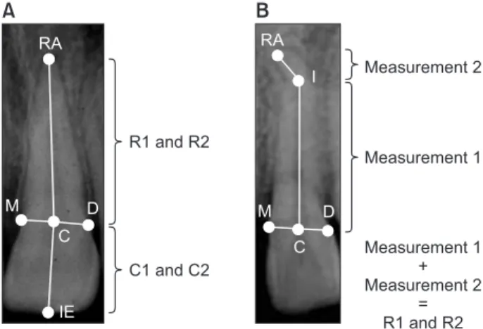
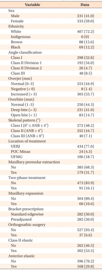
Methods: The root length of 2,173 maxillary incisors was measured on periapical radiographs of 564 patients who received orthodontic treatment.

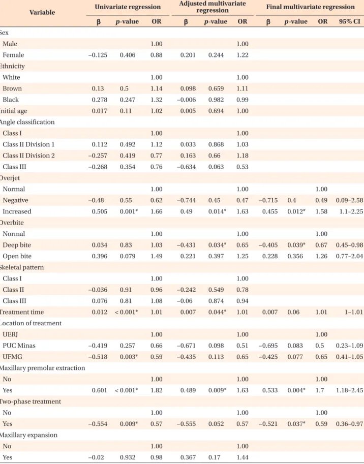
The Kappa test was performed to evaluate intraexaminer and interexaminer reproducibility. Multiple binary logistic regression was used to determine the association between EARR and various factors. Odds ratios and 95% confidence intervals were reported. Results: The risk of developing EARR was 70% higher in orthodontic treatment with maxillary premolar extraction (p = 0.004), 58%

higher in patients with increased overjet (p = 0.012), 41% lower in two-phase orthodontic treatment (p = 0.037), and 33% lower in patients with deep bite (p

= 0.039). The lateral incisors were 54% more likely to develop EARR (p < 0.001), dilacerated roots were 2.26 times more likely to develop EARR (p < 0.001), and for each additional millimeter of root length, the risk of EARR increased by 29%

(p < 0.001). Conclusions: The potential risk factors for EARR after orthodontic treatment included treatment with maxillary premolar extraction, increased overjet at the beginning of treatment, and dilacerated roots.

[Korean J Orthod 2019;49(5):310-318]

Key words: Root resorption, Orthodontic treatment, Incisor Luciana Quintanilha Pires

Fernandes

a

Natália Couto Figueiredo

b

Carina Cristina Montalvany Antonucci

c

Elizabeth Maria Bastos Lages

d

Ildeu Andrade Jr

b

Jonas Capelli Junior

a

a

Department of Orthodontics, School of Dentistry, State University of Rio de Janeiro, Rio de Janeiro, Brazil

b

Department of Orthodontics, School of Dentistry, Pontifical Catholic University of Minas Gerais, Belo Horizonte, Brazil

c

Department of Cell Biology, Institute of Biological Science, Federal University of Minas Gerais, Belo Horizonte, Brazil

d

Department of Orthodontics, School of Dentistry, Federal University of Minas Gerais, Belo Horizonte, Brazil

Received February 21, 2019; Revised May 5, 2019; Accepted May 24, 2019.

Corresponding author: Jonas Capelli Junior.

Chairman, Department of Orthodontics, School of Dentistry, State University of Rio de Janeiro, Boulevard 28 de Setembro, 157, Vila Isabel - Rio de Janeiro, RJ 20551-030, Brazil.

Tel +55-21-2868-8288 e-mail [email protected]

How to cite this article: Fernandes LQP, Figueiredo NC, Antonucci CCM, Lages EMB, Andrade Jr I, Capelli Junior J. Predisposing factors for external apical root resorption associated with orthodontic treatment. Korean J Orthod 2019;49:310-318.

© 2019 The Korean Association of Orthodontists.

This is an Open Access article distributed under the terms of the Creative Commons Attribution Non-Commercial License (http://creativecommons.org/licenses/by-nc/4.0) which permits unrestricted non-commercial use, distribution, and reproduction in any medium, provided the original work is properly cited.

pISSN 2234-7518 • eISSN 2005-372X

https://doi.org/10.4041/kjod.2019.49.5.310

(2)

INTRODUCTION

External apical root resorption (EARR) has been as- sociated with orthodontic treatment.

1-3

Its etiology is multifactorial,

1,2,4-7

and some of the possible orthodontic risk factors evaluated are treatment duration, orthodon- tic forces, extraction treatment, two-phase versus one- phase treatment, maxillary expansion, use of elastics, and bracket prescription. Some malocclusion character- istics were also studied, such as the Angle classification and the severity of overjet and overbite. Many studies have agreed that maxillary incisors are the most fre- quently resorbed teeth,

8-10

and teeth with root shapes other than rhomboid are more affected by EARR.

8,11

Extraction treatment is considered a risk factor for EARR, but the findings of studies on this association have been very controversial.

9,10,12-15

Differences in the evaluated teeth showing EARR, different extraction pat- terns, distinct mechanics, and applied forces for space closure may justify this divergence. However, accord- ing to Brin et al.,

16

two-phase treatment reduces the risk of EARR at the end of orthodontic treatment, since the early modification of growth in patients with Angle Class II malocclusions reduces the severity of overjet, which has been described as a risk factor for EARR. In addition, a pause in the treatment of these patients may contribute to this result, since the disruption of dental movement may favor the cement healing process of the teeth that have developed EARR.

17

Among the malocclusion characteristics, increased overjet has been associated with EARR, since this mal- occlusion presumes a greater root displacement of the maxillary incisors during treatment and has been related to the use of rectangular orthodontic arch wires with active torque. A history of dental trauma has also been described as a risk factor for EARR.

18

Similarly, deep bite is associated with the development of EARR in patients treated with incisor intrusion.

4,19

Although many studies have evaluated the association between these variables and the occurrence of EARR, lit- tle consensus exists about the influence of factors such as root morphology, adjunctive treatments, and maloc- clusion traits on EARR. Differences in methodology and small sample sizes may explain this divergence, given that the division into subgroups is limited. Therefore, the interpretation of results is subject to criticism.

7,8,12,20

Another issue that may explain the difference in results is the sample origin. Sameshima and Sinclair

8

found dif- ferences in the degree of EARR among patients with dif- ferent backgrounds. They suggested that differences in the dental development process might exist because of the differences in tooth size and shape.

Considering the inconclusive literature on the pos- sible risk factors associated with EARR after orthodontic

treatment and the scarcity of research evaluating repre- sentative samples, the current study aimed to identify the possible risk factors for EARR in the maxillary inci- sors after orthodontic treatment.

MATERIALS AND METHODS

This multicenter retrospective study was approved by the Research Ethics Committee of the State Univer- sity of Rio de Janeiro (UERJ) (approval no. 1.605.015).

The sample was selected from three institutions: UERJ, Pontifical Catholic University of Minas Gerais, and Fed- eral University of Minas Gerais, where patients received orthodontic treatment between 1980 and 2016. The maxillary central and lateral incisors were evaluated be- cause of their higher susceptibility to root resorption as reported in the literature.

8-10

Patients were included if they had diagnostically ac- ceptable pretreatment and posttreatment records, in- cluding initial and final periapical radiographs of the maxillary incisors acquired no more than 6 months after the end of active orthodontic treatment. The exclusion criteria were incomplete root formation, dental trauma history, endodontic treatment, presence of restoration at the incisal edge, previous or incomplete orthodontic treatment, and systemic disorders or syndromes.

In total, 2,670 orthodontic records were selected for this study. After applying the inclusion and exclu- sion criteria, the final sample consisted of 564 patients.

Eighty-three incisors were not evaluated because of end- odontic treatment (n = 29), incisal edge restorations (n

= 11), extraction (n = 10), orthodontic traction (n = 13) during treatment, and missing incisor at the beginning of treatment (n = 20). Hence, the final sample consisted of 2,173 maxillary incisors.

Evaluated factors

Two examiners (LQPF and NCF) were previously cali- brated for record evaluation and radiographic measure- ments. The variables evaluated were sex, ethnicity, age, Angle malocclusion classification, overjet, overbite, skeletal pattern, treatment time, location of treatment, treatment with or without extraction of the maxil- lary premolar, one- or two-phase treatment, maxillary expansion, bracket prescription, orthognathic surgery, intermaxillary elastics, and root length and shape. All patients received corrective treatment using 0.022-inch bracket slots. The classification used for evaluating the root shape is shown in Figure 1.

Measurement of external apical root resorption

All periapical radiographs were digitalized using a

scanner with 300-dpi resolution and 256 levels of gray

(Scanjet 4890; Hewlett-Packard, Palo Alto, CA, USA) and

(3)

imported via the ImageJ software (National Institutes of Health, Bethesda, MD, USA) to better visualize and perform digital measurements of the incisors. The length of the radiographic film (40 mm) was used as reference to calibrate the image size. At measurement, both the examiners were double-blinded to the treatment time (initial or final) and patient. The points used for mea- surements are shown in Figure 2.

EARR was calculated according to the formula de- scribed by Linge and Linge,

20

i.e., R1−R2 [C1/C2], with R1 being the root length before treatment; R2, the root length after treatment; C1, the crown length before treatment; and C2, the crown length after treatment.

The crown length (C1 and C2) was defined as the dis- tance from the central point of the cementoenamel junction (point C) to the central point of the incisal edge (point IE). For rhomboid, triangular, and pipette roots, the root length (R1 and R2) was defined as the distance from point C to the root apex (point RA). For dilacer- ated roots, two distances were added to define the root length: the distance from point C to the point of inter- section between the long axis of the tooth, from C, and the long axis of the dilacerated root portion, from point RA (point I), and the distance from point I to point RA.

The correction factor C1/C2 was applied, assuming the crown length to remain unchanged during the observa- tion period. Incisors with EARR ≥ 2 mm were considered affected.

Statistical analysis

All statistical tests were performed using IBM SPSS Statistics for Windows, version 21.0 (IBM Corp., Armonk, NY, USA). Charts and tables were generated using Micro- soft Excel

®

2007 (Microsoft Corporation, Redmond, WA, USA). Descriptive analyses yielded the frequency of cat- egorical variables and the mean with standard deviation

of continuous variables.

A total of 120 teeth were randomly selected to be evaluated two times for EARR, by both examiners, with a 15-day interval between the evaluations. To evalu- ate intraexaminer and interexaminer reproducibility, the Kappa test was applied, and the agreement percentage was calculated.

The relationship between the variables was also ana- lyzed. The occurrence of EARR was evaluated using multiple binary logistic regression modeling to estimate the odds ratios (ORs), in association with the generalized estimation equation to consider more than one tooth per patient. Initially, a univariate regression model was used and adjusted for all variables to eliminate possible confounding factors. The final regression model was adjusted only for the variables that showed a significant OR in the two previous steps, considering a significance level of 5%. In the final model, the 95% confidence in- terval for OR values was estimated.

RESULTS

The descriptive analyses of the sample are shown in Tables 1 and 2. The sample included 1,080 central inci- sors and 1,093 lateral incisors. In total, 810 teeth (37.3%) were affected by EARR ≥ 2 mm, and the lateral incisors Figure 1. Initial root shape classification.

Figure 2. References to the measurement of external api- cal root resorption in rhomboid, triangular, pipette (A), and dilacerated roots (B).

RA, Root apex; M, mesial point of the cementoenamel junction (CEJ); D, distal point of the CEJ; C, central point of the line joining M and D; IE, central point of the incisal edge; I, point of intersection between the long axis of the tooth, from C, and the long axis of the dilacerated root portion, from the RA; R1, the root length before treat- ment; R2, the root length after treatment; C1, the crown length before treatment; C2, the crown length after treatment; Measurement 1, distance from point C to I;

Measurement 2, distance from point I to the RA.

(4)

Table 1. Descriptive analysis of categorical data per patient

Variable Data

Sex

Male 231 (41.0)

Female 333 (59.0)

Ethnicity

White 407 (72.2)

Indigenous 0 (0)

Brown 88 (15.6)

Black 69 (12.2)

Angle classification

Class I 298 (52.8)

Class II Division 1 192 (34.0)

Class II Division 2 26 (4.7)

Class III 48 (8.5)

Overjet (mm)

Normal (0–3) 253 (44.9)

Negative (< 0) 8 (1.4)

Increased (> 3) 303 (53.7)

Overbite (mm)

Normal (1–3) 250 (44.3)

Deep bite (> 3) 231 (41.0)

Open bite (< 1) 83 (14.7)

Skeletal pattern (

o

)

Class I (0

o

≤ ANB ≤ 4

o

) 272 (48.2) Class II (ANB > 4

o

) 252 (44.7) Class III (ANB < 0

o

) 40 (7.1) Location of treatment

UERJ 434 (77.0)

PUC Minas 24 (4.3)

UFMG 106 (18.7)

Maxillary premolar extraction

No 385 (68.3)

Yes 179 (31.7)

Two-phase treatment

No 473 (83.9)

Yes 91 (16.1)

Maxillary expansion

No 504 (89.4)

Yes 60 (10.6)

Bracket prescription

Standard edgewise 282 (50.0)

Preadjusted 282 (50.0)

Orthognathic surgery

No 527 (93.4)

Yes 37 (6.6)

Class II elastic

No 262 (46.5)

Yes 302 (53.5)

Anterior elastic

No 396 (70.2)

Yes 168 (29.8)

Values are presented as number (%).

ANB, A point–nasion–B point; UERJ, State University of Rio de Janeiro; PUC Minas, Pontifical Catholic University of Minas Gerais; UFMG, Federal University of Minas Gerais.

Table 2. Descriptive analysis of continuous data Variable Mean ±

standard

deviation Minimum Maximum Root length (mm) 15.75 ± 2.49 6.43 24.59

Initial age (yr) 15.21 ± 7.39 8 57.1

Treatment time (mo) 53.26 ± 27.12 8 182

Figure 3. Frequency of the central and lateral incisors affected by external apical root resorption.

Figure 4. Frequency of external apical root resorption ac- cording to the initial root shape.

Table 3. Results of the intraexaminer and interexaminer evaluations

Evaluation Observed

agreement (%) Kappa Strength of agreement Intra-examiner

Examiner 1 89.2 0.778 Substantial

Examiner 2 93.3 0.863 Almost perfect

Inter-examiner 86.6 0.727 Substantial

(5)

Table 4. Results of multiple binary logistic regression modeling

Variable Univariate regression Adjusted multivariate

regression Final multivariate regression

β p-value OR β p-value OR β p-value OR 95% CI

Sex

Male 1.00 1.00

Female −0.125 0.406 0.88 0.201 0.244 1.22        

Ethnicity

White 1.00 1.00

Brown 0.13 0.5 1.14 0.098 0.659 1.11

Black 0.278 0.247 1.32 −0.006 0.982 0.99        

Initial age 0.017 0.11 1.02 0.005 0.694 1.00        

Angle classification                    

Class I 1.00 1.00

Class II Division 1 0.112 0.492 1.12 0.033 0.868 1.03

Class II Division 2 −0.257 0.419 0.77 0.163 0.66 1.18

Class III −0.268 0.354 0.76 −0.634 0.063 0.53        

Overjet

Normal 1.00 1.00 1.00

Negative −0.48 0.55 0.62 −0.744 0.45 0.47 −0.715 0.4 0.49 0.09–2.58

Increased 0.505 0.001* 1.66 0.49 0.014* 1.63 0.455 0.012* 1.58 1.1–2.25

Overbite

Normal 1.00 1.00 1.00

Deep bite 0.034 0.83 1.03 −0.431 0.034* 0.65 −0.405 0.039* 0.67 0.45–0.98

Open bite 0.396 0.079 1.49 0.221 0.397 1.25 0.228 0.356 1.26 0.77–2.04

Skeletal pattern

Class I 1.00 1.00

Class II −0.036 0.91 0.96 −0.242 0.549 0.78

Class III 0.076 0.81 1.08 −0.06 0.874 0.94        

Treatment time 0.012 < 0.001* 1.01 0.007 0.044* 1.01 0.007 0.06 1.01 1–1.01

Location of treatment

UERJ 1.00 1.00 1.00

PUC Minas −0.419 0.257 0.66 −0.671 0.098 0.51 −0.695 0.083 0.5 0.23–1.09

UFMG −0.518 0.003* 0.59 −0.435 0.113 0.65 −0.425 0.077 0.65 0.41–1.05

Maxillary premolar extraction                    

No 1.00 1.00 1.00

Yes 0.601 < 0.001* 1.82 0.489 0.009* 1.63 0.533 0.004* 1.7 1.18–2.45

Two-phase treatment

No 1.00 1.00 1.00

Yes −0.554 0.009* 0.57 −0.555 0.052 0.57 −0.521 0.037* 0.59 0.36–0.97

Maxillary expansion

No 1.00 1.00

Yes −0.02 0.932 0.98 0.367 0.17 1.44        

(6)

were the most affected (Figure 3). Out of 1,434 rhom- boid, 388 triangular, 110 pipette, and 241 dilacerated roots, the dilacerated ones were the most affected by EARR (Figure 4). Table 3 shows the Kappa test results, according to the classification suggested by Landis and Koch.

21

Among the clinical characteristics, the final multi- variate regression model showed that patients with in- creased overjet had a 58% greater chance of developing EARR during orthodontic treatment than did patients with normal overjet (p = 0.012), and patients with deep bite were 33% less likely to have EARR than were pa- tients with normal overbite (p = 0.039). Among the variables related to orthodontic treatment, extraction of the maxillary premolars increased the chance of EARR by 70% (p = 0.004). However, patients who underwent two-phase treatment had a 41% lower chance of devel- oping EARR than did patients who underwent single- phase treatment (p = 0.037). The maxillary lateral inci-

sors were 54% more likely to be affected by EARR than were the central incisors (p < 0.001). As for root shape, the incisors with dilacerated roots had a 2.26 times greater chance of developing EARR during orthodontic treatment (p < 0.001), and the risk of EARR increased by 29% for each additional millimeter of root length (p

< 0.001). The other variables evaluated in this study did not present statistically significant ORs (Table 4).

DISCUSSION

The aim of this investigation was to identify possible risk factors for the incidence of EARR in the maxillary incisors after orthodontic treatment. Hence, we used periapical radiographs, which are more reliable in esti- mating EARR than are panoramic films.

22

Cone-beam computed tomography (CBCT) studies could be consid- ered more accurate. However, this examination is not part of the routinely requested orthodontic documenta- Table 4. Continued

Variable Univariate regression Adjusted multivariate

regression Final multivariate regression

β p-value OR β p-value OR β p-value OR 95% CI

Bracket prescription

Standard edgewise 1.00 1.00

Preadjusted −0.196 0.186 0.82 −0.006 0.976 0.99        

Orthognathic surgery

No 1.00 1.00

Yes 0.426 0.187 1.53 0.427 0.293 1.53        

Class II elastic

No 1.00 1.00

Yes 0.206 0.166 1.23 0.167 0.33 1.18        

Anterior elastic

No 1.00 1.00

Yes 0.231 0.154 1.26 –0.087 0.665 0.92        

Incisor

Central incisor 1.00 1.00 1.00

Lateral incisor 0.392 < 0.001* 1.48 0.44 < 0.001* 1.55 0.43 < 0.001* 1.54 1.25–1.83 Root shape

Rhomboid 1.00 1.00 1.00

Triangular 0.581 0.001* 1.79 0.322 0.1 1.38 0.362 0.06 1.44 0.98–2.09

Dilacerated 1.186 < 0.001* 3.27 0.808 < 0.001* 2.24 0.817 < 0.001* 2.26 1.53–3.36

Pipette 0.736 0.003* 2.09 0.46 0.088 1.58 0.537 0.052 1.71 0.99–2.94

Root length 0.221 < 0.001* 1.25 0.266 < 0.001* 1.3 0.253 < 0.001* 1.29 1.2–1.37 OR, Odds ratio; CI, 95% confidence interval; UERJ, State University of Rio de Janeiro; PUC Minas, Pontifical Catholic University of Minas Gerais; UFMG, Federal University of Minas Gerais.

*Statistically significant, p < 0.05.

(7)

tion, and the cost and radiation exposure are greater in CBCT than in conventional two-dimensional examina- tions. The method proposed by Linge and Linge

20

was reported as the most accurate one for evaluating EARR in periapical radiographs,

23

thus supporting its use in this study. Moreover, the cutoff point of 2 mm to determine EARR was methodologically chosen to prevent potential overestimation of the occurrence of EARR because of the limitations of radiographic imaging.

24

On the basis of the literature, our study evaluated the variables related to EARR in patients receiving orthodon- tic treatment. According to the descriptive analysis of the sample, we observed that some variables presented a great discrepancy in the number of participants in each group; therefore, these variables (indigenous ethnicity, Angle Class II Division 2 and Class III, negative overjet, Class III skeletal pattern, maxillary expansion, and or- thognathic surgery) should be evaluated with caution.

The following nine variables were evaluated in the final regression model: overjet, overbite, maxillary pre- molar extraction, two-phase treatment, location and time of treatment, central and lateral incisors, and root length and shape. A greater chance of EARR is observed in patients with increased overjet, thus corroborating the findings of other studies.

8,10,16,25

This malocclusion presumes a greater root displacement of the maxillary incisors during treatment, which explains why it is a high-risk factor for EARR. Regarding overbite, patients with deep bite present a less chance of EARR. Although some studies have already associated the development of EARR with incisor intrusion during the correction of deep bite,

4,19

differences in the mechanics used to cor- rect this malocclusion, which are associated with the force magnitude implemented and the required treat- ment time, can promote different degrees of EARR in these patients.

Our findings show that treatment with maxillary pre- molar extraction is the greatest risk factor for the occur- rence of EARR, which is a controversial conclusion in the literature.

9,10,12-15

Differences in evaluated teeth showing EARR, extraction patterns, and the mechanics and forces applied for space closure may justify this divergence. Ac- cording to Artun et al.,

25

treatment with extractions is a factor that represents a greater amount of orthodontic movement; therefore, it is a risk factor for EARR. Nev- ertheless, in cases of severe crowding, a large retraction of the anterior teeth does not always occur; hence, this could be another possible confounding factor in the studies that evaluated the relationship between orth- odontic treatment with extractions and EARR.

The lowest susceptibility to EARR is observed in pa- tients treated in two phases, and this finding corrobo- rates with those of Brin et al.

16

The reduction of the ini- tially increased overjet in these patients and the interval

between the two phases of treatment, which favors the cement healing process of the teeth that have developed EARR, can justify this finding.

16,17

The location of treatment is not a statistically sig- nificant risk factor for EARR, as reported by Artun et al.

25

All three educational institutions involved in this study have similar orthodontic philosophies, which may explain this finding. Despite the long mean treatment time of this study, it does not present a statistically sig- nificant OR in the final regression model. This finding is not in agreement with those of previous studies report- ing that prolonged treatment time is a risk factor for EARR.

10,12,14-16,26

Considering that this sample comprised patients treated at educational institutions, the recess during the academic calendar could increase the total treatment time; however, this does not necessarily mean that the patients were undergoing active orthodontic treatment the entire time. Moreover, these interruptions of some weeks in active treatment could favor the ce- ment healing process. According to Mehta et al.,

17

60%

of the teeth retained for 6 weeks as a rest period showed anatomical repair, and the remaining 40% showed func- tional repair. A systematic review also concluded that a pause in orthodontic treatment for patients experienc- ing root resorption can reduce the severity of this condi- tion.

27

The lateral incisors are more affected by EARR than are the central incisors, as shown in previous studies.

8,9

In general, these teeth are exposed to greater root dis- placement during correction and improvement of mal- occlusion, function, and smile aesthetics. In addition, root morphology tends to transfer the forces mainly to the root apex, thus promoting EARR in these teeth.

5

We observed that longer roots are more likely to develop EARR, thereby confirming the findings of other stud- ies,

13,25

which reported that the greater the distance from the root apex to the point of orthodontic force applica- tion, the greater the tipping displacement. The incisors with root shapes other than rhomboid are more affected by EARR, especially the dilacerated roots, as reported by other studies.

8,11

Although there is no direct proof, stud- ies have suggested that the deviation in development that caused the abnormal shape may be associated with increased susceptibility to EARR in this region.

8

The present study evaluated 564 patients who received orthodontic treatment at three institutions, and evaluat- ed 19 risk factors for EARR after orthodontic treatment quantified in a total of 2,173 incisors. Many studies with a limited number of participants are found in the literature.

7

However, since sample size may influence the results, one of the advantages of the present study is its larger sample size. The retrospective design and use of radiographs can also be cited as limitations of this work.

In addition, it is important to consider that the inherent

(8)

characteristics of each study population may interfere with the EARR process, since differences in the frequen- cy of EARR as well as in the risk factors associated with EARR among populations have already been described in the literature.

8

Recent studies also reported a genetic predisposition that may explain why one patient is more likely to develop EARR during orthodontic treatment than does another

28,29

; therefore, individual variability should also be considered, even though it has not been evaluated in the present study.

CONCLUSION

On the basis of the current results, we can conclude that the risk of developing EARR ≥ 2 mm in the maxil- lary incisors is 70% higher in patients treated with max- illary premolar extraction; 58% higher in patients with increased overjet at the beginning of treatment; 41%

lower in patients treated in two phases; and greater in the lateral incisors, dilacerated roots, and longer roots.

CONFLICTS OF INTEREST

No potential conflict of interest relevant to this article was reported.

ACKNOWLEDGEMENTS

We would like to especially thank Dr Ronir Rag- gio Luiz for assistance with the statistical tests. This work was supported in part by the Coordenação de Aperfeiçoamento de Pessoal de Nível Superior – Brazil (CAPES) – [Finance Code 001].

REFERENCES

1. Brezniak N, Wasserstein A. Orthodontically induced inflammatory root resorption. Part II: the clinical as- pects. Angle Orthod 2002;72:180-4.

2. Hartsfield JK Jr, Everett ET, Al-Qawasmi RA. Ge- netic factors in external apical root resorption and orthodontic treatment. Crit Rev Oral Biol Med 2004;

15:115-22.

3. Brezniak N, Wasserstein A. Orthodontically induced inflammatory root resorption. Part I: the basic sci- ence aspects. Angle Orthod 2002;72:175-9.

4. Weltman B, Vig KW, Fields HW, Shanker S, Kaizar EE. Root resorption associated with orthodontic tooth movement: a systematic review. Am J Orthod Dentofacial Orthop 2010;137:462-76; discussion 12A.

5. Brezniak N, Wasserstein A. Root resorption after orthodontic treatment: part 2. Literature review. Am J Orthod Dentofacial Orthop 1993;103:138-46.

6. Roscoe MG, Meira JB, Cattaneo PM. Association of orthodontic force system and root resorption: a systematic review. Am J Orthod Dentofacial Orthop 2015;147:610-26.

7. Segal GR, Schiffman PH, Tuncay OC. Meta analysis of the treatment-related factors of external apical root resorption. Orthod Craniofac Res 2004;7:71-8.

8. Sameshima GT, Sinclair PM. Predicting and prevent- ing root resorption: part I. Diagnostic factors. Am J Orthod Dentofacial Orthop 2001;119:505-10.

9. de Freitas JC, Lyra OC, de Alencar AH, Estrela C.

Long-term evaluation of apical root resorption after orthodontic treatment using periapical radiography and cone beam computed tomography. Dental Press J Orthod 2013;18:104-12.

10. Maués CP, do Nascimento RR, Vilella Ode V. Severe root resorption resulting from orthodontic treat- ment: prevalence and risk factors. Dental Press J Orthod 2015;20:52-8.

11. Levander E, Malmgren O, Stenback K. Apical root resorption during orthodontic treatment of patients with multiple aplasia: a study of maxillary incisors.

Eur J Orthod 1998;20:427-34.

12. Picanço GV, de Freitas KM, Cançado RH, Valarelli FP, Picanço PR, Feijão CP. Predisposing factors to severe external root resorption associated to orthodontic treatment. Dental Press J Orthod 2013;18:110-20.

13. Fontana ML, de Souza CM, Bernardino JF, Hoette F, Hoette ML, Thum L, et al. Association analysis of clinical aspects and vitamin D receptor gene poly- morphism with external apical root resorption in orthodontic patients. Am J Orthod Dentofacial Or- thop 2012;142:339-47.

14. Lee YJ, Lee TY. External root resorption during orth- odontic treatment in root-filled teeth and contra- lateral teeth with vital pulp: a clinical study of con- tributing factors. Am J Orthod Dentofacial Orthop 2016;149:84-91.

15. Sharab LY, Morford LA, Dempsey J, Falcão-Alencar G, Mason A, Jacobson E, et al. Genetic and treatment- related risk factors associated with external apical root resorption (EARR) concurrent with orthodontia.

Orthod Craniofac Res 2015;18 Suppl 1:71-82.

16. Brin I, Tulloch JF, Koroluk L, Philips C. External api- cal root resorption in Class II malocclusion: a retro- spective review of 1- versus 2-phase treatment. Am J Orthod Dentofacial Orthop 2003;124:151-6.

17. Mehta SA, Deshmukh SV, Sable RB, Patil AS. Com- parison of 4 and 6 weeks of rest period for repair of root resorption. Prog Orthod 2017;18:18.

18. Linge L, Linge BO. Patient characteristics and treat-

ment variables associated with apical root resorption

during orthodontic treatment. Am J Orthod Dento-

facial Orthop 1991;99:35-43.

(9)

19. Aras I, Tuncer AV. Comparison of anterior and pos- terior mini-implant-assisted maxillary incisor intru- sion: root resorption and treatment efficiency. Angle Orthod 2016;86:746-52.

20. Linge BO, Linge L. Apical root resorption in upper anterior teeth. Eur J Orthod 1983;5:173-83.

21. Landis JR, Koch GG. The measurement of ob- server agreement for categorical data. Biometrics 1977;33:159-74.

22. Sameshima GT, Asgarifar KO. Assessment of root resorption and root shape: periapical vs panoramic films. Angle Orthod 2001;71:185-9.

23. Brezniak N, Goren S, Zoizner R, Dinbar A, Arad A, Wasserstein A, et al. A comparison of three methods to accurately measure root length. Angle Orthod 2004;74:786-91.

24. Bastos Lages EM, Drummond AF, Pretti H, Costa FO, Lages EJ, Gontijo AI, et al. Association of functional gene polymorphism IL-1beta in patients with exter- nal apical root resorption. Am J Orthod Dentofacial Orthop 2009;136:542-6.

25. Artun J, Van 't Hullenaar R, Doppel D, Kuijpers-

Jagtman AM. Identification of orthodontic patients at risk of severe apical root resorption. Am J Orthod Dentofacial Orthop 2009;135:448-55.

26. Jiang F, Chen J, Kula K, Gu H, Du Y, Eckert G.

Root resorptions associated with canine retrac- tion treatment. Am J Orthod Dentofacial Orthop 2017;152:348-54.

27. Currell SD, Liaw A, Blackmore Grant PD, Esterman A, Nimmo A. Orthodontic mechanotherapies and their influence on external root resorption: a sys- tematic review. Am J Orthod Dentofacial Orthop 2019;155:313-29.

28. Aminoshariae A, Aminoshariae A, Valiathan M, Kulild JC. Association of genetic polymorphism and external apical root resorption. Angle Orthod 2016;86:1042-9.

29. Nowrin SA, Jaafar S, Ab Rahman N, Basri R, Alam

MK, Shahid F. Association between genetic poly-

morphisms and external apical root resorption: a

systematic review and meta-analysis. Korean J Or-

thod 2018;48:395-404.

수치

Figure 2. References to the measurement of external api- api-cal root resorption in rhomboid, triangular, pipette (A),  and dilacerated roots (B)
Figure 3. Frequency of the central and lateral incisors affected  by external apical root resorption.
Table 4. Results of multiple binary logistic regression modeling

참조

관련 문서