Original Article
Intraocular Lens Power Calculation Using IOLMaster and Various Formulas in Short Eyes
Young Rae Roh 1 , Sang Mok Lee 1-3 , Young Keun Han 1,2 , Mee Kum Kim 1 , Won Ryang Wee 1 , Jin Hak Lee 1,4
1
Department of Ophthalmology, Seoul National University College of Medicine, Seoul, Korea
2
Department of Ophthalmology, Seoul National University Boramae Hospital, Seoul, Korea
3
Department of Ophthalmology, The Armed Forces Capital Hospital, Seongnam, Korea
4
Department of Ophthalmology, Seoul National University Bundang Hospital, Seongnam, Korea
Purpose: To evaluate the predictability of intraocular lens (IOL) power calculations using the IOLMaster and four dif- ferent IOL power calculation formulas (Haigis, Hoffer Q, SRK II, and SRK/T) for cataract surgery in eyes with a short axial length (AL).
Methods: The present study was a retrospective comparative analysis which included 25 eyes with an AL shorter than 22.0 mm that underwent uneventful phacoemulsification with IOL implantation from July 2007 to December 2008 at Seoul National University Boramae Hospital. Preoperative AL and keratometric power were measured by the IOLMaster, and power of the implanted IOL was determined using Haigis, Hoffer Q, SRK II, and SRK/T formulas. Postoperative refractive errors two months after surgery were measured using automatic refracto-kera- tometry (Nidek) and were compared with the predicted postoperative power. The mean absolute error (MAE) was defined as the average of the absolute value of the difference between actual and predicted spherical equiv- alences of postoperative refractive error.
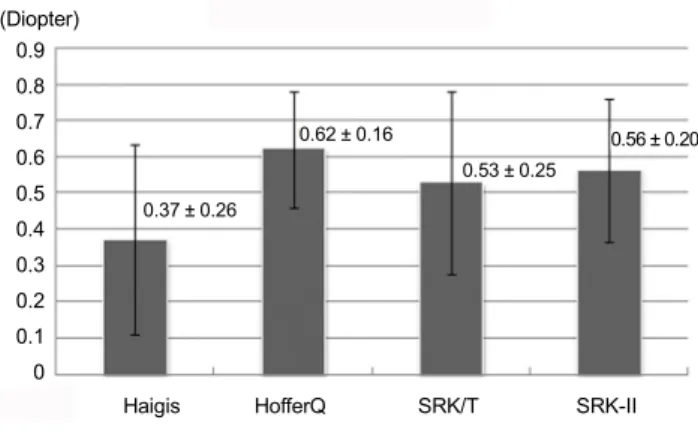
Results: The MAE was smallest with the Haigis formula (0.37 ± 0.26 diopter [D]), followed by those of SRK/T (0.53
± 0.25 D), SRK II (0.56 ± 0.20 D), and Hoffer Q (0.62 ± 0.16 D) in 25 eyes with an AL shorter than 22.0 mm. The proportion with an absolute error (AE) of less than 1 D was greatest in the Haigis formula (96%), followed by those in the SRK II (88%), SRK-T (84%), and Hoffer Q (80%).
Conclusions: The MAE was less than 0.7 D and the proportion of AE less than 1 D was more than 80% in all formulas. The IOL power calculation using the Haigis formula showed the best results for postoperative power prediction in short eyes.
Key Words: Intraocular lens power calculation, IOLMaster, Short eyes
ⓒ2011 The Korean Ophthalmological Society
This is an Open Access article distributed under the terms of the Creative Commons Attribution Non-Commercial License (http://creativecommons.org/licenses /by-nc/3.0/) which permits unrestricted non-commercial use, distribution, and reproduction in any medium, provided the original work is properly cited.
Received: June 22, 2010 Accepted: January 6, 2011
Corresponding Author: Sang Mok Lee, MD. Department of Ophthalmology, The Armed Forces Capital Hospital, San 13-4, Yul-dong, Bundang-gu, Seongnam 463-040, Korea. Tel: 82-16-270-8773, Fax: 82-31-706-0987, E-mail: [email protected]
* This study was previously presented in part as a poster at The American Society of Cataract and Refractive Surgery, Symposium and Congress, San Francisco, California, USA, April 2009.
Accuracy of intraocular lens (IOL) power calculation in cataract surgery is a very important factor associated with postoperative patient satisfaction [1-3]. With the recent de- velopment of multifocal IOL and an accommodative IOL, the accuracy of refractive errors after cataract surgery has
been emphasized [4].
IOL power is predicted preoperatively using several differ- ent formulas and parameters. Measuring errors in these pa- rameters and in the formulas constitute the sources of re- fractive error [5]. Imprecision in measurement of anterior chamber depth (ACD), axial length (AL), and corneal power contribute to 42%, 36% and 22% of errors, respectively [5].
The measurement of AL with partial coherence inter- ferometry (IOLMaster; Carl Zeiss Meditech Inc., Dublin, CA, USA) has been shown to produce significantly more precise IOL power calculation and refractive outcome in cat- aract surgery, thereby avoiding possible compression of the eye with applanation A-scan ultrasound and difficulty with immersion A-scan ultrasound in AL measurements [6,7].
The source of error for postoperative refractive state pre-
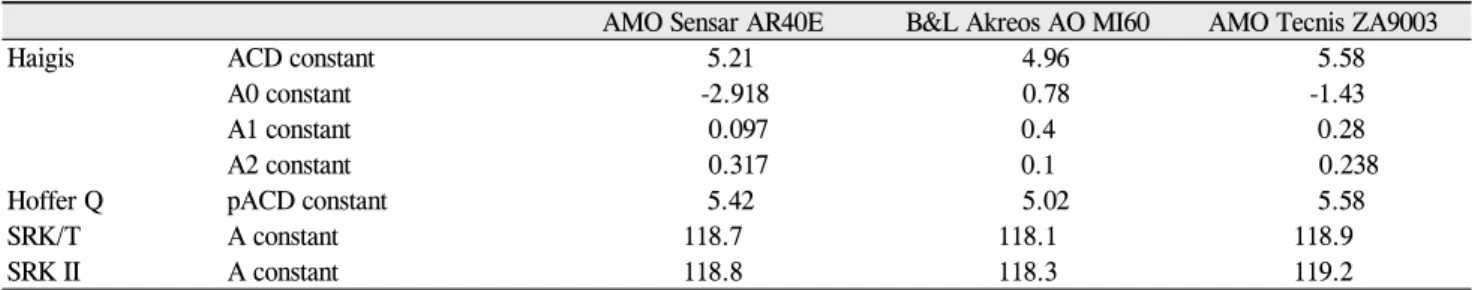
Table 1. The constants used in the four formulas of the IOLMaster in three intraocular lens (IOL) subtypes
AMO Sensar AR40E B&L Akreos AO MI60 AMO Tecnis ZA9003
Haigis ACD constant 5.21 4.96 5.58
A0 constant -2.918 0.78 -1.43
A1 constant 0.097 0.4 0.28
A2 constant 0.317 0.1 0.238
Hoffer Q pACD constant 5.42 5.02 5.58
SRK/T A constant 118.7 118.1 118.9
SRK II A constant 118.8 118.3 119.2
ACD = anterior chamber depth.
diction is due in part to the chosen formula, particularly in eyes with very long or very short AL [8,9].
The Haigis formula incorporated in the IOLMaster pre- dicts effective lens position with improved ACD prediction algorithms and has shown more accurate IOL power pre- diction results, even in extreme eyes [8-10]. However, few reports have compared the accuracies of the various IOL power formulas for cataract surgery using partial coherence interferometry for eyes of short AL less than 22.0 mm.
The purpose of the present study was to evaluate the pre- dictability of IOL power calculations using the IOLMaster and four different IOL power calculation formulas (Haigis, Hoffer Q, SRK II, and SRK/T) for cataract surgery in eyes with a short AL less than 22.0 mm.
Materials and Methods
The present study was a retrospective comparative analy- sis which included 25 eyes from 17 patients with an AL shorter than 22.0 mm and that underwent uneventful phacoe- mulsification with IOL implantation from July 2007 to December 2008. Preoperative AL, keratometric power, and ACD were measured by the IOLMaster version 3.01.0294.
The power of the implanted IOL was determined using Haigis, Hoffer Q, SRK II, and SRK/T formulas calculated by the IOLMaster software. Postoperative refractive errors two months after cataract surgery were measured using automatic refracto-keratometry (RKT-7700; Nidek, Hiroishi, Japan) and were compared with the predicted postoperative power.
The mean absolute error (MAE) was defined as the average of the absolute value of the differences between the actual and predicted spherical equivalences (SE) of the postoperative refractive error.
Cataract surgery was performed by two surgeons (YKH and SML). Topical anesthesia with proparacaine hydro- chloride (Alcaine; Alcon Labs, Fort Worth, TX, USA) or subtenon anesthesia with 3% lidocaine was administered pri- or to the operation. A clear corneal incision 2.75 mm in width was made using a microkeratome at the superior or temporal cornea according to the axis of astigmatism, and phacoe- mulsification was performed after continuous curvilinear capsulorhexis. Three types of IOLs were used in the present study, Sensar ® (AR40e; Abbott Medical Optics, Los Angeles,
CA, USA; five eyes in three patients), Akreos-AO ® (MI60;
Bausch & Lomb, Rochester, NY, USA; ten eyes in seven pa- tients), and Tecnis ® (ZA9003, Abbott Medical Optics; ten eyes in seven patients); IOL was selected based only on the operation date and was not influenced by any other factors.
Cases were excluded if a posterior capsular rupture oc- curred during cataract surgery, if the IOL was inserted into the sulcus, or if the AL could not be measured using the IOLMaster. Also excluded from the present study were pa- tients who could not be observed for at least two months after surgery.
The differences in the MAE according to the four IOL cal- culation formulas in the three IOL groups were analyzed.
Furthermore, the proportions with absolute errors (AE) less than 0.5 diopters (D) and 1 D of the four IOL calculation for- mulas were estimated.
SPSS ver. 15.0 (SPSS Inc., Chicago, IL, USA) was used for statistical analysis. The Mann-Whitney U-test was used to compare differences in the AEs of the formulas. The ANOVA test was used for comparison of the AEs of the for- mulas according to the type of IOL. A statistically significant difference was defined as a p-value <0.05.
Results
A total of 25 eyes from 17 patients were included in the present study. One patient (one eye) was male and 16 patients (24 eyes) were female. The mean age was 70.6 ± 5.5 years (range, 61 to 80 years), and the mean follow-up period was 53.40 ± 15.71 months. The mean AL was 21.60 ± 0.41 mm (range, 20.41 to 21.94 mm). The ACD was 2.70 ± 0.36 mm (range, 2.07 to 3.34 mm).
The constants applied in the four formulas of the IOLMaster
in three IOL subtypes are shown in Table 1. The MAE was
smallest in the Haigis formula (0.37 ± 0.26 D), followed by
those of the SRK/T (0.53 ± 0.25 D), SRK II (0.56 ± 0.20 D),
and Hoffer Q (0.62 ± 0.16 D) formulas (Fig. 1). The pro-
portion of AE less than 0.5 D was greatest in the Haigis for-
mula (76%), followed by those in the SRK II (60%), SRK-T
(60%), and Hoffer Q (48%) formulas. Additionally, the pro-
portion of AE less than 1 D was greatest in the Haigis formula
(96%), followed by those in the SRK II (88%), SRK-T
(84%), and Hoffer Q (80%) formulas (Fig. 2). No statistically
(Diopter)
Haigis HofferQ SRK/T SRK-II (Formulas) 0.9
0.8 0.7 0.6 0.5 0.4 0.3 0.2 0.1 0
0.37 ± 0.26
0.62 ± 0.16
0.53 ± 0.25
0.56 ± 0.20
Fig. 1. Means and standard deviations of the absolute errors the four intraocular lens calculation formulas.
0 20 40 60 80 100 (Proportions of patients, %) (Formulas)
SRK-II
SRK/T
Hoffer Q
Haigis
AE<0.5 D 0.5 D ≤ AE <1.0 D 1.0 D ≤ AE
Fig. 2. Proportion of the absolute errors (AE) less than 1 diopter (D) according to the intraocular lens formulas.
Table 2. Discrepancies in the absolute error in different intraocular lens power calculation formulas
p-value *
Haigis vs. Hoffer Q 0.103
Haigis vs. SRK/T 0.347
Haigis vs. SRK-II 0.028
†Hoffer Q vs. SRK/T 0.415
Hoffer Q vs. SRK-II 0.727
SRK/T vs. SRK-II 0.382
*
The numbers given in the table indicate the p-values for the differences between AE calculated with different IOL power calculation formulas (Mann-Whitney U-test).
†