Article Info
Received June 28, 2021 Revised July 18, 2021 Accepted July 19, 2021 Corresponding Author Oh-yun Kwon
E-mail: [email protected] https://orcid.org/0000-0002-9699-768X
Key Words Movement Rotation
Serratus posterior inferior
Background: The serratus posterior inferior (SPI) muscle originates from the spinous pro- cess of T11-L2 and inserts at the lower border of the 9–12th ribs. This muscle is involved in thoracolumbar rotation and stability. Several positions can be used to improve trunk stability;
the quadruped position is a good position for easily maintaining a neutral spine. In particular, during one arm lifting, various muscles act to maintain a neutral trunk position, and the SPI is one of these muscles. If trunk stability is weakened, uncontrolled trunk rotation may occur at this time. Tape can be used to increase trunk stability. There have been no studies on the effect of taping applied to the SPI muscle on thoracolumbar junction (TLJ) stability.
Objects: This study compared the TLJ rotation angle between three different conditions (without taping, transverse taping, and SPI muscle direction taping).
Methods: Thirty subjects were recruited to the study (18 males and 12 females). The TLJ rota- tion angle was measured during one arm lifting in a quadruped position (ALQP). Two taping methods (transverse and SPI muscle direction taping) were applied, and the TLJ rotation angle was measured in the same movement.
Results: SPI muscle direction taping significantly reduced TLJ rotation compared to that with- out taping (p < 0.001) and with transverse taping (p < 0.001). There was a significant dif- ference in the TLJ rotation angle between transverse taping and SPI muscle direction taping (p < 0.017).
Conclusion: SPI muscle direction taping reduces the TLJ rotation angle during ALQP. There- fore, SPI muscle direction taping is one method to improve TLJ stability and reduce uncon- trolled TLJ rotation during ALQP.
INTRODUCTION
The thoracolumbar junction (TLJ) is a section from the tho- racic spine to the lumbar spine. The facet joints of the tho- racic vertebrae that are frontally oriented are changed to the lumbar vertebrae in the sagittal direction [1]. During rotation, the thoracic vertebrae are restricted by the ribs, and the upper lumbar vertebrae are restricted by facet joints that are sagittally oriented [1,2]. Therefore, trunk rotation occurs more frequently in the thoracic spine than in the lumbar spine, and the TLJ is in a relatively unstable portion [3-5]. Several authors have emphasized the stability of the TLJ to protect against injury or
fracture [6,7]. The stability of the spine can be enhanced by intrinsic supports, such as muscle isometric contraction, and extrinsic supports, such as belts or taping [8-10]. Taping is an extrinsic support device that is a cheap and convenient meth- od for improving stability and muscle function [11]. Taping can provide biomechanical support by improving neuromuscular facilitation and proprioception of the affected joint [12-14]. It can also increase specific joint stability according to the direc- tion of tape attachment [15]. When taping is applied parallel to the muscle fiber, it can facilitate muscle function underneath the tape; when applied perpendicular to the muscle fiber, it can inhibit muscle action [16-18]. Taping is divided into elastic
Copyright ⓒ Korean Research Society of Physical Therapy
This is an Open Access article distributed under the terms of the Creative Commons Attribution Non-Commercial License (http://creativecommons.org/licenses/by-nc/4.0) which permits unrestricted non-commercial use, distribution, and reproduction in any medium, provided the original work is properly cited.
Physical Therapy Korea
PTK https://doi.org/10.12674/ptk.2021.28.3.227 pISSN: 1225-8962 eISSN: 2287-982X Phys Ther Korea. 2021;28(3):227-234
Original Article
Immediate Effect of Serratus Posterior Inferior Muscle Direction Taping on Thoracolumbar Junction Rotation Angle During One Arm Lifting in the Quadruped Position
Nu-ri Kim
1,2, BPT, PT, Sun-hee Ahn
2,3, PhD, PT, Gyeong-tae Gwak
2,3, PhD, PT, Hwa-ik Yoo
1,2, BPT, PT, Oh-yun Kwon
2,3, PhD, PT
1
Department of Physical Therapy, The Graduate School, Yonsei University,
2Laboratory of Kinetic Ergocise Based on Movement Analysis,
3
Department of Physical Therapy, College of Health Science, Yonsei University, Wonju, Korea
and non-elastic taping (NET). NET has stronger fixation than elastic taping because of its material properties [19]. Previous studies have investigated the benefits of NET for stability of the spine to motion, such as trunk flexion and extension; however, no study has numerically identified the effect of NET on trunk rotation [9,19,20]. Kang et al. [9] found that NET applied to the lumbar region provided lumbar stability and increased the range of motion of the hip during lumbar flexion. Kim et al. [12]
reported that NETs in the lumbar spine promote propriocep- tor awareness, provide spinal stiffness, prevent lumbar flexion, and maintain neutral trunk posture when sitting.
Trunk stability is improved in various positions, such as the double leg bridge in hook-lying, front plank, and alternat- ing opposite arm-leg lifting in a quadruped position [21]. In particular, the quadruped position, in which hands and knees are placed on the table, can easily maintain a neutral spine position; therefore, it is recommended as an early stage of re- habilitation exercises [22-24]. In addition, trunk stability can be strengthened through various movements, such as lifting one arm or alternating opposite arm-leg lifting in a quadruped position [25-27]. When trunk rotational torque is generated during one arm lifting, various muscles maintain a neutral trunk position [28]. However, if trunk stability is insufficient to maintain the spine in a neutral position during arm lifting in a quadruped position (ALQP), uncontrolled movement, such as TLJ rotation, may occur. Uncontrolled movement is a dysfunc- tion of movement that may result from muscle weakness [29].
This can lead to tissue stress, spinal loading, and pain [29].
Therefore, ALQP is a good test for assessing trunk stability [30].
The trunk rotation muscles of the back include the latis- simus dorsi (LD), multifidus, longissimus, iliocostalis, rotatores breves and longi, and serratus posterior inferior (SPI) [28,31,32].
These muscles are located from the superficial to the deep lay- er of the back, respectively. Superficial muscles are primarily responsible for movement, while deep muscles are responsible for body stability [33,34]. There are some studies on the effect of taping applied to the erector spinae or multifidus located in deeper layers, but there are no studies on the effect of taping applied to the SPI on trunk stability [35,36]. The SPI, which is directed from the spinous processes of T11-L2 to the lower border of the 9–12th ribs, can lead to TLJ rotation by generat- ing rotational torque [37]. In addition, the SPI is located in a deeper layer, passes through the TLJ, stabilizing the rib cage [38]. Therefore, we designed this study to identify whether SPI
muscle direction taping can reduce uncontrolled TLJ rotation by improving stability during ALQP. If SPI muscle direction taping demonstrates an effect on reducing uncontrolled TLJ ro- tation, it may provide useful information for the management of TLJ instability.
Therefore, the purpose of this study was to compare the TLJ rotation angle between three different conditions (without tap- ing, transverse taping, and SPI muscle direction taping). We hypothesized that SPI muscle direction taping would improve TLJ stability during ALQP.
MATERIALS AND METHODS
1. Subjects
For this cross-sectional study and repeated measures, 30 subjects (18 males and 12 females) participated in this study (Table 1). A total of subjects were volunteers and were recruited from advertisements. Based on pilot data (average trunk rota- tion angle between the two taping methods) gathered from five subjects, G*power software (ver. 3.1; University of Trier, Trier, Germany) was used to calculate the sample size of 21 or more needed to achieve a power of 0.80 and an effect size of 0.608 with an α level of 0.05. We used a post-hoc analysis to inves- tigate the actual power of the sample size. When 30 subjects were recruited, post-hoc analysis indicated an effect size of 0.998. Subjects were included if they met the following criteria:
(1) no history of low back pain, and (2) as per Comerford, dur- ing one arm lifting at 150° in a quadruped position, thoracic rotation was observed [29]. The exclusion criteria were as fol- lows: (1) history of injury to or surgery in the upper extremity;
(2) neurological, psychiatric, or musculoskeletal disorders; (3) allergic to tape [12,16,39]. The research protocol was approved by Yonsei University, Mirae Institutional Review Board (ap- proval NO. 1041849-202101-BM-002-01). Before this study, the subjects were explained the study process and signed an informed consent form.
Table 1.
Table 1. Subject characteristics
Variables Subjects
Age (y) 29.0 ± 8.2
Height (cm) 170.8 ± 7.0
Mass (kg) 70.0 ± 13.5
Values are presented as mean ± standard deviation.
2. Instrumentation
1) Rotation measurement system
Kinematic data of the rotation were measured using a Smart KEMA motion sensor (KOREATECH Co., Ltd., Seoul, Korea) (Figure 1A). The motion sensor contained a tri-axillar gyro- scope, magnetometer, accelerometer, signal converter, and signal transmission sensor. The data from the motion sensor were recorded at sampling frequencies of 25 Hz and transmit- ted to an Android tablet using Smart KEMA software (KORE- ATECH Co., Ltd.) [40-42]. For the measurement, the motion sensor was attached to the holder of the rotation measurement system (RMS) (Figure 1B) [43]. We used the RMS to measure the TLJ rotation in the transverse plane during ALQP. During the operation, the motion sensor indicated a rotation angle in the y-axis of the transverse plane.
3. Taping Methods
A 20-cm long 3M Soft Cloth Tape (5 cm × 10 m; Daemyung Co., Ltd., Yangju, Korea) was used for taping [44]. Two dif- ferent NET methods were applied to reduce uncontrolled TLJ rotation during ALQP. The two taping methods were transverse
and SPI muscle direction taping. Transverse taping was ap- plied to the TLJ (Figure 2A). The SPI muscle direction taping was applied in parallel to the SPI muscle fiber direction (from the lower border of the 9–12th ribs to the spinous processes of T11-L2) of the lifting arm side (Figure 2B). We applied taping in two layers to increase fixation.
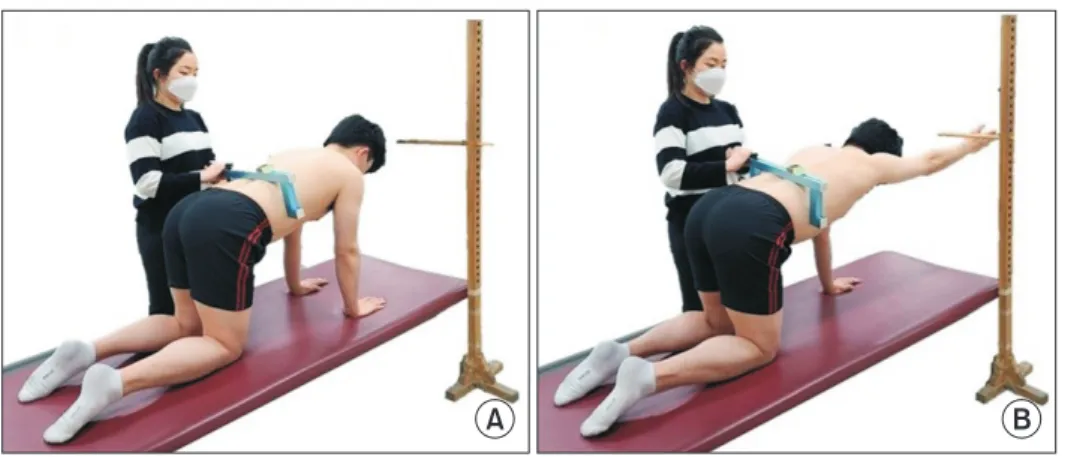
4. Procedures
First, the subjects took a quadruped position with their hands under the shoulder and knees under the hip joint (Figure 3A). A target bar was placed on one individual arm, and the height was adjusted to 180° of shoulder flexion [39]. Before measurement, the RMS attached to the motion sensor was placed on the floor and calibrated to 0°. Next, the bars of the RMS were adjusted to the width of the TLJ for each subject. We then identified that the angle of TLJ rotation was 0° through the motion sensor. Finally, the subjects lifted one arm. When their arm touched the target bar, they held this position for three seconds (Figure 3B). By holding the test position, the motion sensor data were recorded at the TLJ rotation angle and represented numerically. Before testing, the trunk rota-
A B
Figure 2.
Figure 2. Taping methods. (A) Transverse
taping, and (B) right serratus posterior in- ferior muscle direction taping.
Bar
A B
Motion sensor
Figure 1.
Figure 1. Instruments of thoracolumbar
joint rotation angle measurement.
tion angle during ALQP was measured on both sides, and we determined the side with a greater TLJ rotation angle as the test side. Two taping methods were applied in a random order because the taping method sequence may affect TLJ rota- tion during ALQP. The order of the two taping methods was randomized using an online site (https://www.random.org/
sequences). In addition, we provided 5 minutes of rest between taping methods to prevent a learning effect.
5. Statistical Analysis
The data were analyzed using SPSS (version 25.0; IBM Co., Armonk, NY, USA), and the Kolmogorov –Smirnov test was performed to determine whether the data were normally dis- tributed. The intraclass correlation coefficients (ICCs) (3,1) and 95% confidence intervals (CIs) were used to identify the intra- rater reliability of the three trunk rotation angle measurements using the Smart KEMA motion sensor. Data are presented as mean ± standard deviation. One-way analysis of variance with repeated measurements was used to compare the TLJ rotation angle between the three different conditions (without taping, transverse taping, SPI muscle direction taping). The signifi- cance level was set at p < 0.05. If a significant effect was found, Bonferroni correction was used with a statistical significance level of α = 0.05/3 = 0.017.
RESULTS
The ICCs for measuring the TLJ rotation angle were 0.974 (95% CI, 0.946 –0.987) without taping, 0.973 (95% CI, 0.944–
0.987) for transverse taping, and 0.988 (95% CI, 0.975–0.994) for SPI muscle direction taping (Table 2).
Table 3 shows the values and standard deviations of the TLJ rotation angle under three different conditions. Three different
conditions had a significant effect on the rotation angle of the TLJ (Table 3). Transverse taping (12.29° ± 4.35°) did not signif- icantly reduce the TLJ rotation angle during ALQP compared to without taping (13.34° ± 4.83°) (p > 0.017; Figure 4). The SPI muscle direction taping method (8.18° ± 4.00°) showed a sig- nificant effect on reducing the TLJ rotation angle during ALQP compared to without taping (13.34° ± 4.83°) (p < 0.017; Figure 4). In addition, the reduction of the TLJ rotation angle when SPI muscle direction taping was applied was significantly more effective than that with transverse taping (p < 0.017; Figure 4).
DISCUSSION
We investigated the immediate effect of two taping methods on uncontrolled TLJ rotation during ALQP. We found that SPI muscle direction taping was effective for reducing uncontrolled TLJ rotation angle and was more effective than transverse tap- ing. There are some possible reasons for these results.
NET can increase joint stability through a fixation force.
During ALQP, the trunk rotates in the direction of the lifting arm side because the supporting force is removed. In the case of taping attachment, SPI muscle direction taping was applied along the direction of the SPI muscle fibers on the lifting arm side, and transverse taping was applied from the lifting arm side to the opposite side. If a subject had more TLJ rotation on the right during ALQP, the SPI muscle direction taping was
A B Figure 3.
Figure 3. Measurement of thoracolumbarjunction rotation angle.
Table 2.
Table 2. ICCs and 95% CIs of three taping conditions
Taping conditions ICC (95% CI) p-value
Without taping 0.97 (0.95–0.99) < 0.001
Transverse taping 0.97 (0.94–0.99) < 0.001
SPI muscle direction taping 0.99 (0.98–0.99) < 0.001
ICC, intraclass correlation coefficient; CI, confidence interval; SPI, ser-
ratus posterior inferior.
along the right SPI muscle fibers (from the spinous processes of T11-L2 to the lower border of the 9 –12th ribs), and the transverse taping was applied from right to left. By applying taping in a relatively oblique direction, the SPI muscle direc- tion taping may reduce uncontrolled TLJ rotation due to the mechanical effect of counter rotation through the L2 –9th rib direction in contrast to transverse taping.
In addition, NET can increase joint stability by improving proprioception to control joint alignment [19,45,46]. When TLJ rotation occurs, a neutral position is maintained by stabiliz- ers and rotators, such as the LD, multifidus, and SPI [47]. This is consistent with the fact that if taping is applied parallel to the muscle fibers, it can facilitate muscle function [16-18]. SPI muscle direction taping may facilitate the SPI muscle, which is below the tape. In addition, uncontrolled TLJ rotation was reduced by promoting the LD, which is located close to the SPI muscle direction taping. The LD originates from the spinous process of T7-L5, thoracolumbar fascia, and iliac crest and in- serts in the intertubercular groove of the humerus. In addition, it is located on the surface of the TLJ. Vleeming et al. [5] dem- onstrated that the posterior oblique sling is connected in the following order: gluteus maximus, thoracolumbar fascia, and contralateral LD. During ALQP, a coupled force is generated on the LD, which is the lifting side and gluteus maximus on the opposite side. This may increase TLJ stability during ALQP.
An important point of this study is that we identified a re- duction in uncontrolled TLJ rotation angle with SPI muscle direction taping during ALQP. This study demonstrated that NET applied in the direction of SPI muscle fibers may be used to reduce uncontrolled TLJ rotation and increase TLJ stability during ALQP. In particular, SPI muscle direction taping signifi- cantly reduced uncontrolled TLJ rotation relative to that with transverse taping. Therefore, SPI muscle direction taping could be used to correct uncontrolled TLJ rotation angle during ALQP for TLJ stability exercise in reducing uncontrolled TLJ rotation.
This study had some limitations. The main limitation was the lack of electromyography to measure SPI activation. Therefore, further studies are needed to determine SPI muscle activation using needle electromyography to identify the effect of SPI muscle direction taping. Second, all subjects in this study were relatively young and healthy. The findings of this study might be different if the subjects had low back pain, since patients with low back pain have reduced ability to stabilize the spine than healthy subjects [48]. These results also cannot be gener- alized to older people. Further studies involving older people should be conducted.
CONCLUSIONS
We identified that the NET applied to SPI muscle fibers was effective in reducing the TLJ rotation angle during ALQP.
Therefore, SPI muscle direction taping can reduce uncontrolled TLJ rotation and improve TLJ stability during ALQP. It has po- tential for clinical use as a method to improve TLJ stability.
CONFLICTS OF INTEREST
No potential conflict of interest relevant to this article was reported.
AUTHOR CONTRIBUTIONS
Concepualization: NK, GG, OK. Data curation: NK, OK. For- mal analysis: NK, SA, GG, HY, OK. Funding acquisition: NK,
TLJrotationangle()
Without taping 20
15
10
5
0
Transverse taping
SPI muscle direction taping
* *
Figure 4.
Figure 4. Comparison of thoracolumbar junction (TLJ) rotation angle be-
tween three different taping conditions. SPI, serratus posterior inferior.
Table 3.
Table 3. TLJ rotation angle during one arm lifting in the quadruped position