수정체안에서 유리체절제술 및 동반술기가 백내장의 진행에 미치는 영향
:
수정체 보존 유리체절제술을 시행받은 환자에서 유리체절제술 및 동반술기가 백내장의 발생 및 진행에 미치는 영향에 대해 알아보고자 하였다.:
수정체안에서 평면부 유리체절제술을 시행한 후 백내장이 발생 또는 진행되어 백내장적출술(초음파유화 술 또는 낭외적출술)을 시행하고 최소 3개월 이상 추적관찰이 가능했던 47명 48안을 대상으로 하였으며 평균 경과관 찰기간은 10.5개월이었다. 유리체절제술전, 백내장의 발생 또는 진행이 진단된 시기 및 백내장수술전의 수정체혼탁 정 도를 LOCS Ⅲ 분류방식에 따라 비교하였으며 연령, 원인질환, 수술방법 및 동반술기에 따른 유리체절제술후 백내장수 술까지의 기간을 후향적으로 분석하였다.:
유리체절제술전의 수정체혼탁의 평균값은 핵경화가 0.31, 후낭하혼탁이 0.10이었다. 이후 경과관찰 기간중에 백내장의 발생 또는 진행이 진단된 시기는 평균 2.1개월이었으며 이때의 수정체혼탁 정도는 핵경화가 1.29, 후낭하혼 탁이 0.69이었고 백내장수술전 검사상 측정된 수정체혼탁 정도는 핵경화가 3.44, 후낭하혼탁이 2.00이었다. 또한 유리 체절제술후 백내장수술까지의 기간은 평균 11.3개월이었고, 이 가운데 유리체절제술 도중 가스충전술(C3F8 또는 SF6)을 시행한 군(16안)에서는 평균 10.3개월, 실리콘기름을 충전한 군(16안)에서는 7.7개월로 가스나 실리콘기름을 충전하지 않은 군(16안)의 15.8개월 비해 유의하게 짧은 기간을 나타내었다(P=0.003).:
유리체절제술을 시행한 후 발생 또는 진행하는 백내장은 핵경화가 가장 뚜렷이 나타났으며 유리체절제술 및 동 반술기로 가스 또는 실리콘기름을 충전한 군에서 가스나 실리콘기름을 충전하지 않은 군에 비해 유리체절제술 후 백내 장수술까지의 기간이 유의하게 짧았다.<한안지 44(1):53-59, 2003>
Table 1. Procedures combined with vitrectomy.
Combined procedures (n=48) No (%) Endolaser photocoagulation
Membrane peeling
Gas (C3F8 or SF6) tamponade Silicone oil tamponade Cryotherapy
Scleral buckling
34 (70.8) 34 (70.8) 16 (33.3) 16 (33.3) 8 (16.7) 5 (10.4)
Table 4. Lens opacity by LOCS .
Preoperative examination of vitrectomy
Diagnosis of progression of cataract
Preoperative examination of cataract surgery Nuclear color/opalescence
Cortical opacity
Posterior subcapsular opacity
0.31 0.83 0.10
1.29 1.06 0.69
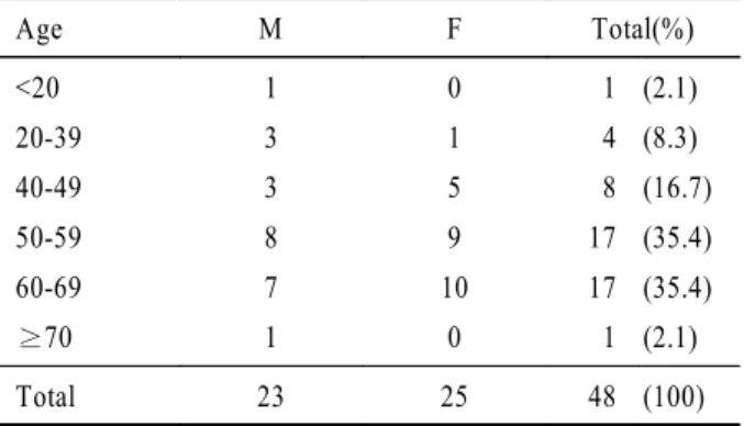
3.44 1.46 2.00 Table 2. Age & Sex Distribution.
Age M F Total(%)
<20 20-39 40-49 50-59 60-69 70
1 3 3 8 7 1
0 1 5 9 10
0
1 4 8 17 17 1
(2.1) (8.3) (16.7) (35.4) (35.4) (2.1)
Total 23 25 48 (100)
Table 3. Etiologic Diseases for Previous Vitrectomy.
Causative disease No (%)
Diabetic retinopathy RRD
BRVO
Other retinal detachment Macular hole
Macular pucker ARMD Others
18 (37.5) 7 (14.6) 6 (12.5) 4 (8.3) 4 (8.3) 3 (6.3) 3 (6.3) 3 (6.3)
Total 48 (100)
RRD : Rhegmatogenous retinal detachment
BRVO : Branched retinal vein occlusion
ARMD : Age-related macular degeneration
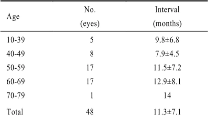
Table 7. Interval between vitrectomy and cataract surgery according to patient’s age.
Age No.
(eyes)
Interval (months) 10-39
40-49 50-59 60-69 70-79
5 8 17 17 1
9.8±6.8 7.9±4.5 11.5±7.2 12.9±8.1
14
Total 48 11.3±7.1
T-test (under 50 vs over 50), P=0.11
Table 9. Interval between vitrectomy and cataract surgery according to operation procedure.
Procedure No.
(eyes)
Interval
(months) P-value PPV
PPCV MP
§only
MP & Gas tamponade 39 9 5 4
10.6±6.7 14.2±8.4 18.6±8.6 8.8±4.4
0.19
0.05
Mann-Whitney test
PPV : Pars plana vitrectomy PPCV : Pars plana core vitrectomy
§
MP : Membrane peeling Table 8. Interval between vitrectomy and cataract surgery
according to combined procedures.
Combined Procedure No.
(eyes)
Interval (months) Gas tamponade
Silicone oil tamponade None
16 16 16
10.3±5.3 7.7±4.6 15.8±8.2 Oneway ANOVA, p=0.003
Table 6. Interval between vitrectomy and cataract surgery according to associated diseases.
Causative diseases No.
(eyes)
Interval (months) Diabetic retinopathy
RRD BRVO
Other retinal detachment Macular hole
Macular pucker ARMD
18 7 6 4 4 3 3
10.3±7.3 9.73±6.7 9.7±6.9 8.0±4.7 11.3±5.1 19.3±11.9 12.7±5.5 RRD : Rhegmatogenous retinal detachment
BRVO : Branched retinal vein occlusion ARMD : Age-related macular degeneration
Table 5. Interval between vitrectomy and cataract surgery.
Interval (months) No of eyes (%) 12
13 24 25 36
30 (62.5)
17 (35.4)
1 (2.1)
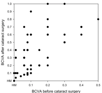
Figure 1. Best corrected visual acuity (BCVA) before and after cataract surgery. Points above the line represent an improvement in visual acuity postoperatively; points below the line, deterioration; and on the line, no change.
BCVA before cataract surgery
HM 0.1 0.2 0.3 0.4 0.5
BCVA after cataract surgery
HM 0.1 0.2 0.3 0.4 0.5 0.6 0.7 0.8 0.9 1.0
1) , , .
. 1998;39:2406-11.
2) , . .
1998;39:3024-8.
3) , , .
. 1999;40:437-44.
4) , .
, .
1999;40:2205-11.
5) , , .
. . 2000;41:2375-80.
6) Schachat AP, Oyakawa RT, Michels RG, Rice TA.
Complications of vitreous surgery for diabetic retinopathy.
. Postoperative complications. Ophthalmol 1983;90:522-30.
7) Hutton WL, Pesicka GA, Fuller DG. Cataract extraction in the diabetic eye after vitrectomy. Am J Ophthalmol 1987; 104:1-4.
8) Cherfan GM, Michels RG, de Bustros S, et al. Nuclear sclerotic cataract after vitrectomy for idiopathic epiretinal membranes causing macular pucker. Am J Ophthalmol 1991;
111:434-8.
9) Novak MA, Rice TA, Michels RG, Auer C. The crystalline lens after vitrectomy for diabetic retinopathy. Ophthalmol 1984;91:1480-4.
10) de Bustros S, Thompson JT, Michels RG, et al. Nuclear sclerosis after vitrectomy for idiopathic epiretinal mem- branes. Am J Ophthalmol 1998;105:160-4.
11) Hsuan JD, Brown NA, Bron AJ, et al. Posterior subcapsular and nuclear cataract after vitrectomy. J Cataract Refract Surg 2001;27:437-44.
12) Blankenship GW, Machemer R. Long-term diabetic vitrec- tomy results. Report of 10 year follow-up. Ophthalmol 1985;92:503-6.
13) Federman JL, Schubert HD. Complications associated with the use of silicone oil in 150 eyes after retina-vitreous surgery. Ophthalmol 1988;95:870-6.
14) Borislav D. Cataract after silicone oil implantation. Doc Ophthalmol 1993;83:79-82.
15) Baer RM, Aylward WG, Leaver PK. Cataract extraction following vitrectomy and silicone oil tamponade. Eye 1995;
9:309-12.
16) Franks WA, Leaver PK. Removal of silicone oil: rewards and penalties. Eye 1991;5:333-7.
17) Poliner LS, Olk RJ, Grand MG, et al. Surgical management of premacular fibroplasia. Arch Ophthalmol 1988;106:761-4.
18) Pesin SR, Olk RJ, Grand MG, et al. Vitrectomy for premacular fibroplasia. Prognostic factors, long-term follow- up, and time course of visual improvement. Ophthalmol 1991;98:1109-14.
19) Thompson JT, Glaser BM, Sjaarda RN, Murphy RP.
Progression of nuclear sclerosis and long-term visual results of vitrectomy with transforming growth factor beta-2 for macular holes. Am J Ophthalmol 1995;119:48-54.
20) Ogura Y, Takanashi T, Ishigooka H, Ogino N. Quantitative analysis of lens changes after vitrectomy by fluoro- photometry. Am J Ophthalmol 1991;111:179-83.
21) Michels RG. Vitrectomy for macular pucker. Ophthalmol 1984;91:1384-8.
22) Blodi BA, Paluska SA. Cataract after vitrectomy in young patients. Ophthalmol 1997;104:1092-5.
23) Margherio RR, Cox MS, Trese MT, Murphy PL, Johnson J, Minor LA. Removal of epimacular membranes. Ophthalmol 1985;92:1075-83.
24) Blankenship G, Cortez R, Machemer R. The lens and pars plana vitrectomy for diabetic retinopathy complications. Arch Ophthalmol 1979;97:1263-7.
25) Haimann MH, Abrams GW. Prevention of lens opacification
during diabetic vitrectomy. Ophthalmol 1984;91:116-21.
26) Saunders DC, Brown A, Jones NP. Extracapsular cataract extraction after vitrectomy. J Cataract Refract Surg 1996;
22:218-21.
27) , , .
. 1999;40:738-43.
28) , , , .
. 1999;40:2481-7.
29) Smiddy WE, Stark WJ, Michels RG, et al. Cataract extraction after vitrectomy. Ophthalmol 1987;94:483-7.
30) McDermott ML, Puklin JE, Abrams GW, Eliott D.
Phacoemulsification for cataract following pars plana vitrec- tomy. Ophthalmic Surg Lasers 1997;28:558-64.
31) Pinter SM, Sugar A. Phacoemulsification in eyes with past pars plana vitrectomy: case-control study. J Cataract Refract Surg 1999;25:556-61.
32) Grusha YO, Masket S, Miller KM. Phacoemulsification and lens implantation after pars plana vitrectomy. Ophthalmol 1998;105:287-94.
33) Melberg NS, Thomas MA. Nuclear sclerotic cataract after vitrectomy in patients younger than 50 years of age. Oph- thalmol 1995;102:1466-71.
34) Kang YH, Lee JH. Phacoemulsification and posterior chamber intraocular lens implantation after scleral buckling, vitrectomy, or both. Ophthalmic Surg Lasers 1998;29:23-7.
35) , , .
. 2002;43:814-8.
The Effect of Vitrectomy and its Combined Procedures on the Progression of Cataract in Phakic Eyes
Hyun-Duck Lee, M.D., Byung-Kyu Kim, M.D., Kwang-Soo Kim, M.D.
Department of Ophthalmology Keimyung University School of Medicine